. 2010 Dec 21;8(12):e1000576.
doi: 10.1371/journal.pbio.1000576.

Phosphorylation of mouse immunity-related GTPase (IRG) resistance proteins is an evasion strategy for virulent Toxoplasma gondii

Affiliations

Phosphorylation of mouse immunity-related GTPase (IRG) resistance proteins is an evasion strategy for virulent Toxoplasma gondii

Tobias Steinfeldt et al. PLoS Biol. .

Erratum in

Abstract

Virulence of complex pathogens in mammals is generally determined by multiple components of the pathogen interacting with the functional complexity and multiple layering of the mammalian immune system. It is most unusual for the resistance of a mammalian host to be overcome by the defeat of a single defence mechanism. In this study we uncover and analyse just such a case at the molecular level, involving the widespread intracellular protozoan pathogen Toxoplasma gondii and one of its most important natural hosts, the house mouse (Mus musculus). Natural polymorphism in virulence of Eurasian T. gondii strains for mice has been correlated in genetic screens with the expression of polymorphic rhoptry kinases (ROP kinases) secreted into the host cell during infection. We show that the molecular targets of the virulent allelic form of ROP18 kinase are members of a family of cellular GTPases, the interferon-inducible IRG (immunity-related GTPase) proteins, known from earlier work to be essential resistance factors in mice against avirulent strains of T. gondii. Virulent T. gondii strain ROP18 kinase phosphorylates several mouse IRG proteins. We show that the parasite kinase phosphorylates host Irga6 at two threonines in the nucleotide-binding domain, biochemically inactivating the GTPase and inhibiting its accumulation and action at the T. gondii parasitophorous vacuole membrane. Our analysis identifies the conformationally active switch I region of the GTP-binding site as an Achilles' heel of the IRG protein pathogen-resistance mechanism. The polymorphism of ROP18 in natural T. gondii populations indicates the existence of a dynamic, rapidly evolving ecological relationship between parasite virulence factors and host resistance factors. This system should be unusually fruitful for analysis at both ecological and molecular levels since both T. gondii and the mouse are widespread and abundant in the wild and are well-established model species with excellent analytical tools available.


Conflict of interest statement

The authors have declared that no competing interests exist.

Figures

Figure 1
Figure 1. Irga6 is phosphorylated by type I virulent T. gondii.
(A) A novel phosphorylated Irga6 band (black arrowhead) was immunoprecipitated with the monoclonal anti-Irga6 antibody, 10E7, from RH-YFP-infected, IFNγ-induced mouse L929 cells metabolically labelled with 35S-methionine/cysteine or 33P-phosphoric acid. Nonphosphorylated Irga6 is indicated with an open arrowhead. The figure is a montage from two segments of a single autoradiogram, joined as indicated by black vertical line. (B) Only virulent T. gondii strains phosphorylate Irga6. MEFs were induced with IFNγ, infected with virulent type I and avirulent type II and type III strains, and labelled with 33P-phosphoric acid. A single phosphorylated band (black arrowhead) was immunoprecipitated with 10E7 only from cells infected with type I strains. The figure is a montage of segments from several autoradiograms, joined as indicated by the black vertical lines. Phosphorylated Irga6 from cells infected with RH-YFP served as positive control for all segments (not shown). (C) The electrophoretically more slowly migrating phosphorylated band (black arrowhead), immunoprecipitated with anti-Irga6 165 antiserum from IFNγ-induced L929 cells infected with virulent strain RH-YFP, is Irga6 and not a coprecipitated protein (Western blot with 10E7). Only the faster band of Irga6 is immunoprecipitated from cells infected with avirulent ME49 strain (open arrowhead). (D) Both Irga6 bands were immunoprecipitated with 10E7 from RH-YFP-infected IFNγ-induced L929 cells labelled with 3H-myristic acid (autoradiogram). In the absence of infection only the lower myristoylated band is seen. Black arrowhead: myristoylated phosphorylated Irga6 (myr-(p)Irga6). Open arrowhead: myristoylated Irga6 (myr-Irga6).
Figure 2
Figure 2. Irga6 is phosphorylated at T102 and T108 in the switch I loop.
(A) MALDI-TOF spectra of tryptic digests of immunoprecipitated phosphorylated (upper profile) and nonphosphorylated (lower profile) Irga6 from IFNγ-induced, RH-infected MEFs. The partial spectra on the left are focused on the m/z 2176.1, corresponding to the nonphosphorylated tryptic Irga6 peptide G91IGNEEEGAAKTGVVEVTMER111. The partial spectra to the right are focused on m/z 2256.0, corresponding to the same peptide monophosphorylated on either T102 or T108. The isotopic pattern of m/z 2256.0 in the phosphorylated material is partially overlaid by a pattern of unknown identity at m/z 2259.1. (B) The switch I region (green) occurs in different configurations in Irga6 crystals (ribbon presentation). T102 and T108 are labelled in red. In PDB structure 1TQ6, T108 is turned outwards while T102 points towards the nucleotide. In PDB 1TQ2B, T108 is turned inwards, pointing towards the γ-phosphate, while T102 is rotated away from the nucleotide (GppNHp in stick format; grey sphere: Mg2+ ion). (C) ClustalW alignment of the amino acid sequence between the first (G1) and the third (G3) nucleotide binding motifs (red boxes) of the mouse IRG proteins . Irga5 and Irgb7 are both severely disabled pseudogenes and are not included in the alignment. There is to date no evidence for a protein product from Irga1, Irga2, and Irga8 (indicated by *), while eight of the IRGB units are expressed in the form of four head-to-tail tandem proteins (b2-b1, b5-b4, b5*-b3, and b9-b8), indicated by brackets ( and unpublished data). Note that there are two identical copies of Irgb5 (Irgb5 and Irgb5 *) present in the C57BL/6 genome represented by one sequence in the alignment. The switch I region is indicated below the alignment. The T108 residue of Irga6 is absolutely conserved in the other IRG proteins, while the T102 of Irga6 is largely conserved (green boxes). Amino acid positions indicated above the alignment refer to Irga6, the ones indicated on the left on the alignment refer to the respective IRG. Shading indicates degree of conservation (black: 90%, grey: 60%).
Figure 3
Figure 3. Rabbit anti-pT102 and anti-pT108 peptide antibodies are specific for phosphorylated Irga6.
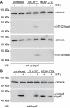
(A) Western blot of detergent lysates of IFNγ-induced L929 cells either uninfected or infected with type I RH-YFP, type II ME49, or type III CTG T. gondii. Only RH-YFP infection caused phosphorylation of Irga6, detectable as a unique band (black arrowhead) with the anti-pT102 (upper blot) or anti-pT108 antibodies (lower blot). (B) Western blot of the same lysates as in Figure 3A in which Irga6 was detected with the monoclonal anti-Irga6 antibody 10E7. The phosphorylated slower-migrating band is detectable only in the lysate from cells infected with virulent RH-YFP. Black arrowhead: phosphorylated Irga6 ((p)Irga6). Open arrowhead: Irga6. Note that the detectability of the phosphorylated bands by the anti-pT102 and anti-pT108 antibodies in (A) is independent of variations in the level of total Irga6.
Figure 4
Figure 4. Phosphomimetic and neutral mutations of T102 and T108 disable Irga6 biochemically.
(A and B) GTP hydrolysis by recombinant Irga6 is lost by phosphomimetic Thr to Asp (A) and neutral Thr to Ala (B) mutations. (C and D) GTP-dependent oligomerisation of recombinant Irga6 is lost by Thr to Asp (C) and Thr to Ala (D) mutations. Very weak oligomerisation is retained by T108D.
Figure 5
Figure 5. Phosphomimetic Irga6 mutants load onto the PVM inefficiently and inhibit loading of wt Irga6.
Wt and phosphomimetic Irga6 mutants T102D and T108D, all tagged with ctag1, were transfected into IFNγ-induced C57BL/6 MEFs. Cells were infected 24 h later with T. gondii strain ME49 for 2 h and vacuoles assayed for loading of the transfected proteins (A and B) or total Irga6 (C). (A) Frequency of vacuoles loaded with transfected wt or phosphomimetic mutant Irga6 proteins (mean and standard deviation of four independent experiments, 100 vacuoles counted per experiment). (B) Loading intensity of transfected wt or phosphomimetic mutant Irga6 proteins (40 vacuoles analysed; *** p<0.0001; horizontal lines indicate means). (C) Frequency of vacuoles loaded with total Irga6 detected with the monoclonal anti-Irga6 antibody 10D7. The transfected phosphomimetic mutant proteins reduced the frequency of vacuoles loaded with endogenous Irga6 (results of two independent experiments, 100 vacuoles counted per experiment).
Figure 6
Figure 6. Phosphorylated Irga6 is detected at the PVM in cells infected with virulent T. gondii.
IFNγ-induced MEFs were infected with the indicated T. gondii strains for 2 h and immunostained with the anti-pT102 and anti-pT108 antibodies. (A) Staining intensities at virulent RH and avirulent ME49 strain vacuoles by anti-pT102 and anti-pT108 antibodies. (*** p<0.0001; horizontal lines indicate means). Weak but significant staining of ME49 vacuoles was recorded from the pT108-specific antibody. One representative out of three independent experiments is shown. (B) Representative immunofluorescence images of total active Irga6 (first column, 10D7) and phosphorylated Irga6 (second column, anti-pT102 and anti-pT108 antibodies) on RH (upper two rows) and ME49 (lower two rows) strain parasitophorous vacuoles. Intracellular T. gondii are indicated by arrows and individual exposure times are indicated in brackets. Corresponding phase contrast images are also given (third column).
Figure 7
Figure 7. Avirulent CTG strain transgenic for virulent ROP18 phosphorylates Irga6 at T102 and T108.
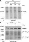
(A) Irga6 immunoprecipitated from 33P-phosphoric acid–labelled, IFNγ-induced L929 cells infected with CTG strain transgenic for empty vector (CTG Ble), kinase-dead ROP18-D394A (CTG L1) or active ROP18 from GT-1 strain (CTG V1) (autoradiogram). Only CTG V1 can phosphorylate Irga6 (black arrowhead). Virulent RH-YFP is a positive control. (B) Detergent lysates from IFNγ-induced L929 cells infected with CTG Ble, CTG L1, CTG V1, or RH-YFP were stained in a Western blot with rabbit anti-pT108 or anti-T102 antibodies. Only CTG V1 and positive control RH-YFP infected cells show the typical band of phosphorylated Irga6 (black arrowhead).
Figure 8
Figure 8. Avirulent CTG strain transgenic for virulent ROP18 phosphorylates Irga6 at the PVM.
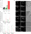
(A) Frequency of vacuoles positive for phosphorylated Irga6 detected by immunofluorescence with anti-pT102 and anti-pT108 antibodies in IFNγ-induced MEFs infected with CTG Ble (black), CTG L1 (green), or CTG V1 (red) transgenic T. gondii strains (results of two independent experiments, 100 vacuoles counted per experiment). (B) Intensity of phosphorylated Ira6 detected by immunofluorescence with anti-pT102 and anti-pT108 antibodies in IFNγ-induced MEFs infected with CTG Ble (black), CTG L1 (green), or CTG V1 (red) transgenic T. gondii strains (20 vacuoles counted; horizontal lines indicate mean signal intensities; *** p<0.0001). (C) Immunofluorescent images of total active Irga6 (first column, detected with monoclonal anti-Irga6 antibody 10D7) and phosphorylated Irga6 (second column, detected with anti-pT102 and anti-pT108 antibodies) on CTG V1, Ble, and L1 vacuoles in IFNγ-induced MEFs. Intracellular T. gondii are indicated by arrows and individual exposure times are indicated in brackets. Corresponding phase contrast images are given (third column). (D) Intensity of Irga6 loaded onto parasitophorous vacuoles in IFNγ-induced MEFs infected with CTG Ble (black), CTG L1 (green), or CTG V1 (red) transgenic T. gondii strains, detected with monoclonal anti-Irga6 antibody 10D7. Loading of Irga6 is inhibited by CTG V1; the effect is greatest at low IFNγ concentrations (0.3 U/ml) (horizontal lines indicate mean signal intensities; *** p<0.0001, * p<0.025). One representative out of two independent experiments is shown.
Figure 9
Figure 9. ROP18 directly phosphorylates Irga6 at T102 and T108 in vitro.
(A) Bacterially expressed, purified wt or T102A and T108A mutant Irga6 proteins (300 ng) were coincubated with bacterially expressed, purified GST-ROP18-Ty (10 µl, 5 µl, 1.7 µl respectively; 1 µl ∼20 ng) in the presence of γ32P-ATP in vitro. The autoradiogram reveals efficient phosphorylation of wt Irga6 and a decrease in signal intensity for all Irga6 alanine mutants. This effect is most apparent at low ROP18 concentrations (1.7 µl). Autophosphorylation of ROP18 ((p)ROP18) is visible at higher kinase concentrations (10 µl and unpublished data) by the appearance of two bands (as previously observed for immunoprecipitated endogenous ROP18 [55]). (B) Quantification of signal intensities observed in (A) for two different ROP18 concentrations (5 µl and 1.7 µl) by phosphorimaging. Black bars represent two independent experiments. (C) Bacterially expressed purified wt or T102A and T108A mutant Irga6 proteins (300 ng) were coincubated with GST-ROP18-Ty (10 µl) in the presence of 1 mM unlabelled ATP in vitro. One-third of each kinase reaction was resolved by SDS-PAGE and subjected to Western blot analysis using the anti-pT102 and anti-pT108 antibodies (upper two blots) or a monoclonal anti-Irga6 antibody, 10E7 (lower blot). Alanine mutation of the target threonines in Irga6 (T102 and T108) specifically blocks recognition by the corresponding antibody.

Similar articles

Cited by

References

    1. Dubey J. The life cycle of Toxoplasma gondii. In: Ajioka J. W, Soldati D, editors. Toxoplasma Molecular and Cellular Biology. Norfolk, UK: Horizon Bioscience; 2007. pp. 3–16.
    1. Howe D. K, Sibley L. D. Toxoplasma gondii comprises three clonal lineages: correlation of parasite genotype with human disease. J Infect Dis. 1995;172:1561–1566. - PubMed
    1. Sibley L. D, Ajioka J. W. Population structure of Toxoplasma gondii: Clonal expansion driven by infrequent recombination and selective sweeps. Annu Rev Microbiol. 2008;62:329–351. - PubMed
    1. Sibley L. D, Boothroyd J. C. Virulent strains of Toxoplasma gondii comprise a single clonal lineage. Nature. 1992;359:82–85. - PubMed
    1. El Hajj H, Lebrun M, Arold S. T, Vial H, Labesse G, et al. ROP18 is a rhoptry kinase controlling the intracellular proliferation of Toxoplasma gondii. PLoS Pathog. 2007;3:e14. doi: 10.1371/journal.ppat.0030014. - DOI - PMC - PubMed

Publication types

MeSH terms