. 2010 Dec;22(12):4009-30.
doi: 10.1105/tpc.110.080697. Epub 2010 Dec 30.

EXPO, an exocyst-positive organelle distinct from multivesicular endosomes and autophagosomes, mediates cytosol to cell wall exocytosis in Arabidopsis and tobacco cells

Affiliations

EXPO, an exocyst-positive organelle distinct from multivesicular endosomes and autophagosomes, mediates cytosol to cell wall exocytosis in Arabidopsis and tobacco cells

Juan Wang et al. Plant Cell. 2010 Dec.

Abstract

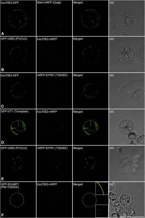
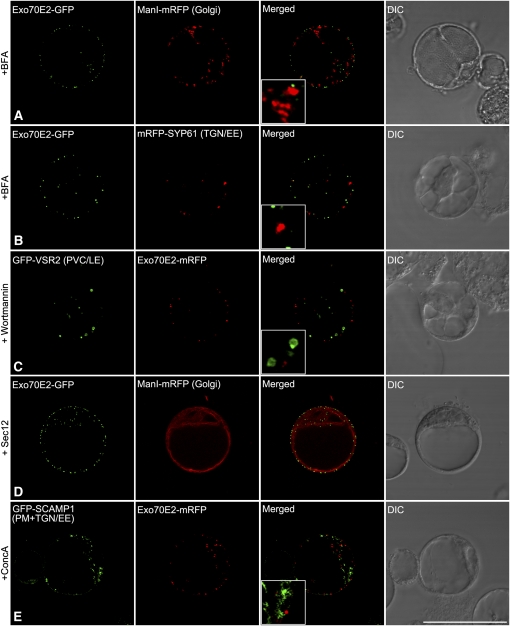
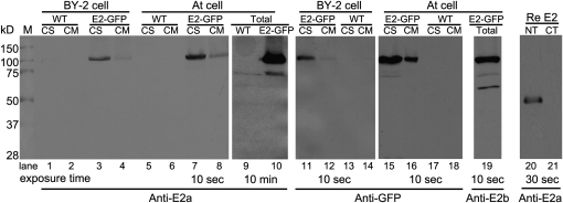
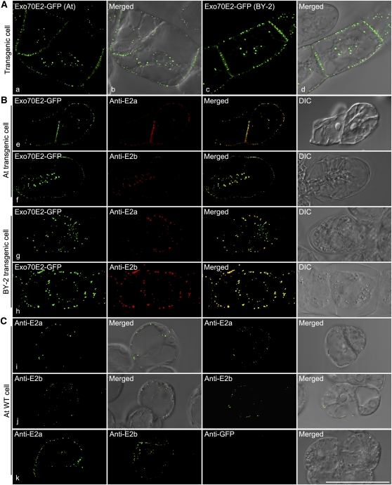
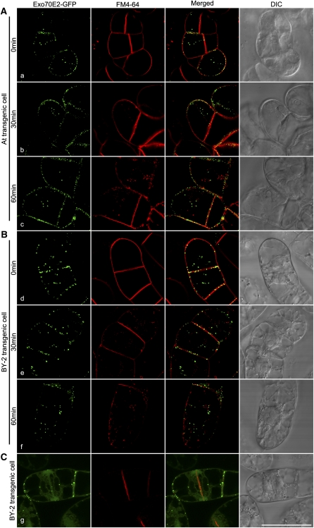
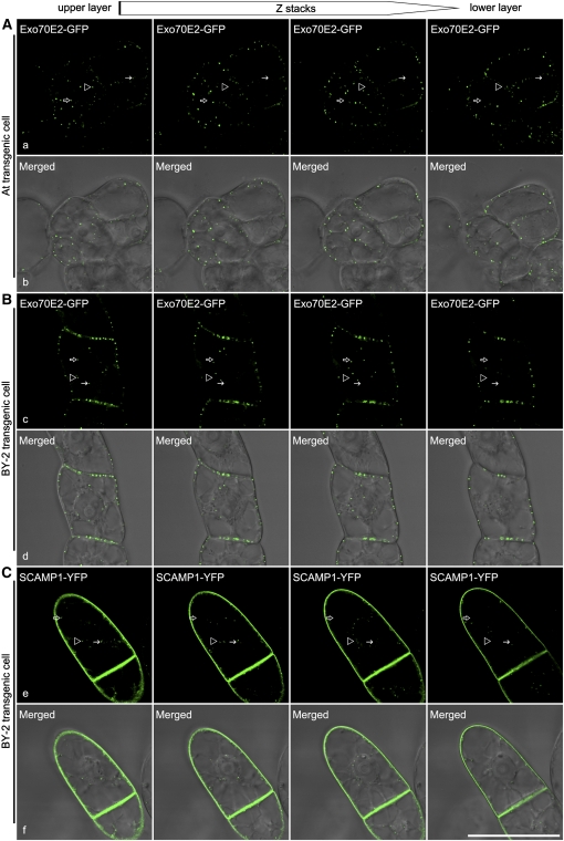
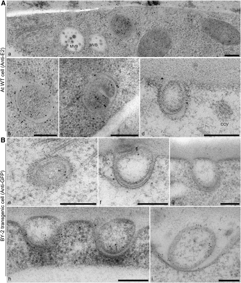
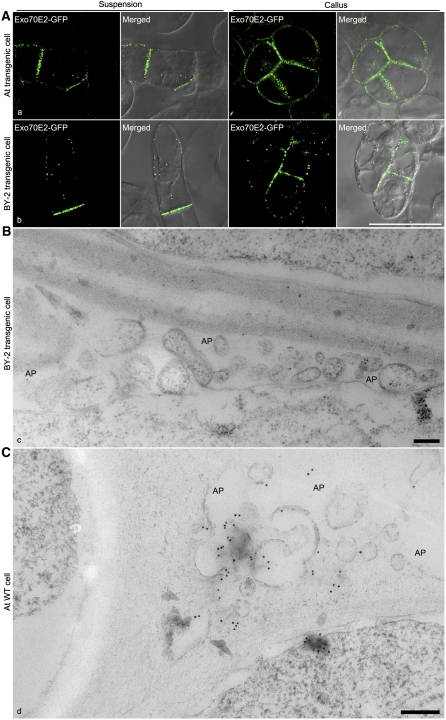
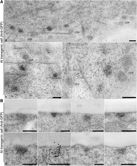
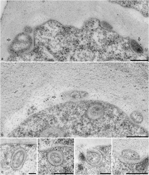
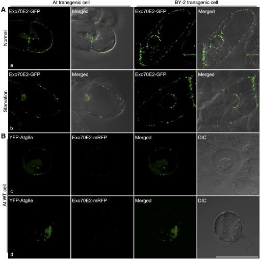
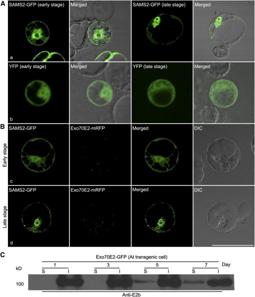
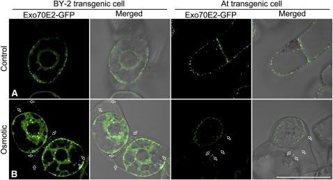
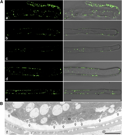
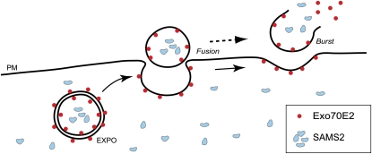
The exocyst protein complex mediates vesicle fusion with the plasma membrane. By expressing an (X)FP-tagged Arabidopsis thaliana homolog of the exocyst protein Exo70 in suspension-cultured Arabidopsis and tobacco (Nicotiana tabacum) BY-2 cells, and using antibodies specific for Exo70, we detected a compartment, which we term EXPO (for exocyst positive organelles). Standard markers for the Golgi apparatus, the trans-Golgi network/early endosome, and the multivesicular body/late endosome in plants do not colocalize with EXPO. Inhibitors of the secretory and endocytic pathways also do not affect EXPO. Exo70E2-(X)FP also locates to the plasma membrane (PM) as discrete punctae and is secreted outside of the cells. Immunogold labeling of sections cut from high-pressure frozen samples reveal EXPO to be spherical double membrane structures resembling autophagosomes. However, unlike autophagosomes, EXPOs are not induced by starvation and do not fuse with the lytic compartment or with endosomes. Instead, they fuse with the PM, releasing a single membrane vesicle into the cell wall. EXPOs are also found in other cell types, including root tips, root hair cells, and pollen grains. EXPOs therefore represent a form of unconventional secretion unique to plants.


Figures

Figure 1.
Figure 1.
Exo70E2 Localizes as Discrete Punctate Signals at the PM and in the Cytosol but Does Not Colocalize with Standard Organelle Markers. Arabidopsis protoplasts were coelectroporated with Exo70E2-(X)FP, and the DNA of a single organelle marker as indicated. After 13 to 16 h of expression, the protoplasts were observed by CLSM. Bar = 50 μm.
Figure 2.
Figure 2.
EXPOs Are Not Affected by Secretory and Endocytosis Inhibitors in Protoplasts. Arabidopsis protoplasts were coelectroporated with Exo70E2-(X)FP and marker organelle DNA as indicated and treated with either BFA, wortmannin (1 h), or ConcA (13 h) before observing by CLSM. For inhibition of ER export, protoplasts expressing both Exo70E2-GFP and ManI-mRFP were additionally electroporated with Sec12. DIC, differential interference contrast. Bar = 50 μm.
Figure 3.
Figure 3.
EXPOs Are Not Affected by Secretory and Endocytosis Inhibitors in Transgenic Arabidopsis and BY-2 Cells. Transgenic Arabidopsis (a, d, and g) and BY-2 (b, e, and h) cells expressing Exo70E2-GFP were treated with either BFA (A), wortmannin (1 h) (B), or ConcA (2 h) (C) before observing by CLSM. As controls, transgenic BY-2 cell lines expressing either the Golgi marker ManI-GFP (c), the PVC/LE marker GFP-BP-80 (f), or the TGN/EE marker SCAMP1-YFP (i) were used in treatments with these three drugs, respectively. Bar = 50 μm. [See online article for color version of this figure.]
Figure 4.
Figure 4.
Immunoblot Characterization of Transgenic Arabidopsis or Tobacco BY-2 Cell Lines Expressing Exo70E2-GFP and Exo70E2. Immunoblot analysis of proteins isolated from either transgenic cell lines (Arabidopsis and BY-2) expressing Exo70E2-GFP (E2-GFP) or their wild-type (WT) cells using E2a/b or GFP antibodies as indicated, showing the specificity of rabbit polyclonal anti-E2a/b antibodies generated against the N-terminal synthetic peptide or recombinant 300–amino acid N terminus of At Exo70E2, respectively. The predicted molecular masses for Exo70E2-GFP and endogenous At Exo70E2 are ~100 and 75 kD, respectively. Both anti-E2a/b and anti-GFP recognized the full-length Exo70E2-GFP fusion proteins in both transgenic cell lines (lanes 3 and 4, 7 and 8, 11 and 12, 15 and 16, and 19). Anti-Ea/b also detected the endogenous At Exo70E2 proteins in both wild-type (lane 9) and transgenic (lanes 10 and 19) Arabidopsis cells. CS, cell-soluble proteins; CM, cell membrane proteins; total, total proteins. NT, N terminus of recombinant Exo70E2; CT, C terminus of recombinant Exo70E2.
Figure 5.
Figure 5.
Confocal Characterization of Transgenic Arabidopsis or Tobacco BY-2 Cell Lines Expressing Exo70E2-GFP and Exo70E2. (A) The expression patterns of Exo70E2-GFP in both cell lines are similar to that seen by transient expression in protoplasts. DIC, differential interference contrast. (B) At Exo70E2a/b antibodies specifically recognize the Exo70E2-GFP signals in both transgenic Arabidopsis and BY-2 cell lines. (C) At Exo70E2a/b antibodies recognize the endogenous At Ex070E2 proteins in wild-type Arabidopsis cells with similar patterns as transgenic cells expressing Exo70E2-GFP. Bar = 50 μm.
Figure 6.
Figure 6.
Time Course of FM4-64 Uptake in Transgenic Arabidopsis and BY-2 Cell Lines Stably Expressing Exo70E2-GFP. FM4-64 (red) uptake study was performed in transgenic Arabidopsis or BY-2 cells expressing Exo70E2-GFP (green), followed by confocal image collection at indicated times. At no time is there a colocalization to be seen between internalized FM4-64 and the Exo70E2-GFP signals in Arabidopsis (A) or BY-2 (B). During cytokinesis, whereas FM4-64 intensively labels the developing cell plate, an equivalent distribution of Exo70E2 signal is not observed (C). Arabidopsis cell line (A); tobacco BY-2 cell lines ([B] and [C]). Bar = 50 μm.
Figure 7.
Figure 7.
EXPOs Are Cytosolic and Not Associated with the PM. Shown are examples of series optical sections of confocal images from top to bottom of transgenic Arabidopsis (A) or tobacco BY-2 cells (B) expressing Exo70E2-GFP or BY-2 cells (C) expressing rice (Oryza sativa) SCAMP1-YFP (Lam et al., 2007), which is located in TGN and PM, as a control. Arrows and arrowheads indicated examples of EXPO not associated with the PM. Bar = 50 μm. [See online article for color version of this figure.]
Figure 8.
Figure 8.
EXPOs Have Two Membranes and Fuse with the PM Expelling a Single Membrane Vesicle into the Apoplast. Immunogold labeling of sections cut from high-pressure frozen/freeze-substituted samples of wild-type Arabidopsis ([A], a to d), and transgenic tobacco BY-2 ([B], e to i) cells. Bars = 200 nm. (A) EXPOs are distinct in morphology from MVBs, which do not label with Exo70E2a antibodies. Label is distributed over both membranes of the EXPO. (B) Gallery of fusion profiles. Label is found on the inner surface of the inner vesicle and is also present in the neighboring vicinity, suggesting that the released vesicle ultimately bursts, releasing Exo70E2 into the cell wall.
Figure 9.
Figure 9.
Likely Nature of EXPO Release in Plant Cells. (A) GFP-positive EXPOs are more abundant in PM/cell wall areas connecting two cells in transgenic Arabidopsis and BY-2 cells expressing Exo70E2-GFP. Bar = 50 μm. (B) Immuno-EM detection of released EXPOs in the apoplasts (AP; as indicated) of adjacent transgenic BY-2 cells expressing Exo70E2-GFP using high-pressure freezing. Bar = 200 nm. (C) Immuno-EM detection of released EXPOs in the apoplasts of adjacent wild-type Arabidopsis cells using high-pressure freezing. Bar = 200 nm. [See online article for color version of this figure.]
Figure 10.
Figure 10.
Immunogold Labeling of EXPO/PM Fusions. (A) Tangential sections through the PM–cell wall interface showing that clathrin-coated pits (CCPs) are separate from GFP antibody–labeled electron opaque patches. (B) Cross sections through the PM–cell wall interface. The Exo70E2-positive patches do not reveal clearly fusion profiles. We interpret these as being the remains of fusions, where exocyst molecules are still attached to the PM. Bars = 200 nm.
Figure 11.
Figure 11.
EXPOs Are Also Labeled by Exo70A1 Antibodies in Wild-Type Arabidopsis Cells. Immunogold labeling of sections cut from high-pressure frozen/freeze-substituted samples of Arabidopsis cells using Exo70A1 antibodies, with similar labeling patterns as that of Exo70E2a. Bars = 200 nm.
Figure 12.
Figure 12.
EXPOs Are Not Induced by Sucrose Starvation and Are Distinct from an Autophagy Marker. (A) Shown are representative examples of transgenic Arabidopsis and BY-2 cells expressing Exo70E2-GFP under normal and starvation cultured conditions after 24 h. (B) Separation of Exo70E2-mRFP from the autophagy marker YFP-Atg8e in wild-type Arabidopsis protoplasts. Bar = 50 μm.
Figure 13.
Figure 13.
EXPO Sequesters Cytosolic SAMS2-GFP in Arabidopsis Protoplasts. (A) When transiently expressed in Arabidopsis protoplasts, SAMS2-GFP showed cytosol and nucleus patterns at early stages (24 h) but became punctate at later stages (a; 48 h). By contrast, YFP remained cytosolic in both stages (b). (B) When coexpressed with Exo70E2-mRFP in Arabidopsis protoplasts, SAMS2-GFP showed cytosol and nucleus patterns at early stages (c), but at later stages, SAMS2-GFP became colocalized with Exo70E2-mRFP in EXPO (d). Bar = 50 μm. (C) Immunoblot detection of Exo70E2-GFP proteins in the culture medium of transgenic Arabidopsis cells expressing Exo70E2-GFP. S, secretion medium proteins; I, intracellular proteins; day, days after subculture.
Figure 14.
Figure 14.
EXPOs Are Detected Outside the PM in Transgenic Cells. Transgenic tobacco BY-2 or Arabidopsis cells expressing Exo70E2-GFP were first subjected to osmotic treatment with 250 mM NaCl for 10 min, followed by confocal imaging. Untreated cells were included as controls. Bars = 50 μm. [See online article for color version of this figure.]
Figure 15.
Figure 15.
Immunofluorescent and Structural EM Detection of EXPO in Other Cell Types. (A) Shown are examples of EXPO detected by anti-Exo70E2a antibodies in wild-type Arabidopsis root tip cells (a) and root hair cells (b to e). Bars = 50 μm. (B) Structural EM detection of EXPO in an ultrathin section prepared from high-pressure freezing/frozen-substituted wild-type tobacco pollen grains. Arrows indicated examples of EXPO. Bars = 2 μm. [See online article for color version of this figure.]
Figure 16.
Figure 16.
Working Model of EXPO in Plant Cells. Illustration depicting the relationship between Exo70E2, and double-membrane EXPO, their fusion with the PM, and release and bursting of the single-membrane EXPO in the harsh environment to complete the unconventional secretion. Meanwhile, cytosolic protein SAMS2 is sequestered by EXPO and finally reaches the cell wall. [See online article for color version of this figure.]

Similar articles

Cited by

References

    1. An Q., van Bel A.J., Hückelhoven R. (2007). Do plant cells secrete exosomes derived from multivesicular bodies? Plant Signal. Behav. 2: 4–7 - PMC - PubMed
    1. Anders N., Jürgens G. (2008). Large ARF guanine nucleotide exchange factors in membrane trafficking. Cell. Mol. Life Sci. 65: 3433–3445 - PMC - PubMed
    1. Baba M., Osumi M., Ohsumi Y. (1995). Analysis of the membrane structures involved in autophagy in yeast by freeze-replica method. Cell Struct. Funct. 20: 465–471 - PubMed
    1. Baba M., Osumi M., Scott S.V., Klionsky D.J., Ohsumi Y. (1997). Two distinct pathways for targeting proteins from the cytoplasm to the vacuole/lysosome. J. Cell Biol. 139: 1687–1695 - PMC - PubMed
    1. Baba M., Takeshige K., Baba N., Ohsumi Y. (1994). Ultrastructural analysis of the autophagic process in yeast: detection of autophagosomes and their characterization. J. Cell Biol. 124: 903–913 - PMC - PubMed

Publication types