. 2010 Dec 16;6(12):e1001231.
doi: 10.1371/journal.ppat.1001231.

NleC, a type III secretion protease, compromises NF-κB activation by targeting p65/RelA

Affiliations

NleC, a type III secretion protease, compromises NF-κB activation by targeting p65/RelA

Hilo Yen et al. PLoS Pathog. .

Abstract

The NF-κB signaling pathway is central to the innate and adaptive immune responses. Upon their detection of pathogen-associated molecular patterns, Toll-like receptors on the cell surface initiate signal transduction and activate the NF-κB pathway, leading to the production of a wide array of inflammatory cytokines, in attempt to eradicate the invaders. As a countermeasure, pathogens have evolved ways to subvert and manipulate this system to their advantage. Enteropathogenic and enterohemorrhagic Escherichia coli (EPEC and EHEC) are closely related bacteria responsible for major food-borne diseases worldwide. Via a needle-like protein complex called the type three secretion system (T3SS), these pathogens deliver virulence factors directly to host cells and modify cellular functions, including by suppressing the inflammatory response. Using gain- and loss-of-function screenings, we identified two bacterial effectors, NleC and NleE, that down-regulate the NF-κB signal upon being injected into a host cell via the T3SS. A recent report showed that NleE inhibits NF-κB activation, although an NleE-deficient pathogen was still immune-suppressive, indicating that other anti-inflammatory effectors are involved. In agreement, our present results showed that NleC was also required to inhibit inflammation. We found that NleC is a zinc protease that disrupts NF-κB activation by the direct cleavage of NF-κB's p65 subunit in the cytoplasm, thereby decreasing the available p65 and reducing the total nuclear entry of active p65. More importantly, we showed that a mutant EPEC/EHEC lacking both NleC and NleE (ΔnleC ΔnleE) caused greater inflammatory response than bacteria carrying ΔnleC or ΔnleE alone. This effect was similar to that of a T3SS-defective mutant. In conclusion, we found that NleC is an anti-inflammatory bacterial zinc protease, and that the cooperative function of NleE and NleC disrupts the NF-κB pathway and accounts for most of the immune suppression caused by EHEC/EPEC.


Conflict of interest statement

The authors have declared that no competing interests exist.

Figures

Figure 1
Figure 1. TOB02 is a functional tool for studying the effects of individual effectors.
(A) Induction of actin polymerization by infection with TOB02. HeLa cells were infected with TOB02 for 3 hours. The cells were then fixed with 4% PFA and stained with Rhodamine-Phalloidin (red) and DAPI (blue). Arrow indicates the location of bacterial attachment. (B) Secretion of LEE-encoded virulence factor by TOB02. Overnight-grown TOB02 was inoculated and cultured in fresh DMEM until O.D600 ∼1.0. The cell pellet (Whole cell) and culture supernatant (Supernatant) were collected and analyzed with anti-EspB and anti-DnaK antibodies. (C) Cellular response to infection with reconstituted strains. HeLa cells were infected with TOB02, E2348/69 WT (EPEC wild type), E2348/69 ΔescF (ΔT3SS), B171-8 WT (EPEC wild type), and B171-8 ΔescC (ΔT3SS) for 3 hours. Then cells were rinsed and incubated in fresh DMEM containing 0.1 mg/ml gentamicin and heat-killed bacteria (HKE) (108/ml). Cells were further cultured for 8 hours, and the culture supernatants were analyzed for IL-8 by ELISA. Negative and positive controls were non-infected/non-stimulated (N.I/N.S) and non-infected/stimulated (N.I/S) cells, respectively.
Figure 2
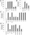
Figure 2. NleC can inhibit NF-κB activation.
Two days prior to infection, HeLa cells were transfected with NF-κB and Luciferase reporter plasmids. On the day of infection, HeLa cells were infected with various bacterial strains for 3 hours. The medium was replaced with fresh DMEM containing gentamicin and HKE. The cells were further cultured for 8 hours, then the IL-8 or reporter activities of NF-κB and Luciferase were determined. The positive and negative controls were non-infected/non-stimulated (N.I/N.S) and non-infected/stimulated (N.I/S) HeLa cells. In the reporter assays, the NF-κB activity was normalized to the luciferase activity. All experiments were performed in triplicate and repeated three times. Representative results from the repeated experiments are shown. Significance was calculated by student's t-test, where p<0.05 was considered significant. (A) IL-8 secretion by cells infected with an nleC- or nleE-expressing strain. Cells were infected with TOB02/HA (HA), TOB02/nleF-HA (nleF), TOB02/nleC-HA (nleC), or TOB02/nleE-HA (nleE). IL-8 was analyzed by ELISA. (* p<0.05, compared to HA). (B) NF-κB activation in cells infected with an nleC-expressing strain. Cells were infected with TOB02/HA or TOB02/nleC-HA (nleC). (* p<0.05, compared to HA) (C) NF-κB activity in cells infected with EPEC mutants. For infection, wild-type EPEC (WT), a ΔT3SS mutant (ΔescF), and a series of isogenic mutants was used: A1 (deletion of IE2 region), A2 (A1 plus PP2 region deletion), A3 (A2 plus PP4 region deletion), A4 (A3 plus IE6 region deletion), A5 (A4 plus PP6 region deletion), A6 (A5 plus IE5 region deletion), and A7 (A6 plus espG deletion). (* p<0.05, compared to WT) (D) NF-κB activity in cells infected with an EPEC mutant expressing non-LEE effectors. Cells were infected with wild-type EPEC (WT), ΔT3SS mutant (ΔescF), A4, or A4 harboring a plasmid with nleB (A4/nleB), nleE (A4/nleE), espL (A4/espL), or nleC (A4/nleC). (* p<0.05, compared to A4).
Figure 3
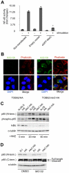
Figure 3. NleC interferes with NF-κB activation by downregulating p65.
(A) NF-κB activity in cells transfected with nleC. HeLa cells were all transfected with NF-κB and luciferase reporter plasmids. In addition, either eGFP plasmid (Empty vector) or eGFP-NleC-expressing plasmid was introduced into the cells. “Non-transfected” indicates cells transfected with only the reporter plasmid. Two days post-transfection, the cells were stimulated with HKE. The culture medium and cell lysates were then analyzed for NF-κB SEAP and Luciferase activity. (* p<0.05, compared to stimulated Empty Vector). (B) Localization of NleC in infected cells. HeLa cells were infected with either TOB02/HA or TOB02/nleC-HA. After 3 hours of infection, cells were washed and fixed with 4% PFA, then stained with anti-HA (FITC), phalloidin (red), and DAPI (blue). (C) Decrease in the IκBα and p65 proteins in cells infected with the nleC-expressing strain. HeLa cells were infected with TOB02/HA (HA) or TOB02/nleC-HA (nleC-HA). After 3 hours of infection, the cells were rinsed and incubated in fresh DMEM containing 0.1 mg/ml gentamicin and a 1/10 volume of HKE. HKE stimulation proceeded for 20 and 40 min, and cells were collected for western blot analysis. Cell extracts were probed with anti-p65 (N-term.), anti-phospho-p65, anti-IκBα, and anti-α-tubulin (as a loading control) antibodies. Non-infected/Non-stimulated cells at 0 min. (N.I/N.S at 0 min.) served as a negative control. Non-infected/stimulated (N.I/S) cells at 20 and 40 min were positive controls. (D) Appearance of a cleaved fragment of p65 in cells infected with the NleC-expressing strain. Two hours prior to infection, HeLa cells were pretreated with either DMSO or MG132 (5 µM). The cells were infected with TOB02/HA (HA) or TOB02/nleC-HA (nleC-HA) for 3 hours. The cells were then rinsed and lysates were prepared for analysis. Anti-p65 antibodies specific to either the N- or C- terminal regions were used. Non-infected (N.I) cells served as a negative control.
Figure 4
Figure 4. NleC cleaves p65 by its zinc protease domain.
(A) Activity of purified GST-NleC protein. For lysates, 1×10⁶ of unstimulated HeLa cells were lysed by NET150 (10 mM Tris-Cl pH 8.0, 0.5% Triton-X100, 150 mM NaCl, 10% glycerol) without EDTA or protease inhibitors. GST protein or GST-NleC protein expressed in bacteria was purified and mixed with the HeLa lysates. The reaction mixtures were incubated at 25°C for 8 hours and then analyzed with antibodies against p65 (N-terminal or C-terminal specific) and α-tubulin (as a loading control). (B) Cleavage of p65 by NleC alone. p65, GST, and GST-NleC were expressed in bacteria and purified. The p65 was then mixed with either GST or GST-NleC in the reaction buffer. The mixtures were incubated at 25°C for 8 hours and analyzed with anti-p65 (N-term.) and anti-p65 (C-term.) antibodies. (C) Zinc protease motif in NleC. Schematic diagram showing the zinc protease domain in NleC. A Histidine (H) at 187 was replaced with Tyrosine (Y) by site-directed mutagenesis. (D) NleC's cleavage activity required the zinc protease motif and divalent metal ions. p65, GST, GST-NleC wild type (GST-NleC), and GST-NleC mutant (GST-NleCmut) were expressed in bacteria and purified. The p65 was mixed with GST, GST-NleC, or GST-NleCmut in the reaction buffer. The cleavage reaction was performed at 25°C for 8 hours. Samples were analyzed with anti-GST and anti-p65 antibodies against either the N- or C-terminal region. For the EDTA inhibition, EDTA was included in the reaction buffer at a final concentration of 10 mM. (E) NF-κB activity in cells infected with the nleCmut-expressing strain. Two days prior to infection, HeLa cells were transfected with NF-κB and luciferase reporter plasmids. Forty-eight hours post-transfection, cells were infected with TOB02/HA, TOB02/nleC-HA (nleC), or TOB02/nleCmut-HA (nleCmut) for 3 hours. Cells were rinsed and incubated in fresh DMEM containing 0.1 mg/ml gentamicin and HKE (108/ml). Cells were stimulated for 8 hours, then the medium and cell lysates were analyzed for NF-κB SEAP and Luciferase reporter activities. Non-infected/non-stimulated (N.I/N.S) and non-infected/stimulated (N.S/S) HeLa cells served as negative and positive controls, respectively.
Figure 5
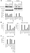
Figure 5. Concurrent deficiency in nleE and nleC impedes the immune-suppressive ability of EPEC/EHEC.
(A) Amount of cytoplasmic and nuclear p65 protein in cells infected with nleC mutants. HeLa cells were infected with EPEC wild type (WT), ΔescF mutant, ΔnleC mutant, and ΔnleC/nleC complemented strains for 2 hours. At the end of the infection, TNF-α (50 ng/ml) was added, and the cells were further cultured for 40 min before being collected. The cytoplasmic and nuclear fractions were analyzed by anti-p65 (C-term.), anti-Lamin A/C and α-tubulin antibodies. This experiment was repeated three times and one of the representative blot is shown. For quantification, the total cytoplasmic p65 and the total nuclear p65 were normalized to the α-tubulin and Lamin A/C using ImageJ software. NI/NS (Non-infected/Non-stimulated) and NI/S (Non-infected/Stimulated) served as the negative and positive controls for p65 translocation in response to the TNF-α stimulation (* p<0.05, compared to cytoplasmic NI/NS; ** p<0.05, compared to nuclear NI/S). (B) IL-8 secretion by cells infected with EPEC or EHEC mutants. HeLa cells were infected with wild type, ΔT3SS mutant (ΔescF for EPEC; ΔescD for EHEC), ΔnleC, ΔnleE, and ΔnleC ΔnleE of EPEC and EHEC. Infections were proceeded for 3 hours for EPEC and 4 hours for EHEC strains. Cells were then rinsed and incubated in fresh DMEM containing 0.1 mg/ml gentamicin and HKE. Culture medium were collected 8 hours of stimulation and amount of IL-8 analyzed by ELISA. Non-infected/non-stimulated (N.I/N.S) and non-infected/stimulated (N.I/S) cells served as negative and positive controls (* p<0.05, compared to WT). Values of ΔnleC ΔnleE double KO mutant are not significantly different from ΔT3SS (p>0.05). (C) IL-8 secretion by cells infected with EHEC compound mutants. HeLa cells were infected with wild-type EHEC (WT), ΔT3SS mutant (ΔescD), ΔnleC ΔnleE mutant, or ΔnleC ΔnleE mutant harboring nleCwt or nleCmut plasmid for 4 hours. The cells were then rinsed and incubated in fresh DMEM containing 0.1 mg/ml gentamicin and HKE. After 8 hours of HKE stimulation, the culture supernatants were collected and analyzed for IL-8 by ELISA. Non-infected/non-stimulated (N.I/N.S) and non-infected/stimulated (N.I/S) cells served as negative and positive controls. (* p<0.05, compared to ΔnleC ΔnleE).
Figure 6
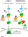
Figure 6. Schematic diagram illustrating the modes of action of NleC and NleE in host NF-κB suppression.
Detection of PAMPs by cell surface receptors triggers the onset of the NF-κB signaling cascade, leading to a chain of events involving protein modifications and degradations; ultimately, activated p65/p50 NF-κBs enter the nucleus to transcribe various inflammatory genes, including IL-8. EPEC/EHEC utilizes two distinct T3SS-dependent non-LEE effectors to subdue this response. This is accomplished by the coordinated targeting of upstream and downstream components of the NF-κB pathway by NleE and NleC, respectively. NleE, by an unknown mechanism, retards the activation of IKK, thus retaining p65/p50 in a complex with IκB. NleC digests activated p65 and to a lesser extent the IκB-bound p65, to decrease the amount of NF-κB available to enter the nucleus, hence reducing the overall host response to the infection.

Similar articles

Cited by

References

    1. Clarke SC. Diarrhoeagenic Escherichia coli—an emerging problem? Diagn Microbiol Infect Dis. 2001;41:93–98. - PubMed
    1. Kaper JB, Nataro JP, Mobley HL. Pathogenic Escherichia coli. Nat Rev Microbiol. 2004;2:123–140. - PubMed
    1. Moon HW, Whipp SC, Argenzio RA, Levine MM, Giannella RA. Attaching and effacing activities of rabbit and human enteropathogenic Escherichia coli in pig and rabbit intestines. Infect Immun. 1983;41:1340–1351. - PMC - PubMed
    1. Jerse AE, Yu J, Tall BD, Kaper JB. A genetic locus of enteropathogenic Escherichia coli necessary for the production of attaching and effacing lesions on tissue culture cells. Proc Natl Acad Sci USA. 1990;87:7839–7843. - PMC - PubMed
    1. Knutton S, Baldwin T, Williams PH, McNeish AS. Actin accumulation at sites of bacterial adhesion to tissue culture cells: basis of a new diagnostic test for enteropathogenic and enterohemorrhagic Escherichia coli. Infect Immun. 1989;57:1290–1298. - PMC - PubMed

Publication types

MeSH terms