. 2011 Mar 4;286(9):6929-39.
doi: 10.1074/jbc.M110.162289. Epub 2010 Dec 23.

RNA sequences and structures required for the recruitment and activity of the dengue virus polymerase

Affiliations

RNA sequences and structures required for the recruitment and activity of the dengue virus polymerase

Claudia V Filomatori et al. J Biol Chem. .

Abstract

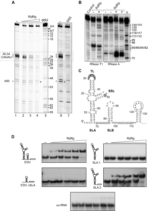
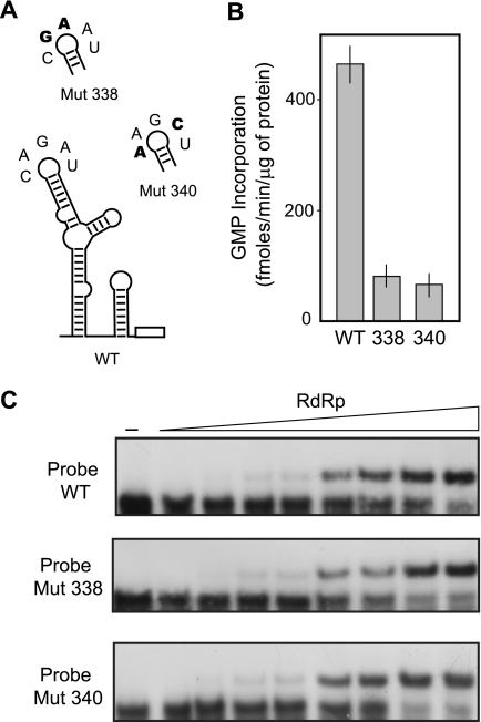
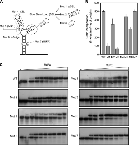
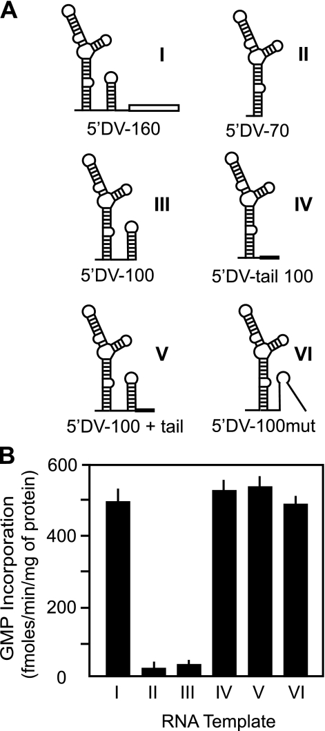
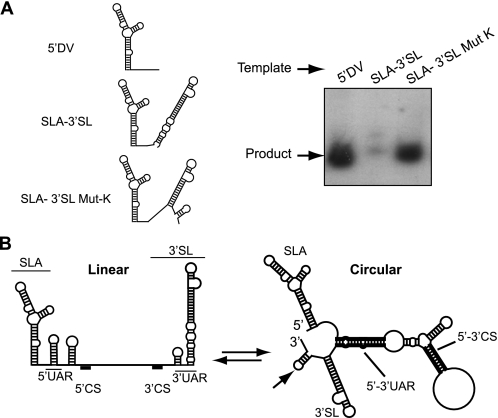
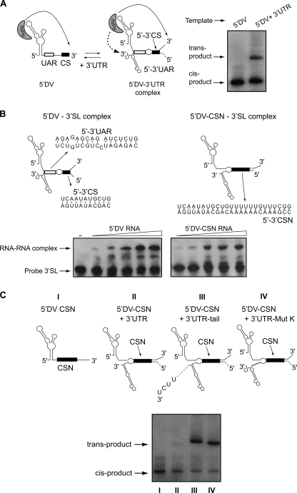
Dengue virus RNA-dependent RNA polymerase specifically binds to the viral genome by interacting with a promoter element known as stem-loop A (SLA). Although a great deal has been learned in recent years about the function of this promoter in dengue virus-infected cells, the molecular details that explain how the SLA interacts with the polymerase to promote viral RNA synthesis remain poorly understood. Using RNA binding and polymerase activity assays, we defined two elements of the SLA that are involved in polymerase interaction and RNA synthesis. Mutations at the top of the SLA resulted in RNAs that retained the ability to bind the polymerase but impaired promoter-dependent RNA synthesis. These results indicate that protein binding to the SLA is not sufficient to induce polymerase activity and that specific nucleotides of the SLA are necessary to render an active polymerase-promoter complex for RNA synthesis. We also report that protein binding to the viral RNA induces conformational changes downstream of the promoter element. Furthermore, we found that structured RNA elements at the 3' end of the template repress dengue virus polymerase activity in the context of a fully active SLA promoter. Using assays to evaluate initiation of RNA synthesis at the viral 3'-UTR, we found that the RNA-RNA interaction mediated by 5'-3'-hybridization was able to release the silencing effect of the 3'-stem-loop structure. We propose that the long range RNA-RNA interactions in the viral genome play multiple roles during RNA synthesis. Together, we provide new molecular details about the promoter-dependent dengue virus RNA polymerase activity.


Figures

FIGURE 1.
FIGURE 1.
Expression, purification, and characterization of DENV full-length NS5 and the two domains RdRp and MTase. A, SDS-PAGE analysis of purified recombinant proteins expressed in E. coli. Mobilities of molecular mass markers (MW) are indicated on the left (in kDa). B, in vitro polymerase and methyltransferase activities of recombinant proteins. The RdRp assay was carried out as described under “Experimental Procedures,” using an in vitro transcribed 5′-DV RNA as template. The RdRp activity is expressed in fmol of [α-32P]GMP incorporated into acid-insoluble RNA/min and per μg of protein. The MTase assay was carried out as described under “Experimental Procedures,” using as template an in vitro transcribed GpppA-5′-DV RNA or an uncapped 5′-DV RNA. The MTase activity is expressed in cpm of [3H]methyl-incorporated. C, binding of the recombinant proteins to the 5′-DV RNA. The interaction of the three proteins with the 5′-DV was evaluated by EMSA. The RNA corresponding to the first 160 nucleotides of the viral genome was uniformly 32P-labeled and titrated (0.1 nm, 30,000 cpm) with increasing concentrations of RdRp, MTase, or full-length NS5 (0, 0.2, 0.5, 1, 5, 10, 30, and 240 nm).
FIGURE 2.
FIGURE 2.
Interaction of the dengue virus RdRp with the viral RNA. A, footprinting assay indicates the interaction of the viral RdRp or full-length NS5 with elements of the SLA. The viral RNA was incubated with increasing concentrations of the RdRp (0, 30, 60, and 120 nm; left panel) or the full-length NS5 (0 and 500 nm; right panel) and subjected to RNase PhyM treatment. The cleaved RNAs were analyzed after primer extension in a sequencing gel along with a sequencing ladder (ddU). Regions showing protection from RNase cleavage upon protein binding are indicated on the left. B, the interaction of the RdRp with the viral RNA alters RNA structures downstream of the promoter element. A 5′ end-labeled RNA of the first 160 nucleotides of the viral genome was incubated with 120 nm RdRp and subjected to RNase T1 or RNase A treatment (as indicated at the bottom). The cleaved RNAs were analyzed along with a control sample without treatment (Control). The labeled RNA was also digested with RNase T1 under denaturing conditions to generate a G ladder (G). Regions showing exposure to RNase cleavages upon RdRp binding are indicated with dashed boxes. C, MFOLD-predicted RNA secondary structure of 5′-DV that includes a summary of protected (shaded gray) and exposed (underlined) nucleotides upon RdRp binding. The relevant elements are indicated: SLA, SLB, TL, and SSL. D, binding of the RdRp to different RNA molecules corresponding to the viral 5′-UTR. The interaction of four RNA probes with the RdRp was evaluated by EMSA. The RNA corresponding to the first 160 nucleotides of the viral genome (5′DV), the first 70 nucleotides (SLA 1), the 5′-DV RNA with a deletion of the SLA (5′DV ΔSLA), the SLA 1 followed by unstructured 32 nucleotides (SLA 2), or an unstructured unrelated RNA (uu-RNA) were uniformly 32P-labeled and titrated (0.1 nm, 30,000 cpm) with increasing concentrations of the RdRp (0, 5, 10, 15, 20, and 240 nm).
FIGURE 3.
FIGURE 3.
Specific nucleotides at the top loop of the SLA promoter impair RNA synthesis without altering the RNA-RdRp complex formation. A, schematic representation of specific mutations within the TL of SLA. The nucleotide sequence of the WT and the mutants Mut 338 and Mut 340 are shown. The nucleotide changes are indicated in bold. B, in vitro polymerase activity using WT and mutated RNA templates. The activity is expressed in fmol of [α-32P]GMP incorporated into acid-insoluble RNA/min and per μg of protein. The reaction was carried out as described under “Experimental Procedures.” The error bars indicate the standard deviations of results from three experiments. C, RNA mobility shift assays showing the interaction between the purified RdRp and the WT or mutated RNA probes. Uniformly 32P-labeled RNAs corresponding to the first 160 nucleotides of the viral genome (0.1 nm, 30,000 cpm) were titrated with increasing concentrations of RdRp (0, 0.5, 1, 3, 5, 10, 15, 50, and 160 nm).
FIGURE 4.
FIGURE 4.
RNA elements of the SLA promoter required for RdRp binding and activity. A, schematic representation of specific mutations in the viral RNA. The nucleotide changes are indicated for each case: in Mut 1, the SSL was deleted, and a UUC sequence was inserted; in Mut 2, the stem of the SSL was stabilized by 5 GC base pairs; in Mut 3, a 2-base pair deletion in the stem of the SSL was included; in Mut 4, the TL was deleted; in Mut 5, the sequence of the GA bulge was replaced by a single U residue; in Mut 6, the GGA bulge was deleted; and in Mut 7 the sequence of the UU bulge was replaced by a single A residue. B, in vitro polymerase activity using WT and mutated RNA templates shown in A. The activity is expressed in fmol of [α-32P]GMP incorporated into acid-insoluble RNA/min and per μg of protein. The reaction was carried out as described under “Experimental Procedures.” The error bars indicate the standard deviations of results from three experiments. C, RNA mobility shift assays showing the interaction between the WT or the mutated RNA probes and the purified RdRp. Uniformly 32P-labeled RNA probes (0.1 nm, 30,000 cpm) were titrated with increasing concentrations of RdRp (0, 1, 5, 10, 15, 20, 25, 80, 160, and 240 nm).
FIGURE 5.
FIGURE 5.
Template activity of RNA molecules carrying different structures at the 3′ end. A, schematic representation of the structures of the 5′-DV RNAs used as templates. Template I (5′DV-160), template II (5′DV-70), and template III (5′DV-100) correspond to the first 160, 70, and 100 nucleotides of the viral genome, respectively. In templates IV (5′DV-tail 100) and V (5′DV-100 + tail), the first 77 or 100 nucleotides of the genome, respectively, were fused to a 26-nucleotide-long unstructured sequence. Template VI (5′DV-100mut) was generated introducing three point mutations in template III that opens the stem of SLB. B, in vitro polymerase activity for templates I–VI, expressed in fmol of [α-32P]GMP incorporated into acid-insoluble RNA/min and per μg of protein.
FIGURE 6.
FIGURE 6.
Effect of the 3′-SL structure on the SLA-dependent RdRp activity. A, analysis of radiolabeled RNA products in a denaturing 5% polyacrylamide gel synthesized by the recombinant RdRp. Schematic representations of the RNAs used are indicated on the left: 5′DV, 5′ end 160 nucleotides of DENV genome; SLA-3′SL, RNA containing the first 76 nucleotides fused to the last 107 nucleotides of DENV genome; SLA-3′SL Mut-K, SLA-3′-SL with seven point mutations at the 3′-SL, mimicking the long range interaction. B, schematic representation of linear and circular conformations of DENV genome. Relevant cis-acting elements are indicated: SLA, the cyclization sequences (5′-3′UAR and 5′-3′CS), and the 3′-SL. Note that in the circular conformation, the predicted SLA and the upper part of the 3′-SL are maintained after 5′-3′ hybridization, whereas the bottom part of the 3′-SL is disrupted to form an unstable hairpin (indicated by an arrow).
FIGURE 7.
FIGURE 7.
The SLA and changes in the 3′-SL structure are necessary for the RdRp activity using the 3′-UTR as template. A, trans-initiation assay. Left panel, schematic representation of the interaction of two RNA molecules representing the 5′-DV (160 nucleotides) and 3′-UTR (454 nucleotides) and the viral polymerase initiating at the 3′ end of the two RNA molecules. Right panel, a denaturing 5% polyacrylamide gel showing the radiolabeled products from an in vitro trans-initiation assay. B, schematic representation of the hybridized 5′-DV/3′-SL and the 5′-DV-CSN/3′-SL RNAs. The interacting sequences for both complexes are indicated below the structures. On the bottom, EMSA assays showing the interaction between the 3′-SL WT probe, corresponding to the last 106 nucleotides of the viral genome (nucleotides 10617–10723), and the 5′ DV or the 5′ DV-CSN RNAs. The mobility of the 3′-SL probe is indicated on the left. C, analysis of radiolabeled RNA products in a denaturing 5% polyacrylamide gel using as templates the molecules schematized at the top of the gel: 5′DV-CSN, 5′ end of DENV genome carrying a deletion of the SLB and an insertion of 16 nucleotides downstream 5′-CS; and the combination of 5′DV-CSN with the complete 3′-UTR of 454 nucleotides (5′DV-CSN + 3-UTR), with the 3′-UTR carrying a duplication of the last 10 nucleotides of the viral genome (5′DV-CSN + 3UTR-tail) or with the 3′-UTR with seven point mutations at the 3′-SL that mimic the long range interaction (5′DV-CSN + 3′-UTR-Mut K).

Similar articles

Cited by

References

    1. Gubler D., Kuno G., Markoff L. (2007) in Fields Virology, Vol. 1, pp. 1153–1252, Lippincott-Raven, Philadelphia
    1. Gamarnik A. V. (2010) in Frontiers in Dengue Virus Reserach (Hanley K. A., Weaver S. C. eds) pp. 55–78, Caister Academic Press, Norfolk, UK
    1. Filomatori C. V., Lodeiro M. F., Alvarez D. E., Samsa M. M., Pietrasanta L., Gamarnik A. V. (2006) Genes Dev. 20, 2238–2249 - PMC - PubMed
    1. Guzmán Tirado M. G. (1980) Rev. Cubana Med. Trop. 32, 123–130 - PubMed
    1. Gritsun T. S., Gould E. A. (2007) Virology 366, 8–15 - PubMed

Publication types

MeSH terms

LinkOut - more resources