

PARP-1 cleavage fragments: signatures of cell-death proteases in neurodegeneration
- PMID: 21176168
- PMCID: PMC3022541
- DOI: 10.1186/1478-811X-8-31
PARP-1 cleavage fragments: signatures of cell-death proteases in neurodegeneration
Abstract
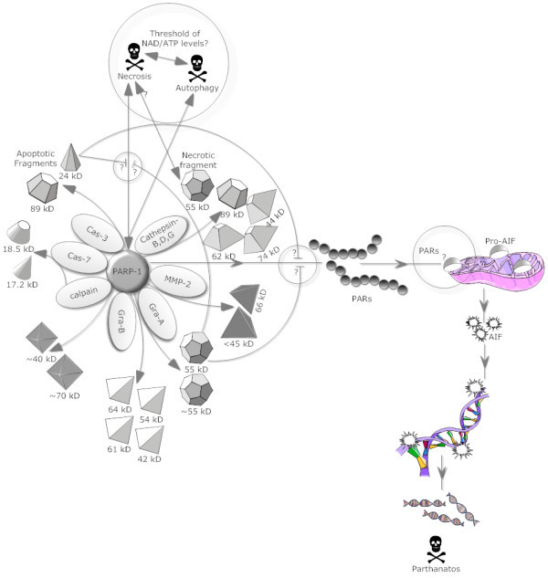
The normal function of poly (ADP-ribose) polymerase-1 (PARP-1) is the routine repair of DNA damage by adding poly (ADP ribose) polymers in response to a variety of cellular stresses. Recently, it has become widely appreciated that PARP-1 also participates in diverse physiological and pathological functions from cell survival to several forms of cell death and has been implicated in gene transcription, immune responses, inflammation, learning, memory, synaptic functions, angiogenesis and aging. In the CNS, PARP inhibition attenuates injury in pathologies like cerebral ischemia, trauma and excitotoxicity demonstrating a central role of PARP-1 in these pathologies. PARP-1 is also a preferred substrate for several 'suicidal' proteases and the proteolytic action of suicidal proteases (caspases, calpains, cathepsins, granzymes and matrix metalloproteinases (MMPs)) on PARP-1 produces several specific proteolytic cleavage fragments with different molecular weights. These PARP-1 signature fragments are recognized biomarkers for specific patterns of protease activity in unique cell death programs. This review focuses on specific suicidal proteases active towards PARP-1 to generate signature PARP-1 fragments that can identify key proteases and particular forms of cell death involved in pathophysiology. The roles played by some of the PARP-1 fragments and their associated binding partners in the control of different forms of cell death are also discussed.
Figures
Similar articles
-
Differential PARP cleavage: an indication of heterogeneous forms of cell death and involvement of multiple proteases in the infarct of focal cerebral ischemia in rat.Cell Mol Neurobiol. 2009 Jun;29(4):563-73. doi: 10.1007/s10571-009-9348-8. Epub 2009 Feb 19. Cell Mol Neurobiol. 2009. PMID: 19225880
-
Cleavage of poly(ADP-ribose) polymerase: a sensitive parameter to study cell death.Biochem Cell Biol. 1997;75(4):337-49. Biochem Cell Biol. 1997. PMID: 9493956 Review.
-
Characterization of the necrotic cleavage of poly(ADP-ribose) polymerase (PARP-1): implication of lysosomal proteases.Cell Death Differ. 2001 Jun;8(6):588-94. doi: 10.1038/sj.cdd.4400851. Cell Death Differ. 2001. PMID: 11536009
-
Gain-of-function of poly(ADP-ribose) polymerase-1 upon cleavage by apoptotic proteases: implications for apoptosis.J Cell Sci. 2001 Oct;114(Pt 20):3771-8. doi: 10.1242/jcs.114.20.3771. J Cell Sci. 2001. PMID: 11707529
-
Poly(ADP-Ribose) polymerase-1 in acute neuronal death and inflammation: a strategy for neuroprotection.Ann N Y Acad Sci. 2003 May;993:217-28; discussion 287-8. doi: 10.1111/j.1749-6632.2003.tb07532.x. Ann N Y Acad Sci. 2003. PMID: 12853316 Review.
Cited by
-
Quantitative spectrofluorometric assay detecting nuclear condensation and fragmentation in intact cells.Sci Rep. 2021 Jun 7;11(1):11921. doi: 10.1038/s41598-021-91380-3. Sci Rep. 2021. PMID: 34099803 Free PMC article.
-
ATR and p-ATR are emerging prognostic biomarkers and DNA damage response targets in ovarian cancer.Ther Adv Med Oncol. 2020 Dec 23;12:1758835920982853. doi: 10.1177/1758835920982853. eCollection 2020. Ther Adv Med Oncol. 2020. PMID: 33854565 Free PMC article.
-
How Do Post-Translational Modifications Influence the Pathomechanistic Landscape of Huntington's Disease? A Comprehensive Review.Int J Mol Sci. 2020 Jun 16;21(12):4282. doi: 10.3390/ijms21124282. Int J Mol Sci. 2020. PMID: 32560122 Free PMC article. Review.
-
Time-Point Dependent Activation of Autophagy and the UPS in SOD1G93A Mice Skeletal Muscle.PLoS One. 2015 Aug 5;10(8):e0134830. doi: 10.1371/journal.pone.0134830. eCollection 2015. PLoS One. 2015. PMID: 26244336 Free PMC article.
-
Sarcoma stratification by combined pH2AX and MAP17 (PDZK1IP1) levels for a better outcome on doxorubicin plus olaparib treatment.Signal Transduct Target Ther. 2020 Sep 23;5(1):195. doi: 10.1038/s41392-020-00246-z. Signal Transduct Target Ther. 2020. PMID: 32963243 Free PMC article.
References
-
- Benjamin RC, Gill DM. Poly(ADP-ribose) synthesis in vitro programmed by damaged DNA. A comparison of DNA molecules containing different types of strand breaks. J Biol Chem. 1980;255:10502–10508. - PubMed
LinkOut - more resources
Full Text Sources
Other Literature Sources
Miscellaneous
