. 2010 Dec 13;191(6):1127-39.
doi: 10.1083/jcb.201005046.

Molecular architecture of a dynamin adaptor: implications for assembly of mitochondrial fission complexes

Affiliations

Molecular architecture of a dynamin adaptor: implications for assembly of mitochondrial fission complexes

Sajjan Koirala et al. J Cell Biol. .

Abstract

Recruitment and assembly of some dynamin-related guanosine triphosphatases depends on adaptor proteins restricted to distinct cellular membranes. The yeast Mdv1 adaptor localizes to mitochondria by binding to the membrane protein Fis1. Subsequent Mdv1 binding to the mitochondrial dynamin Dnm1 stimulates Dnm1 assembly into spirals, which encircle and divide the mitochondrial compartment. In this study, we report that dimeric Mdv1 is joined at its center by a 92-Å antiparallel coiled coil (CC). Modeling of the Fis1-Mdv1 complex using available crystal structures suggests that the Mdv1 CC lies parallel to the bilayer with N termini at opposite ends bound to Fis1 and C-terminal β-propeller domains (Dnm1-binding sites) extending into the cytoplasm. A CC length of appropriate length and sequence is necessary for optimal Mdv1 interaction with Fis1 and Dnm1 and is important for proper Dnm1 assembly before membrane scission. Our results provide a framework for understanding how adaptors act as scaffolds to orient and stabilize the assembly of dynamins on membranes.


Figures

Figure 1.
Figure 1.
Mdv1 self-assembles via a dimeric, antiparallel CC. (A, top) Domain structure of Mdv1, including the NTE, predicted CC, and WD repeats predicted to form a β-propeller. (bottom) The construct used for purification of the Mdv1 CC domain includes the MBP fused to 10xHis, the PreScission protease cleavage site (PPCS), and Mdv1 residues 231–299 (CC; MBP-10xHis-PPCS-CC). (B) SDS-PAGE analysis of purified MBP-10xHis-PPCS-CC fusion protein stained with Coomassie brilliant blue (lane 1), PreScission protease–cleaved MBP-10xHis-PPCS + CC (lane 2), and purified CC (lane 3). (C) Sedimentation equilibrium profile of the Mdv1 CC fragment at the indicated initial loading concentrations (open symbols) with the corresponding fit of 16,263 D (MWobs/MWmonomer = 1.95). Residuals for the nonlinear least-squared fits are shown below. (D) CD wavelength scan of CC231–299 dimer. (E) Crystal structure of CC231–292 dimer at a 2.6-Å resolution. Hydrophobic side chains of residues L233, L237, I251, I254, L268, I272, and I275 are shown in magenta.
Figure 2.
Figure 2.
Disruption of Mdv1 CC formation blocks mitochondrial fission. (A) Schematic of the mdv1CC7 mutant protein. Substitutions introduced at a and d positions in the CC to generate the mdv1CC7 mutant protein are listed below the diagram. (B) Steady-state abundance of C-terminal HA-tagged Mdv1 and mdv1CC7 mutant proteins expressed in adaptor Δ cells. The mdv1CC7 protein sometimes migrates at a higher molecular mass than WT Mdv1 (Figs. 3 A, 4 A, and 6). WCEs separated by SDS-PAGE were immunoblotted with anti-HA and anti-porin antibodies. (C) Lysates from cells expressing the indicated C-terminal Myc- and HA-tagged Mdv1 proteins were used for IP with anti-Myc agarose beads. Lysates (bottom) and immunoprecipitated fractions (top) were analyzed by SDS-PAGE and Western blotting with anti-Myc and anti-HA antibodies. Asterisks mark protein breakdown products. (D) Cartoons depicting mitochondrial morphologies scored in quantification experiments as WT or fission mutant. (E and F) Quantification of mitochondrial morphologies in adaptor Δ (E) or WT (F) cells expressing Mdv1 and mdv1CC7 mutant proteins. Black and gray bars and error bars represent the mean and standard deviation of at least three independent experiments (n = 100).
Figure 3.
Figure 3.
CC formation promotes mitochondrial recruitment and assembly of the Mdv1 fission adaptor. (A) Subcellular localization of N-terminal GFP-tagged Mdv1 and mdv1CC7 proteins imaged in adaptor Δ cells. (B) Mitochondrial distribution of GFP-tagged Mdv1 and mdv1CC7 proteins imaged in adaptor Δ cells. Bars and error bars represent the mean and standard deviation of at least three independent experiments (n = 100). (C) Representative images of GFP-Mdv1 and GFP-mdv1CC7 localization and distribution quantified in A and B. DIC, mitochondrial matrix–targeted dsRed (mito-RFP), and merged RFP and GFP images are shown. Bar, 5 µm.
Figure 4.
Figure 4.
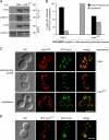
Efficient Dnm1–Mdv1 interaction and Dnm1 assembly into functional mitochondrial fission complexes requires Mdv1 CC formation. (A) Lysates from cells expressing the indicated C-terminal HA-tagged Mdv1 proteins were used for IP with anti-HA agarose beads. Lysate (bottom) and immunoprecipitated fractions (top) were analyzed by SDS-PAGE and Western blotting with anti-HA and anti-Dnm1 antibodies. Asterisks mark protein breakdown products. (B) Localization of genomically expressed GFP-Dnm1 in cells expressing Mdv1 or mdv1CC7. Bars and error bars represent the mean and standard deviation of at least three independent experiments (n = 100). (C) Representative images of GFP-Dnm1 localization quantified in B. DIC, mitochondrial matrix–targeted RFP (mito-RFP), GFP-Dnm1, and merged RFP and GFP images are shown. (D) Colocalization of RFP-mdvCC7 and GFP-Dnm1. DIC and merged RFP with GFP images are shown. Bars, 5 µm.
Figure 5.
Figure 5.
Dimerization via a heterologous antiparallel CC partially restores Mdv1 adaptor function. (A) Schematic representation of the mdv1HR2 chimera. Residues 230–294 encompassing the Mdv1 antiparallel CC are replaced by residues 674–734 of the Mfn1 HR2 domain. (B) Steady-state abundance of C-terminal HA-tagged Mdv1 and mdv1HR2 mutant proteins expressed in fission adaptor Δ cells. WCEs separated by SDS-PAGE were immunoblotted with anti-HA and anti-porin antibodies. (C) Differential sedimentation and Western blot analysis of cytoplasmic 3PGK, mitochondrial porin, and HA-tagged Mdv1 and mdv1HR2 proteins in WCE, postmitochondrial supernatant (PMS), and mitochondrial (Mito) fractions. (D) Lysates from cells expressing the indicated C-terminal Myc- and HA-tagged Mdv1 and mdv1HR2 proteins were used for IP with anti-Myc agarose beads. Lysate (bottom) and immunoprecipitated fractions (top) were analyzed by SDS-PAGE and Western blotting with anti-HA and anti-Myc antibodies. (E) Quantification of mitochondrial morphologies in adaptor Δ cells expressing Mdv1, mdv1CC7 mutant, or mdv1HR2 chimeric proteins. Bars and error bars represent the mean and standard deviation of at least three independent experiments (n = 100). (F) Lysates from cells expressing HA-tagged Mdv1 or mdv1HR2 were used for IP with anti-HA agarose beads. Lysates (bottom) and immunoprecipitated fractions (top) were analyzed by SDS-PAGE and Western blotting with anti-HA and anti-Dnm1 antibodies. Asterisks mark protein breakdown products.
Figure 6.
Figure 6.
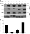
The Mdv1 CC sequence contributes to efficient Fis1 binding. (A) Lysates from cells expressing the indicated Mdv1-HA variants and Myc-Fis1 were used for coIP with anti-Myc agarose beads. Lysate (bottom) and IP fractions (top) were analyzed by SDS-PAGE and ECL Western blotting with anti-HA and anti-Myc antibodies. Lower molecular mass bands in all lanes are protein breakdown products. (B) Quantification of coIPs shown in A. Myc- and HA-tagged proteins were detected using a fluorescent secondary antibody followed by scanning on an imaging system. The mean intensity of each Mdv1-HA protein band was normalized to the Myc-Fis1 signal in the same coIP. Bars represent the abundance of Mdv1-HA signal in each coIP relative to the WT. Error bars represent the mean and standard deviation from three independent experiments.
Figure 7.
Figure 7.
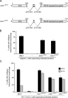
Effect of CC shortening on Mdv1 function. (A) Schematic of the mdv1Δ2HR and mdv1Δ4HR proteins. Residues corresponding to HR deletions are shown below each diagram. (B) Quantification of mitochondrial morphologies in adaptor Δ cells expressing Mdv1, mdv1Δ2HR, and mdv1Δ4HR proteins. (C) Quantification of mitochondrial morphologies at 25 and 37°C in fis1-3 mdv1Δ cells expressing WT and mutant Mdv1 proteins. Black and gray bars and error bars represent the mean and standard deviation of at least three independent experiments (n = 100).
Figure 8.
Figure 8.
Molecular architecture of the mitochondrial dynamin-related receptor. A structural model of the Mdv1 dimer (green/purple) bound to two Fis1 cytoplasmic domains (red) anchored in the outer mitochondrial membrane (gray). The model was generated from crystal structures of the Fis1 cytoplasmic domain in complex with a fission adaptor NTE (Protein Data Bank [PDB] accession no. 2PQR) and the Mdv1 antiparallel CC (PDB accession no. 2XU6). The C-terminal WD40 domain of Mdv1 is shown as a homology model based on the known structure of the Cdc4 WD40 repeat (PDB 1NEX). Loops of variable length and unknown structure connect these two regions to the central CC dimer.

Similar articles

Cited by

References

    1. Amiott E.A., Cohen M.M., Saint-Georges Y., Weissman A.M., Shaw J.M. 2009. A mutation associated with CMT2A neuropathy causes defects in Fzo1 GTP hydrolysis, ubiquitylation, and protein turnover. Mol. Biol. Cell. 20:5026–5035 10.1091/mbc.E09-07-0622 - DOI - PMC - PubMed
    1. Bhar D., Karren M.A., Babst M., Shaw J.M. 2006. Dimeric Dnm1-G385D interacts with Mdv1 on mitochondria and can be stimulated to assemble into fission complexes containing Mdv1 and Fis1. J. Biol. Chem. 281:17312–17320 10.1074/jbc.M513530200 - DOI - PubMed
    1. Bleazard W., McCaffery J.M., King E.J., Bale S., Mozdy A., Tieu Q., Nunnari J., Shaw J.M. 1999. The dynamin-related GTPase Dnm1 regulates mitochondrial fission in yeast. Nat. Cell Biol. 1:298–304 10.1038/13014 - DOI - PMC - PubMed
    1. Collaborative Computational Project 4 1994. The CCP4 suite: programs for protein crystallography. Acta Crystallogr. D. Biol. Crystallogr. D50:760–763 - PubMed
    1. Cerveny K.L., Jensen R.E. 2003. The WD-repeats of Net2p interact with Dnm1p and Fis1p to regulate division of mitochondria. Mol. Biol. Cell. 14:4126–4139 10.1091/mbc.E03-02-0092 - DOI - PMC - PubMed

Publication types

MeSH terms

Substances