. 2011 Mar 18;286(11):9382-92.
doi: 10.1074/jbc.M110.203638. Epub 2010 Dec 9.

The BH3 alpha-helical mimic BH3-M6 disrupts Bcl-X(L), Bcl-2, and MCL-1 protein-protein interactions with Bax, Bak, Bad, or Bim and induces apoptosis in a Bax- and Bim-dependent manner

Affiliations

The BH3 alpha-helical mimic BH3-M6 disrupts Bcl-X(L), Bcl-2, and MCL-1 protein-protein interactions with Bax, Bak, Bad, or Bim and induces apoptosis in a Bax- and Bim-dependent manner

Aslamuzzaman Kazi et al. J Biol Chem. .

Abstract

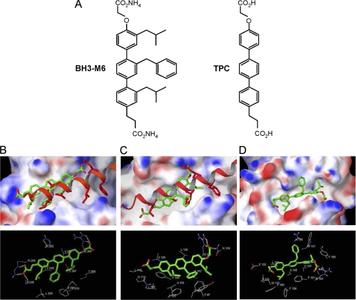
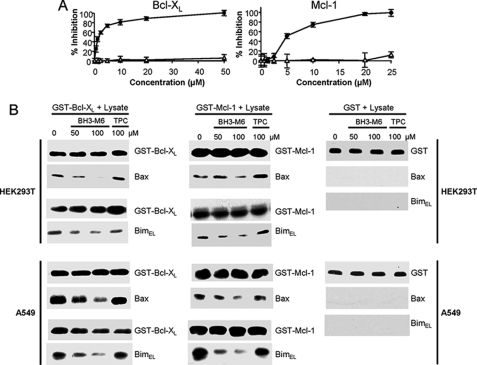
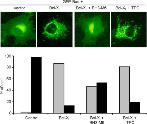
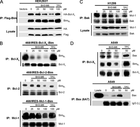
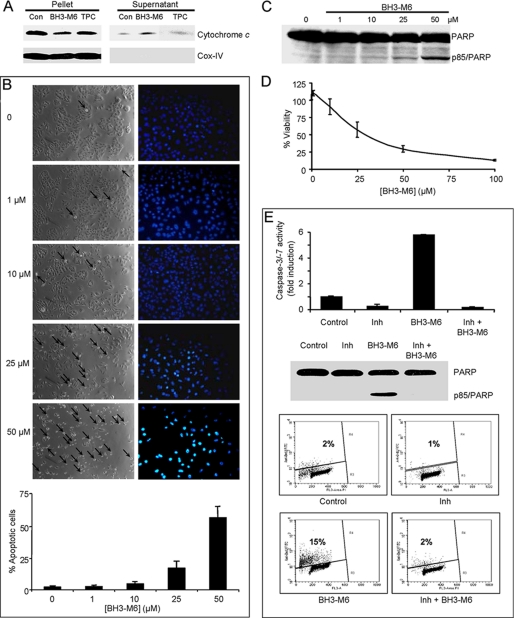
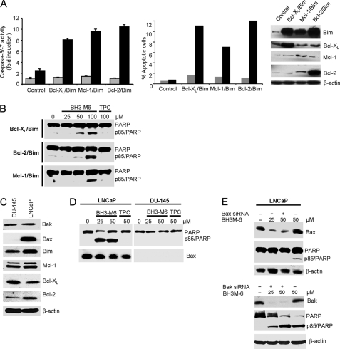
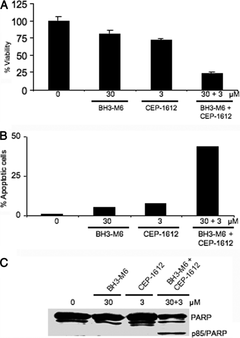
A critical hallmark of cancer cell survival is evasion of apoptosis. This is commonly due to overexpression of anti-apoptotic proteins such as Bcl-2, Bcl-X(L), and Mcl-1, which bind to the BH3 α-helical domain of pro-apoptotic proteins such as Bax, Bak, Bad, and Bim, and inhibit their function. We designed a BH3 α-helical mimetic BH3-M6 that binds to Bcl-X(L) and Mcl-1 and prevents their binding to fluorescently labeled Bak- or Bim-BH3 peptides in vitro. Using several approaches, we demonstrate that BH3-M6 is a pan-Bcl-2 antagonist that inhibits the binding of Bcl-X(L), Bcl-2, and Mcl-1 to multi-domain Bax or Bak, or BH3-only Bim or Bad in cell-free systems and in intact human cancer cells, freeing up pro-apoptotic proteins to induce apoptosis. BH3-M6 disruption of these protein-protein interactions is associated with cytochrome c release from mitochondria, caspase-3 activation and PARP cleavage. Using caspase inhibitors and Bax and Bak siRNAs, we demonstrate that BH3-M6-induced apoptosis is caspase- and Bax-, but not Bak-dependent. Furthermore, BH3-M6 disrupts Bcl-X(L)/Bim, Bcl-2/Bim, and Mcl-1/Bim protein-protein interactions and frees up Bim to induce apoptosis in human cancer cells that depend for tumor survival on the neutralization of Bim with Bcl-X(L), Bcl-2, or Mcl-1. Finally, BH3-M6 sensitizes cells to apoptosis induced by the proteasome inhibitor CEP-1612.


Figures

FIGURE 1.
FIGURE 1.
Molecular docking studies of the interactions between BH3-M6 and Mcl-1, Bcl-XL, and Bcl-2. A, chemical structures of BH3-M6 and TPC. B–D, BH3-M6 docked to Mcl-1, Bcl-XL, and Bcl-2. B, crystal structure of human Bim BH3 helix bound to human Mcl-1 with the side chains of Bim Glu-55, Ile-58, Leu-62, Ile-65, Asp-67, and Phe-69, relative to the BH3-M6 position. The protein Mcl-1 is shown by its molecular surface. Area with positive electrostatic potential is colored blue and that with negative electrostatic potential colored red (the same presentation is used for proteins Bcl-XL and Bcl2). B, lower panel, residues of Mcl-1 interacting with BH3-M6. Yellow dotted lines indicate hydrogen bonds. C, crystal structure of mouse Bim BH3 helix bound to mouse Bcl-XL with the side chains of Bim Ile-90, Leu-94, Ile-97, and Asp-99, relative to the BH3-M6 position. C, lower panel, residues of Bcl-XL interacting with BH3-M6. Yellow dotted lines indicate hydrogen bonds. D, BH3-M6 docked to the original NMR structure of Bcl-2. D, lower panel, residues of Bcl-2 that interact with BH3-M6. The alignment between human and mouse Bim BH3 α-helix is as follows: Human (58-I A Q E L R R I G D E F N A Y-72) Mouse (90-I A Q E L R R I G D E F N E T-104).
FIGURE 2.
FIGURE 2.
BH3-M6, but not TPC, inhibits the binding of anti-apoptotic proteins to pro-apoptotic proteins in vitro. A, BH3-M6 blocks the binding of Bak BH3 α-helix to Bcl-XL (left) or Bim BH3 α-helix to Mcl-1 (right) as measured by fluorescence polarization assay, black circles: BH3-M6, open triangles: TPC. B, BH3-M6 inhibits the interaction of Bcl-XL or Mcl-1 with Bax and Bim as measured by GST pull-down assay. Purified GST, GST-Bcl-XL, and GST-Mcl-1 fusion proteins immobilized on glutathione-Sepharose beads were incubated for 1 h with 0, 50, and 100 μm BH3-M6 or 100 μm TPC, followed by addition of cell extracts from HEK293T or A549 cells for 1 h. Proteins associated with beads were eluted and analyzed by Western blotting with Bax and Bim antibodies. We used GST antibodies either for negative controls (in lysates incubated with GST beads) or to demonstrate equal input (in lysates incubated with GST-Bcl-XL or GST-Mcl-1 beads).
FIGURE 3.
FIGURE 3.
BH3-M6 inhibits the Bcl-XL interaction with Bad in intact cells. BH3-M6 inhibits Bcl-XL-induced GFP-Bad mitochondrial localization. COS-7 cells were grown in DMEM supplemented with 10% FBS and antibiotics, transfected with the indicated constructs, treated and analyzed as described under “Experimental Procedures.” The upper panel shows cells expressing pEGFP-Bad and pcDNA3-HA-Bcl-XL. The lower panel represents the quantification of the fluorescence data (black: diffuse, gray: punctate cells). At least 85 transfected cells per sample were counted in three different fields of view.
FIGURE 4.
FIGURE 4.
BH3-M6 inhibits the interaction between Bcl-XL, Bcl-2, and Mcl-1 with Bim, Bak, or Bax in intact cells. A, co-immunoprecipitation from HEK293T cells. HEK293T cells were co-transfected with HA-Bcl-XL and Flag-BimEL for 18 h. Cells were exposed to 0, 25, 50, and 100 μm of BH3-M6 or 100 μm of TPC for 2 h at 37 °C, lysed, and subjected to immunoprecipitation with anti-FLAG-M2 beads. The resulting immune complexes, as well as total lysates, were analyzed by Western blotting with the indicated antibodies. B, co-immunoprecipitation from MDA-MB-468 cells expressing Bcl-XL-IRES-BimEL, Bcl-2-IRES-BimEL, and Mcl-1-IRES-BimEL. Cells were grown in 100-mm plates and treated with 0, 25, 50, and 100 μm BH3-M6 or 100 μm TPC for 24 h at 37 °C, lysed, and subjected to immunoprecipitation with Bcl-XL, Bcl-2, and Mcl-1 antibodies. The resulting immune complexes were analyzed by Western blotting with Bim, Bcl-XL, Bcl-2, and Mcl-1 antibodies. C, co-immunoprecipitation from H1299 cells. H1299 non-small lung cancer cells were grown in RPMI 1640 medium plus 10% FBS, antibiotics and 1% sodium pyruvate, 1% HEPES, and 1.1% glucose. They were seeded in 100-mm plates and treated with 0, 25, 50, and 100 μm BH3-M6 or 100 μm TPC for 24 h at 37 °C, lysed and subjected to immunoprecipitation with Bak antibody. The resulting immune complexes were analyzed by Western blotting with the indicated antibodies. D, co-immunoprecipitation from A549 cells. A549 cells were grown in F-12K medium plus 10% FBS and antibiotics and then serum-starved for 20 h, followed by treatment with 0, 25, and 50 μm of BH3-M6 or 50 μm of TPC for 1 h at 37 °C. Cells were then lysed and subjected to immunoprecipitation with Bcl-XL antibody (upper panel) and Bax 6A7 antibody (lower panel). The resulting immune complexes were analyzed by Western blotting with the antibodies indicated on the right.
FIGURE 5.
FIGURE 5.
BH3-M6 induces apoptosis in a caspase-dependent manner. A, BH3-M6 releases cytochrome c from mitochondria. Mitochondria from A549 cells were incubated at 37 °C with 0, 50 μm BH3-M6, or TPC for 60 min. Cytochrome c release was determined as described under “Experimental Procedures.” B–D, A549 cells were treated with the indicated BH3-M6 concentrations for up to 72 h and then processed for various assays as described under “Experimental Procedures.” B, BH3-M6 induces apoptosis after 48 h of exposure to BH3-M6, as shown by phase images of cells (left panel) or DAPI nuclear staining (right panel). The arrows point toward cells in the process of rounding. The images from phase and DAPI are from different experiments. The graph below the images shows the percent of apoptotic cells and is the quantification of DAPI nuclear staining. Error bars indicate standard deviations of triplicates. C, BH3-M6 induces PARP cleavage after 48 h of drug treatment. D, BH3-M6 decreases the viability of A549 cells in a dose-dependent manner as measured by MTT assay after 72 h of treatment. Error bars indicate standard deviations of triplicates. E, upper panel, activation of caspase-3/-7 by BH3-M6 is blocked by a pan-caspase inhibitor. DoHH2 cells were pretreated with 50 μm Boc-d-FMK, a pan-caspase inhibitor, followed by a co-treatment with 50 μm BH3-M6 for 24 h. Caspase-3/-7 activity was determined as described under “Experimental Procedures.” Results shown represent one of two independent experiments in triplicate. Error bars indicate standard deviations of triplicates. E, middle panel, PARP cleavage was measured by Western blotting, lower panel, extent of apoptosis was measured by TUNEL assay.
FIGURE 6.
FIGURE 6.
BH3-M6 induces apoptosis in a Bim-, Bcl-2-, Bcl-XL-, and Mcl-1-dependent manner. BH3-M6 induces caspase-3/-7 activities and apoptosis in Bcl-XL/Bim-, Bcl-2/Bim-, and Mcl-1/Bim-overexpressing cells. MDA-MB-468 cells transfected with Bcl-XL-IRES-BimEL, Bcl-2-IRES-BimEL, and Mcl-1-IRES-BimEL were grown in 100-mm plates and treated with 0, 25, 50, or 100 μm BH3-M6 or 100 μm of TPC for 24 h at 37 °C. Cells were then harvested and lysed using 0.2% Nonidet P-40 lysis buffer (without protease and phosphatase inhibitors for cell-free caspase assay and with protease and phosphatase inhibitors for Western blot analysis). A, caspase-3/-7 activity was determined by incubating whole-cell extracts with caspase-3/-7 substrate and measuring free AMCs (left panel), apoptosis was quantified by TUNEL assay (middle panel), gray: TPC, black: BH3-M6. BimEL, Bcl-XL, Bcl-2, and Mcl-1 expression levels were determined by Western blotting (right panel). B, PARP cleavage by Western blotting. C, expression levels of different anti- and pro-apoptotic proteins in DU-145 and LNCaP cells as determined by Western blotting. D, BH3-M6 induces apoptosis in LNCaP human prostate cancer cells expressing Bax, but not in DU-145 human prostate cancer cells lacking detectable Bax expression. LNCaP and DU-145 cells were treated with 0, 25, and 50 μm of BH3-M6 or 50 μm of TPC for 24 h. Cells were lysed and subjected to Western blot analysis. E, depletion of Bax, but not Bak by siRNA renders LNCaP cells resistant to apoptosis. LNCaP cells were transfected with 10 nm Bax or Bak siRNA or control siRNA for 48 h, followed by 24 h of treatment with or without 25 or 50 μm BH3-M6. Cells were lysed and subjected to Western blot analysis with the indicated antibodies.
FIGURE 7.
FIGURE 7.
BH3-M6 sensitizes A549 cells to the proteasome inhibitor CEP-1612 to induce apoptosis. A549 cells were treated with the indicated concentrations of either drug alone or in combination and then processed for various assays as described under “Experimental Procedures.” A, cell viability as measured by MTT assay after 72 h of treatment, error bars indicate standard deviations of triplicates. B, apoptosis as determined by TUNEL assay after 48 h of treatment. C, PARP cleavage after 24 h of drug treatment.

Similar articles

Cited by

References

    1. Green D. R., Evan G. I. (2002) Cancer Cell 1, 19–30 - PubMed
    1. Wei M. C., Zong W. X., Cheng E. H., Lindsten T., Panoutsakopoulou V., Ross A. J., Roth K. A., MacGregor G. R., Thompson C. B., Korsmeyer S. J. (2001) Science 292, 727–730 - PMC - PubMed
    1. Korsmeyer S. J. (1999) Cancer Res. 59, 1693s–1700s - PubMed
    1. Hengartner M. O. (2000) Nature 407, 770–776 - PubMed
    1. Adams J. M., Cory S. (2007) Oncogene 26, 1324–1337 - PMC - PubMed

Publication types

MeSH terms

Substances