. 2011 Feb;85(4):1706-17.
doi: 10.1128/JVI.02268-10. Epub 2010 Dec 8.

Hepatitis C virus NS2 coordinates virus particle assembly through physical interactions with the E1-E2 glycoprotein and NS3-NS4A enzyme complexes

Affiliations

Hepatitis C virus NS2 coordinates virus particle assembly through physical interactions with the E1-E2 glycoprotein and NS3-NS4A enzyme complexes

Kenneth A Stapleford et al. J Virol. 2011 Feb.

Abstract

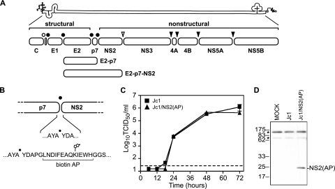
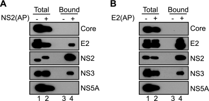
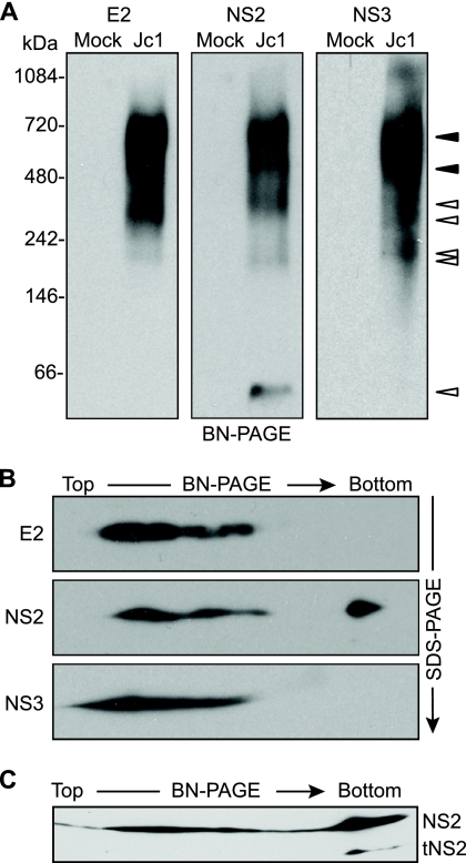
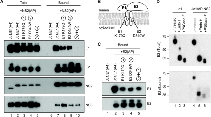
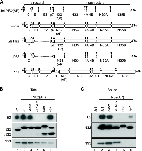
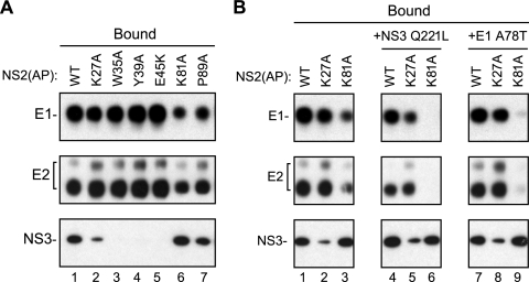
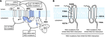
The hepatitis C virus (HCV) NS2 protein is essential for particle assembly, but its function in this process is unknown. We previously identified critical genetic interactions between NS2 and the viral E1-E2 glycoprotein and NS3-NS4A enzyme complexes. Based on these data, we hypothesized that interactions between these viral proteins are essential for HCV particle assembly. To identify interaction partners of NS2, we developed methods to site-specifically biotinylate NS2 in vivo and affinity capture NS2-containing protein complexes from virus-producing cells with streptavidin magnetic beads. By using these methods, we confirmed that NS2 physically interacts with E1, E2, and NS3 but did not stably interact with viral core or NS5A proteins. We further characterized these protein complexes by blue native polyacrylamide gel electrophoresis and identified ≈ 520-kDa and ≈ 680-kDa complexes containing E2, NS2, and NS3. The formation of NS2 protein complexes was dependent on coexpression of the viral p7 protein and enhanced by cotranslation of viral proteins as a polyprotein. Further characterization indicated that the glycoprotein complex interacts with NS2 via E2, and the pattern of N-linked glycosylation on E1 and E2 suggested that these interactions occur in the early secretory pathway. Importantly, several mutations that inhibited virus assembly were shown to inhibit NS2 protein complex formation, and NS2 was essential for mediating the interaction between E2 and NS3. These studies demonstrate that NS2 plays a central organizing role in HCV particle assembly by bringing together viral structural and nonstructural proteins.


Figures

FIG. 1.
FIG. 1.
HCV genome and NS2 affinity purification system. (A) HCV genome and polyprotein. The open bullet represents a signal peptide peptidase cleavage site; closed bullets represent signal peptidase cleavage sites; the open arrowhead represents the NS2-3 cysteine autoprotease cleavage site; closed arrowheads represent NS3-4A serine protease cleavage sites. (B) Schematic of the AP inserted into the N terminus of NS2. The biotinylated lysine residue is indicated. (C) Time course of Jc1 and Jc1/NS2(AP) virus production after RNA electroporation. Viral infectivities are represented as tissue-culture infectious doses (50% endpoint) per ml (TCID50/ml). Values are averages of results from three independent transfections; errors represent standard deviations from the means. The dotted line represents the limit of detection of our titering assay. (D) HCV NS2(AP) is biotinylated in Huh-7.5(BirA) cells. Huh-7.5(BirA) cells were transfected with Jc1 and Jc1/NS2(AP) RNA and harvested 48 h posttransfection. Cells were lysed in SDS-PAGE buffer, and proteins were separated by SDS-PAGE and transferred to PVDF membranes. Biotinylated proteins were visualized by using the Vectastain ABC kit. Asterisks indicate two endogenously biotinylated mitochondrial proteins, most likely pyruvate carboxylase (upper band) and propionyl coenzyme A carboxylase (lower band) (29).
FIG. 2.
FIG. 2.
HCV NS2 interacts with E2 and NS3 in virus-producing cells. (A) Specific affinity purification of NS2(AP)-containing complexes from cells transfected with Jc1 or Jc1/NS2(AP), as indicated. Biotinylated proteins were captured on streptavidin beads and eluted into SDS-PAGE sample buffer, as described in Materials and Methods. Proteins present in lysates or eluates from streptavidin beads were separated by SDS-PAGE, transferred to PVDF membranes, and immunoblotted for the indicated viral proteins as described in Materials and Methods. “Total” indicates proteins in the clarified lysates; “Bound” indicates proteins captured by strepavidin beads; minus signs indicate untagged Jc1; plus signs Jc1/NS2(AP). (B) Specific affinity purification of E2(AP)-containing complexes from cells transfected with Jc1 or Jc1/E2(AP), as indicated. Samples were prepared and analyzed as in panel A.
FIG. 3.
FIG. 3.
E2, NS2, and NS3 form discrete, membrane-associated high-molecular-mass complexes. (A) BN-PAGE separation of E2-, NS2-, and NS3-containing protein complexes. Microsomes were prepared at 48 h postelectroporation of Huh-7.5 cells with Jc1 RNA or cells transfected without RNA (Mock). Proteins were solubilized in 0.5% (vol/vol) digitonin, separated under native conditions on a 4 to 16% Bis-Tris polyacrylamide gel, and transferred to PVDF. The blot was cut into three strips and immunoblotted for E2, NS2, and NS3. Open arrowheads indicate bands that were specific for a given protein; closed arrowheads indicate bands in which E2, NS2, and NS3 comigrated (see Results). (B) 2D BN/SDS-PAGE analysis of E2-, NS2-, and NS3-containing protein complexes. Microsome-associated protein (20 μg) was solubilized in digitonin and separated in the first dimension by BN-PAGE and in the second dimension on a 10% SDS-PAGE gel. E2, NS2, and NS3 were detected by immunoblotting. (C) Microsome-associated protein (40 μg) was solubilized in digitonin and separated in the first dimension by BN-PAGE and in the second dimension on a 12% SDS-PAGE gel. NS2 was detected by immunoblotting.
FIG. 4.
FIG. 4.
Characterization of the NS2-associated glycoproteins. (A) NS2 interacts with E1-E2 through E2. NS2(AP) was affinity captured from Huh-7.5(BirA) cell lysates at 48 h postelectroporation with the indicated constructs and analyzed as in Fig. 2A. All constructs encoded the A4 epitope in the E1 protein; the indicated constructs also encoded AP-tagged NS2, with or without mutations in E1 and/or E2 that blocked E1-E2 heterodimerization. (B) Schematic of glycoprotein heterodimerization mutations. (C) E1 K179Q and E2 D349W mutations inhibit E1-E2 heterodimerization. E2(AP) was affinity captured from Huh-7.5(BirA) cell lysates at 48 h postelectroporation with the indicated constructs and analyzed as in Fig. 2A. All constructs encoded the A4 epitope in the E1 protein; the indicated constructs also encoded AP-tagged E2; the indicated constructs also contained mutations in E1 and/or E2 that blocked E1-E2 heterodimerization. (D) NS2-interacting E2 contains high-mannose glycans. (Top) Cell lysates from Jc1- or Jc1/NS2(AP)-transfected Huh-7.5(BirA) cells were treated with Endo H, PNGase F, or left untreated. Proteins were separated by SDS-PAGE, transferred to a PVDF membrane, and immunoblotted for E2. (Bottom) NS2(AP)-associated E2 was cocaptured on streptavidin beads as in Fig. 2A, treated with Endo H, PNGase F, or left untreated, separated by SDS-PAGE, transferred to a PVDF membrane, and detected by immunoblotting.
FIG. 5.
FIG. 5.
NS2 complex formation is enhanced by cotranslation as a polyprotein. (A) Schematic of bicistronic HCV constructs containing insertions of a stop codon and ECMV IRES element (indicated by E·I). Protein cleavage sites are labeled as in Fig. 1A; the AP tag is indicated by a black box. (B) Affinity purification of NS2(AP)-containing complexes. Huh-7.5(BirA) cells were transfected with the indicated constructs and samples were prepared and analyzed as in Fig. 2A. (C) Bicistronic HCV constructs replicate efficiently. Huh-7.5(BirA) cells were split at 48 h posttransfection with the indicated viral RNAs, reseeded, and allowed to grow for an additional 48 h. Total cellular RNAs were extracted, and the level of HCV RNA was quantified by quantitative reverse transcription (qRT)-PCR as previously described (37). Jc1 GNN was a replication-defective Jc1 genome containing inactivating mutations in the NS5B RNA polymerase active site. The y axis represents copies of HCV genome per 10 ng of total RNA. (D) Bicistronic HCVcc constructs produced reduced viral titers. Media were collected from Huh-7.5(BirA) cells at 48 h posttransfection with the indicated viral RNAs, and titers were determined on Huh-7.5 cells. Values represent averages of results from three independent experiments; error bars represent the standard deviations from the means.
FIG. 6.
FIG. 6.
HCV p7 is essential for NS2 complex assembly. (A) Schematic of deletion constructs. HCV genomes are annotated as in Fig. 5A. In-frame deletions are indicated by dashed lines. (B) Expression of E2, NS2, and NS3. Cellular lysates were prepared at 48 h posttransfection with the indicated constructs and analyzed by SDS-PAGE and immunoblotting with antibodies specific for E2, NS2, or NS3. (C) Affinity purification of NS2(AP)-associated proteins from deletion constructs. NS2(AP) was affinity captured from the lysates in panel A and analyzed as in Fig. 2A.
FIG. 7.
FIG. 7.
Mutations in NS2 inhibit NS2 protein-protein interactions. (A) NS2(AP) was affinity captured from cell lysates at 48 h postelectroporation of Huh-7.5(BirA) cells with Jc1/NS2(AP), either with or without (WT) the indicated mutations in NS2. NS2(AP)-associated proteins were detected as in Fig. 2A. (B) Affinity purification of NS2(AP)-associated proteins with (K27A, K81A) or without (WT) mutations in NS2 and with (+NS3 Q221L, +E1 A78T) or without second-site suppressor mutations. NS2(AP)-associated proteins were detected as in Fig. 2A. This panel is a composite created from a single exposure; it is representative of additional experiments.
FIG. 8.
FIG. 8.
NS2 plays a central role in coordinating interaction between the E1-E2 glycoprotein and NS3-4A enzyme complexes. (A) Structure of Jc1/E2(AP) and ΔNS2. Constructs are labeled as in Fig. 5A. (B) NS2 is required for E2-NS3 interaction. Protein lysates were prepared from Huh-7.5(BirA) cells transfected with the indicated constructs at 48 h postelectroporation. NS3 present in lysates (Total) or eluates from streptavidin beads (Bound) was separated by SDS-PAGE, transferred to PVDF membranes, and detected by immunoblotting. (C) NS2 mutations inhibit E2-NS3 interaction. Huh-7.5(BirA) cells were transfected with Jc1/E2(AP) or Jc1/E2(AP) containing the indicated NS2 mutations, and lysates were prepared 48 h postelectroporation. E2(AP)-associated NS3 protein (Bound) was detected as in Fig. 2B and 8A.
FIG. 9.
FIG. 9.
Model of HCV NS2 complex assembly. (A) NS2 complex. Proteins within the NS2 complex (E1, E2, NS2, and NS3) are indicated; proteins suspected to be in the NS2 complex (p7, NS4A) are shown with question marks. White arrows denote interactions identified in this work; boxes and thick black lines indicate inhibitors of these interactions. For contrast, NS4A and one monomer of NS2 are shown in blue. (B) NS2 mutations that inhibit NS2-NS3, NS2-E2, or E2-NS3 interaction. The locations of these mutations are mapped onto a model of the NS2 transmembrane domains (23, 55); the cysteine protease domain is not shown.

Similar articles

Cited by

References

    1. Appel, N., U. Herian, and R. Bartenschlager. 2005. Efficient rescue of hepatitis C virus RNA replication by trans-complementation with nonstructural protein 5A. J. Virol. 79:896-909. - PMC - PubMed
    1. Appel, N., T. Pietschmann, and R. Bartenschlager. 2005. Mutational analysis of hepatitis C virus nonstructural protein 5A: potential role of differential phosphorylation in RNA replication and identification of a genetically flexible domain. J. Virol. 79:3187-3194. - PMC - PubMed
    1. Appel, N., et al. 2008. Essential role of domain III of nonstructural protein 5A for hepatitis C virus infectious particle assembly. PLoS Pathog. 4:e1000035. - PMC - PubMed
    1. Bartenschlager, R., L. Ahlborn-Laake, J. Mous, and H. Jacobsen. 1994. Kinetic and structural analyses of hepatitis C virus polyprotein processing. J. Virol. 68:5045-5055. - PMC - PubMed
    1. Beckett, D., E. Kovaleva, and P. J. Schatz. 1999. A minimal peptide substrate in biotin holoenzyme synthetase-catalyzed biotinylation. Protein Sci. 8:921-929. - PMC - PubMed

Publication types

MeSH terms

Substances