. 2010 Dec 1;24(23):2627-39.
doi: 10.1101/gad.1978310.

Constitutive mTORC1 activation by a herpesvirus Akt surrogate stimulates mRNA translation and viral replication

Affiliations

Constitutive mTORC1 activation by a herpesvirus Akt surrogate stimulates mRNA translation and viral replication

Uyanga Chuluunbaatar et al. Genes Dev. .

Abstract

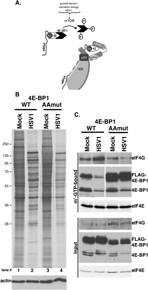
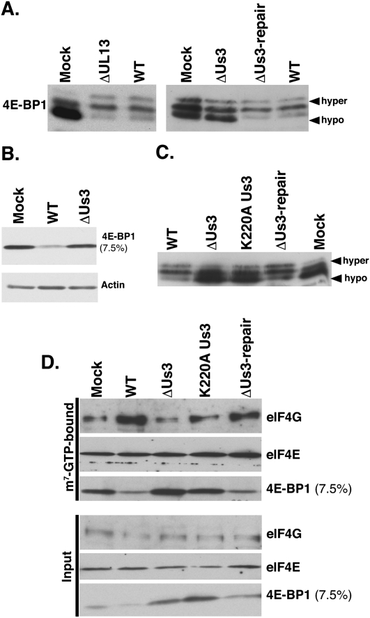
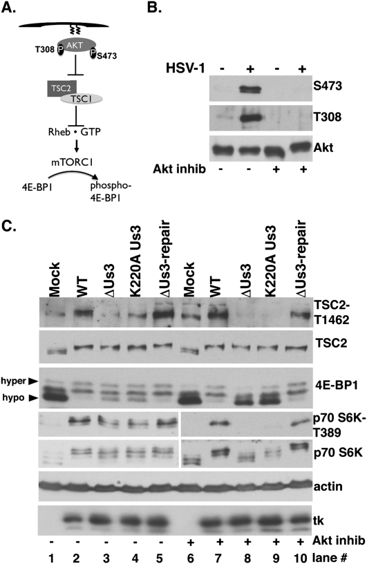
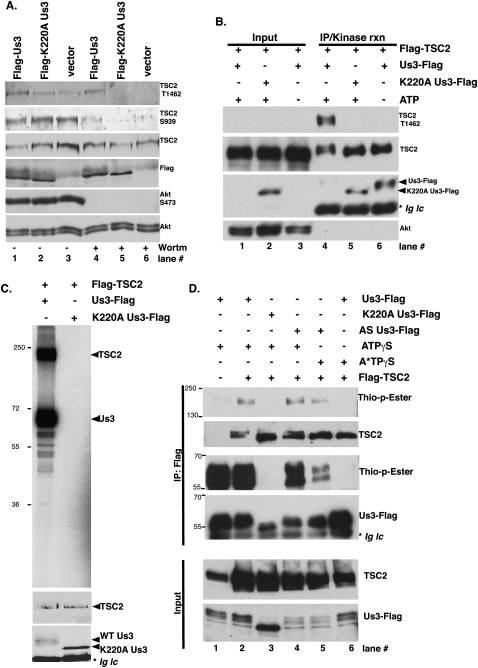
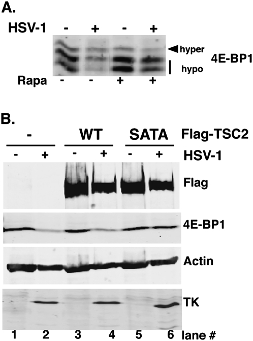
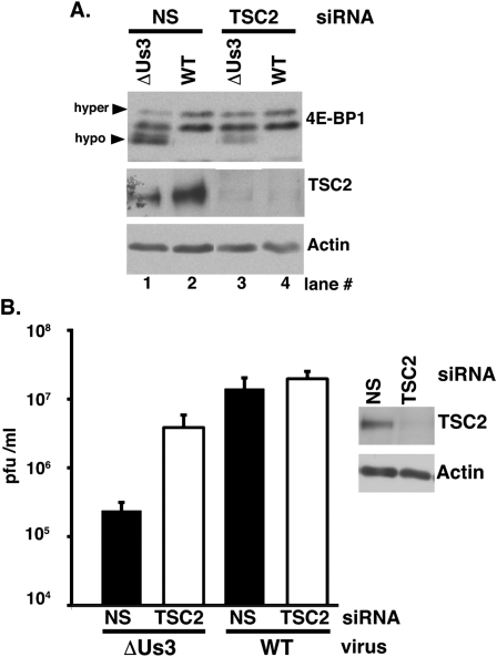
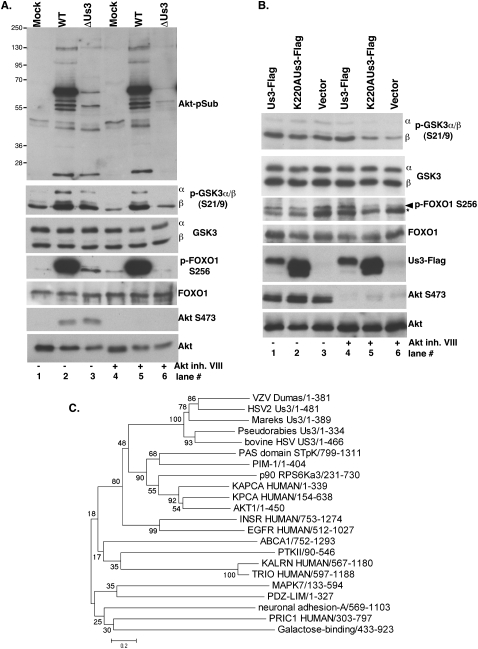
All viruses require cellular ribosomes to translate their mRNAs. Viruses producing methyl-7 (m⁷) GTP-capped mRNAs, like Herpes Simplex Virus-1 (HSV-1), stimulate cap-dependent translation by activating mTORC1 to inhibit the translational repressor 4E-binding protein 1 (4E-BP1). Here, we establish that the HSV-1 kinase Us3 masquerades as Akt to activate mTORC1. Remarkably, Us3 displays no sequence homology with the cellular kinase Akt, yet directly phosphorylates tuberous sclerosis complex 2 (TSC2) on the same sites as Akt. TSC2 depletion rescued Us3-deficient virus replication, establishing that Us3 enhances replication by phosphorylating TSC2 to constitutively activate mTORC1, effectively bypassing S6K-mediated feedback inhibition. Moreover, Us3 stimulated Akt substrate phosphorylation in infected cells, including FOXO1 and GSK3. Thus, HSV-1 encodes an Akt surrogate with overlapping substrate specificity to activate mTORC1, stimulating translation and virus replication. This establishes Us3 as a unique viral kinase with promising drug development potential.


Figures

Figure 1.
Figure 1.
Regulation of eIF4F assembly by site-specific 4E-BP1 phosphorylation in HSV-1-infected cells. (A) Inactivation of the translational repressor 4E-BP1 by mTOR. mTOR activation in response to different stimuli results in 4E-BP1 hyperphosphorylation and release from the cap-binding protein eIF4E. Subsequent eIF4E incorporation into a multisubunit initiation factor complex containing eIF4G and eIF4A allows recruitment of eIF3-bound 40S subunits to the mRNA 5′ end. (B) NHDFs stably expressing epitope-tagged wild-type (WT) or AA 4E-BP1 growth-arrested by serum deprivation were mock-infected or infected with HSV-1 (MOI = 5). Cultures were metabolically pulse-labeled for 1 h with 35S-amino acids at 14 hpi. Total protein was subsequently isolated and fractionated by SDS-PAGE, and the fixed, dried gel was exposed to X-ray film. Migration of molecular mass standards (in kilodaltons) appears to the left. (Bottom panel) Immunoblot loading control probed with anti-actin antibody. (C) Soluble extracts prepared from NHDFs described in B were incubated with m7GTP-Sepharose. After washing, input (bottom panel) and bound (top panel) fractions were separated by SDS-PAGE and analyzed by immunoblotting with the indicated antisera. Both anti-4E-BP1 panels in the m7GTP-bound group were from the same membrane.
Figure 2.
Figure 2.
Inactivation of 4E-BP1 in HSV-1-infected cells requires the virus-encoded Us3 Ser/Thr kinase. (A) Growth-arrested NHDFs were mock-infected (Mock) or infected (MOI = 5) with wild-type HSV-1 (WT), a UL13-deficient mutant (ΔUL13), a Us3-deficient virus (ΔUs3), or a virus where the Us3 deficiency was repaired (ΔUs3 REPAIR). At 18 hpi, total protein was isolated, fractionated by SDS-PAGE (17.5% gel), and analyzed by immunoblotting using anti-4E-BP1. Slow-migrating hyperphosphorylated (hyper) and fast-migrating hypophosphorylated (hypo) 4E-BP1 forms are noted on the right. (B) As in A, except total protein was fractionated using a 7.5% gel, which does not resolve phosphorylated 4E-BP1 isoforms. (C) As in A, except a virus expressing a catalytically inactive Us3 protein was included (Us3 K220A contains a single Lys-to-Ala substitution at residue 220). (D) Soluble extracts prepared from NHDFs described in A were incubated with m7GTP-Sepharose and analyzed as described in Figure 1C.
Figure 3.
Figure 3.
Inhibition of Akt does not suppress Us3-dependent 4E-BP1 and TSC2 phosphorylation in HSV-1-infected cells. (A) Activation of mTORC1 by Akt. Once plasma membrane localized via a lipid-binding plekstrin homology domain, Akt is activated by T308/S473 phosphorylation. Akt inhibits TSC rheb-GAP activity by phosphorylating TSC2 on T1462/S939, promoting rheb•GTP-mediated mTORC1 activation and subsequent 4E-BP1 hyperphosphorylation. (B) Growth-arrested NHDFs were mock-infected (−) or infected (MOI = 5) with wild-type (WT) HSV-1 (+). After 4 h, cells were treated with DMSO or Akt inhibitor VIII (5 μM). At 9 hpi, total protein was fractionated by SDS-PAGE and analyzed by immunoblotting with indicated antibodies (S473, T308-phospho specific Akt antibodies, and Akt total). (C) As in B, except ΔUs3, K220A Us3, and Us3 repair were included. Total protein was isolated at 15 hpi, fractionated by SDS-PAGE, and analyzed by immunoblotting with the indicated antisera: TSC2-T1462 (phospho-specific T1462 antibody), TSC2 (total TSC2), anti-4E-BP1, anti-HSV-1 tk antibody, S6K-T389 (phospho-specific T389 p70 S6K), and total p70 S6K. Different phospho-4E-BP1 forms were resolved (hyper vs. hypo) as described in Figure 2A.
Figure 4.
Figure 4.
Us3 Ser/Thr kinase directly phosphorylates TSC2. (A) U20S cells were transfected with plasmids expressing Flag-Us3, Flag-K220AUs3, or an empty vector control (vector). After 24 h, cells were treated with DMSO or 200 nM wortmannin for 30 min to eliminate PI3K-mediated, Akt-dependent TSC2 phosphorylation. Total protein was isolated, fractionated by SDS-PAGE, and analyzed by immunoblotting with the indicated antibodies. (B) Us3 stimulates TSC2 phosphorylation in vitro. HEK 293 cells were transiently transfected with plasmids expressing Flag-TSC2 or individual Us3 expression plasmids described in A. After 24 h, soluble cell-free fractions from Flag-TSC2 and Flag-Us3 transfected cells were mixed and immunoprecipitated using anti-Flag antibody. After a high-salt wash to reduce nonspecific binding, immune complexes were re-equilibrated in kinase buffer (±ATP) and incubated for 30 min at 30°C. Samples were subsequently fractionated by SDS-PAGE and analyzed by immunoblotting using the indicated antibodies. Wild-type Flag-Us3 autophosphorylates in the presence of ATP and thus migrates slower (Sagou et al. 2009). The band marked with an asterisk represents immunoglobulin light chain (Ig lc). (C, top panel) As in B, except kinase reactions contained [γ32P]-ATP and the fixed dried gel exposed to X-ray film. (Middle panel) TSC2 immunoblot. (Bottom panel) Anti-Flag immunoblot. The band marked with an asterisk represents immunoglobulin light chain. (D) Soluble cell-free fractions from cells transfected and immunoprecipitated as in B were incubated with the indicated ATP derivatives for 60 min at 30°C. Following thiophosphate alkylation using p-nitrobenzylmesylate (PNBM), samples were fractionated by SDS-PAGE and analyzed by immunoblotting using the indicated antibodies. The band marked with an asterisk in the anti-Flag blot represents immunoglobulin light chain (Ig lc).
Figure 5.
Figure 5.
Inactivation of the 4E-BP1 translational repressor in HSV-1-infected cells requires site-specific TSC2 phosphorylation. (A) 293 cells were mock-infected (−) or infected (+) with HSV1 (MOI = 10) and, after 3 h, were treated with DMSO or rapamycin. Total protein was collected at 12 hpi, fractionated by SDS-PAGE (17.5% gel), and immunoblotted with anti-4E-BP1 antibody. (B) 293 cells were transiently transfected with either empty vector (−), pcDNA3-Flag-TSC2 (wild type [WT]), or pcDNA3-Flag-TSC2SATA (SATA). After 6 h, cells were serum-starved for 18 h and subsequently mock-infected or infected (MOI = 10) with HSV-1. At 12 hpi, total protein was fractionated by SDS-PAGE (7.5% gel) and analyzed by immunoblotting with the indicated antibodies.
Figure 6.
Figure 6.
TSC2 restricts 4E-BP1 repressor inactivation and viral replication in cells infected with a Us3-deficient virus. (A) NHDFs treated with nonsilencing (NS) or TSC2-specific siRNAs were either mock-infected or infected (MOI = 5) with Us3-deficient (ΔUs3) or wild-type (WT) HSV-1. After 15 h, total protein was harvested, fractionated by SDS-PAGE, and analyzed by immunoblotting with the indicated antisera. Phospho-4E-BP1 forms were resolved (hyper vs. hypo) as described in Figure 2A. (B) NHDFs treated with siRNAs as in A were infected (MOI = 1 × 10⁻⁴) with the indicated viruses. After 4 d, the virus produced was quantified by plaque assay in Vero cells. (Right panel) Immunoblot illustrating efficiency and persistence of TSC2 siRNA depletion after 4 d.
Figure 7.
Figure 7.
Stimulation of Akt substrate phosphorylation by Us3. (A) NHDFs were mock-infected (Mock) or infected (MOI = 5) with Us3-deficient (ΔUs3) or wild-type HSV-1 (WT) in the presence (+) or absence (−) of Akt inhibitor VIII. At 16 hpi, total protein was isolated, fractionated by SDS-PAGE, and analyzed by immunoblotting with the indicated antisera. (B) U2OS cells transfected with the indicated plasmids (described in Fig. 4A) were treated with DMSO (−) or Akt inhibitor VIII (+). Proteins were analyzed as described in Figure 4A. In the p-FOXO immunoblot, the asterisk represents a nonspecific band insensitive to Akt inhibitor routinely observed in U2OS cells. (C) α Herpesvirus Us3 protein sequences (HSV-1, VZV, HSV-2, bovine herpesvirus, Mareks, and Pseudorabies) were aligned using ClustalW2 (Larkin et al. 2007), with a broad selection of kinases representing the range of subfamilies in the InterPro Protein Kinase superfamily (CL0016), including AKT1 (AAL55732), KAPCA (P17612), KPCA (P17252), p90_RPS6Ka3 (NP_004577), PIM-1 (P11309), ABCA1 (NP_005493), PAS_domain_STpK (NP_055963), PTKII (NP_751911), MAPK7 (O43318), INSR (P06213), EGFR (P00533), KALRN (O60229), TRIO (O75962), neuronal adhesion A (NP_001032209), Galactose-binding (O14786), PDZ-LIM (O00151), and PRIC1 (Q96MT3). The evolutionary history was inferred using the Minimum Evolution (ME) method (Rzhetsky and Nei 1992). The optimal tree with the sum of the branch length = 16.70884448 is shown. The percentage of replicate trees in which the associated taxa clustered together in the bootstrap test (500 replicates) are shown next to the branches (Felsenstein 1985). The tree is drawn to scale, with branch lengths in the same units as those of the evolutionary distances used to infer the phylogenetic tree. The evolutionary distances were computed using the Poisson correction method and are in the units of the number of amino acid substitutions per site. The ME tree was searched using the Close Neighbor Interchange (CNI) algorithm (Nei and Kumar 2000) at a search level of 1. The neighbor-joining algorithm (Saitou and Nei 1987) was used to generate the initial tree. All positions containing gaps and missing data were eliminated from the data set (complete deletion option). There were a total of 137 positions in the final data set. Phylogenetic analyses were conducted in MEGA4 (Tamura et al. 2007).

Comment in

Similar articles

Cited by

References

    1. Allen JJ, Li M, Brinkworth CS, Paulson JL, Wang D, Hübner A, Chou WH, Davis RJ, Burlingame AL, Messing RO, et al. 2007. A semisynthetic epitope for kinase substrates. Nat Methods 4: 511–516 - PMC - PubMed
    1. Arias C, Walsh D, Harbell J, Wilson AC, Mohr I 2009. Activation of host translational control pathways by a viral developmental switch. PLoS Pathog 5: e1000334 doi: 10.1371/journal.ppat.1000334 - PMC - PubMed
    1. Benetti L, Roizman B 2004. Herpes simplex virus protein kinase US3 activates and functionally overlaps protein kinase A to block apoptosis. Proc Natl Acad Sci 101: 9411–9416 - PMC - PubMed
    1. Benetti L, Roizman B 2006. Protein kinase B/Akt is present in activated form throughout the entire replicative cycle of ΔU(S)3 mutant virus but only at early times after infection with wild-type herpes simplex virus 1. J Virol 80: 3341–3348 - PMC - PubMed
    1. Biondi RM, Nebreda AR 2003. Signaling specificity of ser/thr protein kinases through docking-site mediated interactions. Biochem J 372: 1–13 - PMC - PubMed

Publication types

MeSH terms