. 2011 Feb;85(4):1604-14.
doi: 10.1128/JVI.01608-10. Epub 2010 Dec 1.

The Epstein-Barr virus-encoded BILF1 protein modulates immune recognition of endogenously processed antigen by targeting major histocompatibility complex class I molecules trafficking on both the exocytic and endocytic pathways

Affiliations

The Epstein-Barr virus-encoded BILF1 protein modulates immune recognition of endogenously processed antigen by targeting major histocompatibility complex class I molecules trafficking on both the exocytic and endocytic pathways

Jianmin Zuo et al. J Virol. 2011 Feb.

Abstract

Despite triggering strong immune responses, Epstein-Barr virus (EBV) has colonized more than 90% of the adult human population. Successful persistence of EBV depends on the establishment of a balance between host immune responses and viral immune evasion. Here we have extended our studies on the EBV-encoded BILF1 protein, which was recently identified as an immunoevasin that functions by enhancing degradation of major histocompatibility complex class I (MHC-I) antigens via lysosomes. We now demonstrate that disruption of the EKT signaling motif of BILF1 by a K122A mutation impairs the ability of BILF1 to enhance endocytosis of surface MHC-I molecules, while subsequent lysosomal degradation was impaired by deletion of the 21-residue C-terminal tail of BILF1. Furthermore, we identified another mechanism of BILF1 immunomodulation: it targets newly synthesized MHC-I/peptide complexes en route to the cell surface. Importantly, although the diversion of MHC-I on the exocytic pathway caused a relatively modest reduction in cell surface MHC-I, presentation of endogenously processed target peptides to immune CD8(+) effector T cells was reduced by around 65%. The immune-modulating functions of BILF1 in the context of the whole virus were confirmed in cells lytically infected with a recombinant EBV in which BILF1 was deleted. This study therefore extends our initial observations on BILF1 to show that this immunoevasin can target MHC-I antigen presentation via both the exocytic and endocytic trafficking pathways. The results also emphasize the merits of including functional T cell recognition assays to gain a more complete picture of immunoevasin effects on the antigen presentation pathway.


Figures
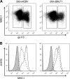
FIG. 1.
FIG. 1.
Surface HLA class I expression in wt 293 cells and ΔBILF1 293 cells expressing late lytic antigens. 293 epithelial cells carrying recombinant wt EBV or ΔBILF1 EBV were induced into the EBV lytic cycle by transfecting a BZLF1 expression plasmid. At 24 h postinduction, these two cell lines were stained for surface HLA class I and intracellular gp110 and then analyzed by flow cytometry. (A) Two-color analysis of surface MHC-I and intracellular viral gp110 expression, showing the gates used to analyze latent (gp110-negative) and lytic (gp110-positive) populations. (B) Histograms of surface MHC-I expression on latent cells (solid line histogram) and lytic cells (dotted line histogram). The shaded histogram shows the isotype control staining.
FIG. 2.
FIG. 2.
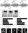
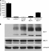
The EKT signaling motif and a C-terminal domain of BILF1 cooperate for degradation of MHC-I. (A) Schematic representation of the mutated BILF1 proteins, showing the seven transmembrane helices, the location of the K122A mutation in the DRY-like EKT motif, and the truncation of the C terminus. (B) HEK293 cells stably transduced with control (PQC), wt BILF1, K122A mutant BILF1, ΔC mutant BILF1, or K122A/ΔC mutant BILF1 retroviruses were analyzed by Western blotting. Total cell lysates from 105 cells were separated by SDS-PAGE and analyzed by Western blotting with MAbs specific for BILF1 (3F10; anti-HA tag), MHC-I (HC10), or with polyclonal antibodies to calregulin as a loading control. (C) Histogram showing the mean results of quantification of Western blotting results by densitometry from three independent experiments. The densities of the HC10 bands were normalized relative to their own calregulin loading control. All results are expressed as amounts of total MHC-I expression as a percentage of the expression in PQC-293 cells, and error bars indicate standard deviations of triplicate experiments. (D) Histogram showing the mean results of quantification of surface MHC-I expression by flow cytometry. Viable cells of the same panel of lines as shown in panel C were stained with PE-labeled W6/32 MAb and analyzed by flow cytometry. All results are expressed as the amount of surface MHC-I staining as a percentage of the staining in PQC-293 cells, and error bars indicate standard deviations of triplicate experiments. (E) Immunofluorescence staining with W6/32 MAb to HLA class I complexes in fixed cell cultures of HEK293 cells transduced with control retrovirus (PQC control) or retroviruses expressing wt BILF1, K122A BILF1, ΔC BILF1, or K122A/ΔC BILF1.
FIG. 3.
FIG. 3.
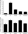
Signaling functions of BILF1 mutants and the requirement of the EKT signaling motif for enhanced endocytosis of surface MHC-I. (A) Control (PQC), wt BILF1, K122A BILF1, ΔC BILF1, or K122A/ΔC BILF1 expression plasmids were transfected into HEK293 cells together with an NF-κB reporter plasmid and a Renilla luciferase reporter construct. The degree of NF-κB activation was measured by detection of luciferase activity. The results are the means ± standard deviations for three independent experiments which were themselves performed in triplicate. (B) Assay for the rate of internalization of cell surface MHC-I complexes. HEK293 cells stably transduced with different recombinant retroviruses were incubated at 0°C with saturating concentrations of W6/32 MAb to MHC-I and then washed and incubated at 37°C for 20 min. The viable cells were then stained with PE-conjugated goat anti-mouse IgG antibody and analyzed by flow cytometry. The mean fluorescence intensities of staining were averaged for triplicate samples. The histogram shows the percentages of internalized MHC-I in 20 min. The results are the means of three independent experiments, and error bars indicate standard deviations of three experiments.
FIG. 4.
FIG. 4.
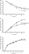
The K122A/ΔC double mutant BILF1 selectively reduces the rate of appearance of surface MHC-I. Internalization, appearance, and recycling assays were performed on HEK293 cells stably transduced with PQC control, wt BILF1, or K122A/ΔC mutant BILF1 retroviruses. (A) Internalization assay. As for Fig. 2B, cells were treated with saturating concentrations of W6/32 MAb to MHC-I and then washed and incubated at 37°C for up to 60 min. At the indicated times, viable cells were stained with PE-conjugated goat anti-mouse IgG antibody and analyzed by flow cytometry. The mean fluorescence intensities of staining were averaged for triplicate samples and then normalized to the time zero samples; error bars indicating standard deviations of triplicate samples are shown for all samples, although the errors were often smaller than the symbols used in the graphs and are therefore not always visible. (B) Appearance assay. Cells were incubated at 0°C with saturating concentrations of W6/32 MAb to MHC-I molecules and then washed and incubated at 37°C for different periods of time. The appearance of new MHC-I molecules was assayed by staining with PE-conjugated W6/32 antibody. The mean fluorescence intensities of staining were averaged for triplicate samples; error bars indicate standard deviations of triplicate samples. (C) Recycling assay. Cells were incubated at 0°C with saturating concentrations of W6/32 MAb to MHC-I and then washed and incubated at 37°C for 30 min before stripping the remaining surface-bound W6/32 MAb and resuming incubation at 37°C for the indicated periods of time. The cells were then stained with PE-conjugated goat anti-mouse IgG antibody and analyzed by flow cytometry. The mean fluorescence intensities of staining were averaged for triplicate samples and are expressed as the percentage of the amount of MHC-I internalized in the initial 30-min incubation; error bars indicate standard deviations of triplicate samples.
FIG. 5.
FIG. 5.
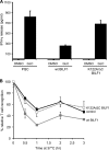
BILF1-mediated enhanced endocytosis of MHC-I from cell surface modulates antigen presentation to CD8+ T cells. HEK293 cells stably transduced with control (PQC), wt BILF1, or K122A/ΔC mutant BILF1 recombinant retroviral vectors were pulsed with synthetic GLCTLVAML peptide and used as targets in T cell assays. (A) Peptide-pulsed cells were extensively washed to remove unbound peptide, and replicate aliquots of cells were fixed with 1% PFA. Following coculture of the fixed peptide-pulsed cells with GLC effector CD8+ T cells for 18 h, the supernatants were tested for the release of IFN-γ as a measure of T cell recognition. All results are expressed as IFN-γ release (in pg/ml), and error bars indicate standard deviations of triplicate cultures. (B) Peptide-pulsed cells (PQC 293 controls, wild-type BILF1-293, and K122A/ΔC BILF1-293) were extensively washed to remove unbound peptide and then incubated at 37°C for up to 3 h. At the indicated periods of time, triplicate aliquots of peptide-pulsed cells were fixed with 1% PFA and were cocultured with GLC effector CD8+ T cells for a further 18 h. The supernatants were tested for the release of IFN-γ as a measure of T cell recognition. All results are expressed as IFN-γ release as a percentage of that observed at time zero. Error bars indicate standard deviations of triplicate cultures.
FIG. 6.
FIG. 6.
Diversion of MHC-I from the exocytic pathway by BILF1 modulates antigen presentation to CD8+ T cells. (A) HEK293 cells were cotransfected with a BMLF1 expression vector together with control (PQC), wt BILF1, K122A BILF1, ΔC BILF1, or K122A/ΔC BILF1 expression plasmids. At 24 h posttransfection, the cells were cocultured with GLC CD8+ effector T cells (specific for a BMLF1-derived peptide) for a further 18 h, and the supernatants were tested for the release of IFN-γ as a measure of T cell recognition. All results are expressed as IFN-γ release (in pg/ml), and error bars indicate standard deviations of triplicate cultures. (B) Total cell lysates were generated from aliquots of the above target cell transfections and were analyzed by Western blotting using antibodies specific for HA-tagged BILF1(3F10; anti-HA tag), BMLF1, or calregulin as a loading control.
FIG. 7.
FIG. 7.
A role for BILF1 in modulating T cell recognition during the lytic cycle in B cells. (A) The proportions of LCLs spontaneously reactivating into the lytic cycle in EBV ΔBILF1 and EBV wt LCLs were assessed by intracellular BZLF1 staining and analysis by flow cytometry. (B) CD8+ T cell recognition of wt, ΔBILF1, and ΔBZLF1 LCLs using three HLA-A*0201-restricted CD8+ effector clones specific for IE, E, or DE lytic cycle antigens. Effector clone YVL was specific for BRLF1; clone GLC was specific for BMLF; clone TLD was specific for BMRF1. T cell recognition of the HLA-A*0201-matched LCL targets was measured in an IFN-γ ELISA. All results are expressed as IFN-γ release (in pg/ml), and error bars indicate standard deviations of triplicate cultures.

Similar articles

Cited by

References

    1. Arenzana-Seisdedos, F., et al. 1993. Phosphatidylcholine hydrolysis activates NF-kappa B and increases human immunodeficiency virus replication in human monocytes and T lymphocytes. J. Virol. 67:6596-6604. - PMC - PubMed
    1. Atkinson, P. G., H. J. Coope, M. Rowe, and S. C. Ley. 2003. Latent membrane protein 1 of Epstein-Barr virus stimulates processing of NF-kappa B2 p100 to p52. J. Biol. Chem. 278:51134-51142. - PubMed
    1. Barnstable, C. J., et al. 1978. Production of monoclonal antibodies to group A erythrocytes, HLA and other human cell surface antigens-new tools for genetic analysis. Cell 14:9-20. - PubMed
    1. Beisser, P. S., et al. 2005. The Epstein-Barr virus BILF1 gene encodes a G protein-coupled receptor that inhibits phosphorylation of RNA-dependent protein kinase. J. Virol. 79:441-449. - PMC - PubMed
    1. Bonifacino, J. S., and L. M. Traub. 2003. Signals for sorting of transmembrane proteins to endosomes and lysosomes. Annu. Rev. Biochem. 72:395-447. - PubMed

Publication types

MeSH terms

Substances

LinkOut - more resources