Identification of new carbohydrate and membrane protein antigens in cardiac xenotransplantation
- PMID: 21119562
- PMCID: PMC10022691
- DOI: 10.1097/TP.0b013e318203c27d
Identification of new carbohydrate and membrane protein antigens in cardiac xenotransplantation
Abstract
Background: α1,3-Galactosyltransferase gene knockout (GTKO) pigs reduced the significance of antibody to galactose alpha 1,3-galactose (Gal) antigens but did not eliminate delayed xenograft rejection (DXR). We hypothesize that DXR of GTKO organs results from an antibody response to a limited number of non-Gal endothelial cell (EC) membrane antigens. In this study, we screened a retrovirus expression library to identify EC membrane antigens detected after cardiac xenotransplantation.
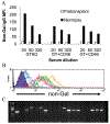
Methods: Expression libraries were made from GT:CD46 and GTKO porcine aortic ECs. Viral stocks were used to infect human embryonic kidney cells (HEK) that were selected by flow cytometry for IgG binding from sensitized cardiac heterotopic xenograft recipients. After three to seven rounds of selection, individual clones were assessed for non-Gal IgG binding. The porcine complementary DNA was recovered by polymerase chain reaction amplification, sequenced, and identified by homology comparisons.
Results: A total of 199 and 317 clones were analyzed from GT:CD46 and GTKO porcine aortic EC complementary DNA libraries, respectively. Sequence analysis identified porcine CD9, CD46, CD59, and the EC protein C receptor. We also identified porcine annexin A2 and a glycosyltransferase with homology to the human β1,4 N-acetylgalactosaminyl transferase 2 gene.
Conclusion: The identified proteins include key EC functions and suggest that non-Gal antibody responses may compromise EC functions and thereby contribute to DXR. Recovery of the porcine β1,4 N-acetylgalactosaminyl transferase 2 suggests that an antibody response to a SD-like carbohydrate may represent a new carbohydrate moiety involved in xenotransplantation. The identification of these porcine gene products may lead to further donor modification to enhance resistance to DXR and further reduce the level of xenograft antigenicity.
Conflict of interest statement
G.W.B. and C.G.A.M. are the inventors of technology related to xenotransplantation that has been licensed by the Mayo Clinic to a commercial entity. The other authors declare no conflict of interest.
Figures


Similar articles
-
Cloning and expression of porcine β1,4 N-acetylgalactosaminyl transferase encoding a new xenoreactive antigen.Xenotransplantation. 2014 Nov-Dec;21(6):543-54. doi: 10.1111/xen.12124. Epub 2014 Sep 1. Xenotransplantation. 2014. PMID: 25176027 Free PMC article.
-
Initial study of α1,3-galactosyltransferase gene-knockout/CD46 pig full-thickness corneal xenografts in rhesus monkeys.Xenotransplantation. 2017 Jan;24(1). doi: 10.1111/xen.12282. Epub 2017 Jan 5. Xenotransplantation. 2017. PMID: 28054735
-
Proteomic identification of non-Gal antibody targets after pig-to-primate cardiac xenotransplantation.Xenotransplantation. 2008 Jul-Aug;15(4):268-76. doi: 10.1111/j.1399-3089.2008.00480.x. Xenotransplantation. 2008. PMID: 18957049 Free PMC article.
-
Cardiac xenotransplantation: progress and challenges.Curr Opin Organ Transplant. 2012 Apr;17(2):148-54. doi: 10.1097/MOT.0b013e3283509120. Curr Opin Organ Transplant. 2012. PMID: 22327911 Free PMC article. Review.
-
Xenotransplantation of solid organs in the pig-to-primate model.Transpl Immunol. 2009 Jun;21(2):87-92. doi: 10.1016/j.trim.2008.10.005. Epub 2008 Oct 26. Transpl Immunol. 2009. PMID: 18955143 Review.
Cited by
-
The immunobiology and clinical use of genetically engineered porcine hearts for cardiac xenotransplantation.Nat Cardiovasc Res. 2022 Aug;1(8):715-726. doi: 10.1038/s44161-022-00112-x. Epub 2022 Aug 9. Nat Cardiovasc Res. 2022. PMID: 36895262 Free PMC article.
-
Bioprosthetic Heart Valves: Upgrading a 50-Year Old Technology.Front Cardiovasc Med. 2019 Apr 11;6:47. doi: 10.3389/fcvm.2019.00047. eCollection 2019. Front Cardiovasc Med. 2019. PMID: 31032263 Free PMC article. Review.
-
Xenotransplantation: Current Status in Preclinical Research.Front Immunol. 2020 Jan 23;10:3060. doi: 10.3389/fimmu.2019.03060. eCollection 2019. Front Immunol. 2020. PMID: 32038617 Free PMC article. Review.
-
B4GALNT2 and xenotransplantation: A newly appreciated xenogeneic antigen.Xenotransplantation. 2018 Sep;25(5):e12394. doi: 10.1111/xen.12394. Epub 2018 Mar 31. Xenotransplantation. 2018. PMID: 29604134 Free PMC article. Review.
-
Carbohydrate antigen microarray analysis of serum IgG and IgM antibodies before and after adult porcine islet xenotransplantation in cynomolgus macaques.PLoS One. 2021 Jun 17;16(6):e0253029. doi: 10.1371/journal.pone.0253029. eCollection 2021. PLoS One. 2021. PMID: 34138941 Free PMC article.
References
-
- Platt JL, Lin SS, McGregor CG. Acute vascular rejection. Xenotransplantation 1998; 5: 169. - PubMed
-
- Cowan PJ. Coagulation and the xenograft endothelium. Xenotransplantation 2007; 14: 7. - PubMed
-
- Byrne GW, Schwarz A, Fesi JR, et al. Evaluation of different alpha-galactosyl glycoconjugates for use in xenotransplantation. Bioconjug Chem 2002; 13: 571. - PubMed
-
- Lai L, Kolber-Simonds D, Park KW, et al. Production of alpha-1,3-galactosyltransferase knockout pigs by nuclear transfer cloning. Science 2002; 295: 1089. - PubMed
-
- Lam TT, Paniagua R, Shivaram G, et al. Anti-non-Gal porcine endothelial cell antibodies in acute humoral xenograft rejection of hDAF-transgenic porcine hearts in cynomolgus monkeys. Xenotransplantation 2004; 11: 531. - PubMed
Publication types
MeSH terms
Substances
Grants and funding
LinkOut - more resources
Full Text Sources
Other Literature Sources
Medical
Miscellaneous

