. 2010 Dec 20;207(13):2959-73.
doi: 10.1084/jem.20092641. Epub 2010 Nov 29.

MCP-induced protein 1 deubiquitinates TRAF proteins and negatively regulates JNK and NF-kappaB signaling

Affiliations

MCP-induced protein 1 deubiquitinates TRAF proteins and negatively regulates JNK and NF-kappaB signaling

Jian Liang et al. J Exp Med. .

Abstract

The intensity and duration of macrophage-mediated inflammatory responses are controlled by proteins that modulate inflammatory signaling pathways. MCPIP1 (monocyte chemotactic protein-induced protein 1), a recently identified CCCH Zn finger-containing protein, plays an essential role in controlling macrophage-mediated inflammatory responses. However, its mechanism of action is poorly understood. In this study, we show that MCPIP1 negatively regulates c-Jun N-terminal kinase (JNK) and NF-κB activity by removing ubiquitin moieties from proteins, including TRAF2, TRAF3, and TRAF6. MCPIP1-deficient mice spontaneously developed fatal inflammatory syndrome. Macrophages and splenocytes from MCPIP1(-/-) mice showed elevated expression of inflammatory gene expression, increased JNK and IκB kinase activation, and increased polyubiquitination of TNF receptor-associated factors. In vitro assays directly demonstrated the deubiquitinating activity of purified MCPIP1. Sequence analysis together with serial mutagenesis defined a deubiquitinating enzyme domain and a ubiquitin association domain in MCPIP1. Our results indicate that MCPIP1 is a critical modulator of inflammatory signaling.


Figures

Figure 1.
Figure 1.
Disruption of MCPIP1 leads to severe inflammatory response and premature death in mice. (a) The structure of the mouse Mcpip1 gene is shown. The targeting strategy deleted exons 4 and 5 and most of 6, which encode the most part of the MCPIP1 protein, including the ZF and Pro domains. (b) Proteins extracted from the intestine, lung, and spleen of mice were analyzed by immunoblotting with MCPIP1-specific antibody. The same membrane was probed with antiactin to show equal loading. The experiment was performed on three independent sets of animals. (c and d) Gross appearance of Mcpip1+/+ and Mcpip1−/− mice (6–8-wk-old male; c), as well as spleens (d, top) and LNs (d, bottom) from these mice. The ruler (left margin) indicates size in centimeters. Three independent sets of animals showed similar results. (e) Survival rate of Mcpip1+/+ and Mcpip1−/− mice (n = 19). (f) ELISA was used to measure serum cytokine levels in WT and KO mice. Data are presented as mean ± SD (n = 6; *, P < 0.05; **, P < 0.01 vs. WT group). (g) BMDMs from WT (open bars) and KO (closed bars) mice were stimulated with PBS or 100 ng/ml LPS for 5 h. ELISA was used to measure cytokine levels in cultured medium. Data are presented as mean ± SD (n = 3; *, P < 0.05; **, P < 0.01 vs. WT group). Results are representative of two independent experiments. (h) Total RNA isolated from BMDMs stimulated with LPS for the indicated time periods was reverse transcribed and analyzed by QPCR with gene-specific primers as indicated. Data are presented as mean ± SD (n = 3). Student’s t test was performed. The experiment was performed on two independent sets of animals with triplicate measurements.
Figure 2.
Figure 2.
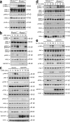
Effect of MCPIP1 on LPS- and IL-1β–induced JNK and NF-κB activation. (a) Proteins extracted from the lung of three pairs of Mcpip1+/+ and Mcpip1−/− mice were analyzed by immunoblot with the indicated antibodies. Results are representative of three independent experiments. (b) BMDMs from Mcpip1−/− or Mcpip1+/+ mice were quiescent for 16 h and then stimulated with 100 ng/ml LPS for the indicated time periods. WCEs were subjected to immunoblot analysis with antibodies as indicated. Similar results were seen in three independent experiments. (c) Raw264.7 cells were transfected with pcDNA3 (vector) or pHA-MCPIP1. After 24 h, the transfected cells were quiescent for 16 h and then stimulated with 100 ng/ml LPS for the indicated time periods. WCEs were subjected to immunoblot analysis with antibodies as indicated. Nuclear fractions (Nuc.) were prepared and subjected to immunoblot analysis using p65-specific antibody. Results are representative of at least three independent experiments. (d) MEFs from Mcpip1−/− or Mcpip1+/+ mice were quiescent for 16 h and stimulated with 10 ng/ml IL-1β for the indicated time periods. WCEs were subjected to immunoblot analysis with antibodies as indicated. (e) HepG2 cells were transfected with pcDNA3 (vector) or pHA-MCPIP1. After 24 h, the transfected cells were quiescent for 16 h and then stimulated with 10 ng/ml IL-1β for the indicated times. WCEs were subjected to immunoblot analysis with antibodies as indicated.
Figure 3.
Figure 3.
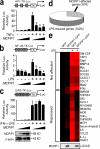
Overexpression of MCPIP1 inhibits NF-κB and AP-1 but not STAT activation. (a–c) Raw264.7 cells were cotransfected AP-1–TK-Luc (a), NF-κB–TK-Luc (b), or GAS-TK-Luc (c) with increasing amounts of pHA-MCPIP1 (0, 25, 50, and 100 ng/well, as indicated by wedges) using Fugene6 (Roche). After quiescence for 16 h, cells were treated with or without 10 ng/ml TNF (a), 100 ng/ml LPS (b), or IFN-γ plus LPS (c). Reporter gene activity was analyzed 24 h later. The data are presented as mean ± SD (n = 4; *, P < 0.01 vs. stimulated without MCPIP1 transfection group). Analysis of variance with Bonferroni correction was performed. Results are representative of at least three independent experiments. Immunoblotting shows expression of HA-MCPIP1. β-Actin served as a loading control. (d) Raw264.7 cells were transfected with pEGFP-N1 or pMCPIP1-GFP plasmids. 16 h later, the transfected cells were treated with or without 1 mg/ml LPS for 8 h. mRNA was labeled and hybridized to the 44K Agilent Mouse Oligo Microarray. 30% of LPS-induced genes were affected by the expression of MCPIP1. (e) 26 selected genes that are induced by LPS >10-fold (some unaffected by MCPIP1 and others affected by MCPIP1) were clustered and presented as a heat map. Red is represented as high expression, whereas black means low expression. The expression of MCPIP1-GFP (93 kD) was detected by immunoblotting with anti-MCPIP1 and shown at the bottom.
Figure 4.
Figure 4.
MCPIP1 acts as a deubiquitinase. (a) HEK293 cells were cotransfected with HA-ubiquitin (Ub) and/or MCPIP1-GFP and/or Flag-TTP. 36 h later, the ubiquitinated proteins in denatured cell lysates were detected by immunoblotting (IB) with HA-specific antibody. The expression of MCPIP1-GFP, Flag-TTP, and actin was also detected by immunoblotting with MCPIP1, Flag, and actin-specific antibodies. Results are representative of at least three independent experiments. (b) Flag-tagged MCPIP1 was immunoprecipitated from transfected HEK293 cells. The purified protein was examined by Coomassie blue staining (Coomas.) and immunoblotting using Flag-specific antibody. (c) 1 µg of purified Flag-MCPIP1 was incubated with K63- or K48-linked pentaubiquitin chains (Ub5) at 37°C for 16 h. Reactions were analyzed by immunoblotting with ubiquitin-specific antibody. Recombinant IsoT was used as a positive control. The blot below the dashed lines had a longer exposure time than the blot above the dashed lines. Long exposure means 2–5 min exposure of the same film. (d) Flag-MCPIP1, 3xFlag elution buffer, immunoprecipitants from empty vector–transfected cells, or Flag-p53 was incubated with K63-linked Ub6 at 37°C for 16 h. Reactions were analyzed by immunoblotting with ubiquitin-specific antibody. HMUb, high molecular weight ubiquitin. (e) In vitro deubiquitination assays using purified Flag-MCPIP1 showing cleavage of high molecular K63-linked polyubiquitin chains (Ub5–8) but not low molecular polyubiquitin chains (Ub2–4) or linear Ub2. (f) In vitro deubiquitination showing that MCPIP1 cleaved Ub8 in time- and dose-dependent manners. (g and h) MCPIP1 deubiquitinase activity was assessed in the presence of the Cys protease inhibitor NEM, 10 mmol/l Zn2+, 10 mmol/l Mn2+, 50 mmol/l EDTA, 10 mmol/l Mg2+, or 10 mmol/l Ca2+. Data in b–h are representative of at least three independent experiments. (i) 0.5, 1, and 2 µg (indicated by wedge) of purified Flag-MCPIP1 was incubated with 80 µM ubiquitin-AFC at 23°C for 5 min. The protease activity was determined by the release of fluorescent AFC (ex: 400 nm, em: 505 nm). 1 µg IsoT was used as a positive control. Values were represented as relative fluorescent units (RFU; mean ± SD; n = 3; *, P < 0.001 vs. none group). Two-way analysis of variance with Bonferroni correction was performed. The top shows a fluorescent image of the reactions in a 96-well plate. Results are representative of two independent experiments. (j) The global cellular ubiquitinated proteins in the splenocytes from Mcpip1+/+ and Mcpip1−/− mice were detected by immunoblotting with ubiquitin-specific antibody. Actin was detected and served as a loading control. Three independent experiments showed similar results.
Figure 5.
Figure 5.
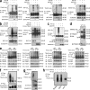
MCPIP1 deubiquitinase targets TRAFs. (a) HEK293 cells were cotransfected with HA-tagged human WT ubiquitin (Ub) with or without MCPIP1-GFP or Flag-TTP. 24 h later, the cells were treated with TNF for 2 h. TRAF2, TRAF6, RIP, and NEMO were immunoprecipitated (IP) and immunoblotted (IB) using anti-HA, anti-TRAF2, anti-TRAF6, anti-RIP, or anti-NEMO as indicated. MCPIP1 expression in WCEs was detected using anti-MCPIP1. IgH, Ig heavy chain. (b) HEK293 cells were cotransfected with HA-ubiquitin, Flag-TRAF2, or Flag-TRAF3 with or without MCPIP1-GFP. 24 h later, the cells were treated with TNF for 2 h. Lysates were subjected to immunoprecipitation and immunoblot with the indicated antibodies. MCPIP1 expression in WCEs was detected using anti-MCPIP1. (c) HEK293 cells were cotransfected with HA-ubiquitin with or without MCPIP1-GFP. After 2 h of TNF and MG132 treatment, the cells were harvested and subjected to immunoprecipitation using IκBα-specific antibody. The immunoprecipitates were examined by immunoblot with anti-HA and anti-IκBα. Data in a–c are representative of at least two independent experiments. (d) Endogenous TRAF2 in the spleens from Mcpip1+/+ and Mcpip1−/− mice was immunoprecipitated with anti-TRAF2, followed by immunoblotting with ubiquitin and TRAF2-specific antibodies. Results are representative of two independent experiments with similar results. (e) Splenocytes from Mcpip1+/+ and Mcpip1−/− mice were stimulated with 100 ng/ml LPS for 30 min. The endogenous TRAF2, TRAF3, TRAF6, and RIP were immunoprecipitated, followed by immunoblotting with antiubiquitin. Results are representative of two independent experiments with similar results. (f and g) Ubiquitinated purified TRAF2 and TRAF3 were incubated with Flag-MCPIP1 at 37°C for 4 h and analyzed by immunoblotting with anti-HA and anti-Flag. The ubiquitin was tagged with HA. Two independent experiments showed similar results. (h) HEK293 cells were cotransfected with Flag-TRAF2 with or without HA-ubiquitin. Flag-TRAF2 protein in cell lysates was purified by anti-Flag matrix and incubated with 3xFlag buffer only, 0.5 µg Flag-BAP, or 0.5 µg IsoT at 37°C for 4 h and analyzed by immunoblotting with anti-HA.
Figure 6.
Figure 6.
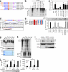
MCPIP1 contains a novel DUB domain. (a) The putative functional domains and conserved regions of MCPIP1 and the deletion strategy are shown. (b) HEK293 cells were cotransfected with HA-ubiquitin (Ub) and with the indicated MCPIP1 constructs. The overall ubiquitination of cellular proteins was examined by immunoblot with HA-specific antibody. Results are representative of at least three independent experiments. (c) The influence of each MCPIP1 construct on NF-κB reporter activity in TNF-stimulated HEK293 cells. Values are expressed as the fold increase in luciferase expression (±SD) compared with the vector alone (n = 4; *, P < 0.01 vs. MCPIP1(1–599) group). Results are representative of three independent experiments. (d) The putative Asp box and Cys box in the NCR are shown. Point mutants of MCPIP1 were generated as marked with asterisks. (e) Alignment of the Cys region of MCPIP1 with that from the UCH-L1 DUB domain. The NCBI Protein database accession numbers for human UCH-L1 and MCPIP1 of human, mouse, rat, zebrafish, and fly are P09936, NP_079355, NP_766373, NP_001071139, XP_001338688, and NP_650863, respectively. Red indicates completely conserved. Blue indicates mostly conserved. (f) The influence of each MCPIP1 construct on NF-κB reporter activity in TNF-stimulated HEK293 cells. Values are expressed as the fold increase in luciferase expression (±SD) compared with the vector alone (n = 4; *, P < 0.01 vs. MCPIP1 WT group). Results are representative of three independent experiments. The expression of each construct was detected by immunoblot (IB) with anti-Flag antibody as shown. (g) Flag-tagged MCPIP1 proteins were purified from HEK293 cells and incubated with K63-linked Ub6 at 37°C for 16 h. The reactions were examined by immunoblotting with ubiquitin-specific antibody. The images of Coomassie blue–stained protein gel and immunoblot with anti-Flag are shown at the bottom. Results are representative of three independent experiments. HMUb, high molecular weight ubiquitin. (h) Flag-tagged MCPIP1 proteins were incubated with 32P-labeled transcripts from the IL-6 3′ UTR at 30°C for 7 h. Samples were run on a 6% polyacrylamide–6M urea gel. The gels were dried and autoradiographed. The proteins loaded were detected by immunoblotting with anti-Flag as shown on the bottom. Two independent experiments showed similar results. (f–h) Vertical black lines indicate that intervening lanes have been spliced out. (i) HEK293 cells were transfected with control vector, Flag-MCPIP1, or Flag-C157A. After 24 h, the transfected cells were quiescent for 16 h and then stimulated with 100 ng/ml LPS for the indicated times. WCEs were subjected to immunoblot analysis with the antibodies as indicated. Two independent experiments showed similar results. (j) Mcpip1−/− MEFs were transfected with Flag-MCPIP1 or Flag-C157A. After 24 h, the transfected cells and untransfected MEFs were stimulated with LPS for 5 h. TNF and MCP-1 in medium were measured by ELISA. Data are represented as mean ± SD (n = 3; *, P < 0.01). The expression of MCPIP1 constructs was detected by immunoblotting with anti-Flag as shown on the bottom.
Figure 7.
Figure 7.
MCPIP1 interacts with ubiquitin through the UBA at the N terminus. (a) The alignment of the putative UBA of MCPIP1 proteins from human, mouse, rat, zebrafish, and fly with UBA consensus sequence. Red indicates completely conserved amino acids. Blue indicates partially conserved amino acids. (b) HEK293 cells were transfected with Flag-tagged MCPIP1 and its C-terminal or N-terminal truncated mutants as indicated. (c) The cell lysates from transfected cells were incubated with ubiquitin-agarose and washed and analyzed by immunoblot with anti-Flag (pull-down). Input levels of each construct in the cell lysates were examined by immunoblot with anti-Flag. Results are representative of two independent experiments.

Similar articles

Cited by

References

    1. Deng L., Wang C., Spencer E., Yang L., Braun A., You J., Slaughter C., Pickart C., Chen Z.J.. 2000. Activation of the IkappaB kinase complex by TRAF6 requires a dimeric ubiquitin-conjugating enzyme complex and a unique polyubiquitin chain. Cell. 103:351–361. 10.1016/S0092-8674(00)00126-4 - DOI - PubMed
    1. Dinarello C.A. 2005. Blocking IL-1 in systemic inflammation. J. Exp. Med. 201:1355–1359. 10.1084/jem.20050640 - DOI - PMC - PubMed
    1. Dong W., Liu Y., Peng J., Chen L., Zou T., Xiao H., Liu Z., Li W., Bu Y., Qi Y.. 2006. The IRAK-1-BCL10-MALT1-TRAF6-TAK1 cascade mediates signaling to NF-kappaB from Toll-like receptor 4. J. Biol. Chem. 281:26029–26040. 10.1074/jbc.M513057200 - DOI - PubMed
    1. Evans P.C., Smith T.S., Lai M.J., Williams M.G., Burke D.F., Heyninck K., Kreike M.M., Beyaert R., Blundell T.L., Kilshaw P.J.. 2003. A novel type of deubiquitinating enzyme. J. Biol. Chem. 278:23180–23186. 10.1074/jbc.M301863200 - DOI - PubMed
    1. Hadari T., Warms J.V., Rose I.A., Hershko A.. 1992. A ubiquitin C-terminal isopeptidase that acts on polyubiquitin chains. Role in protein degradation. J. Biol. Chem. 267:719–727. - PubMed

Publication types

MeSH terms