. 2011 Feb 11;286(6):4443-53.
doi: 10.1074/jbc.M110.100388. Epub 2010 Nov 26.

14S,21R-dihydroxydocosahexaenoic acid remedies impaired healing and mesenchymal stem cell functions in diabetic wounds

Affiliations

14S,21R-dihydroxydocosahexaenoic acid remedies impaired healing and mesenchymal stem cell functions in diabetic wounds

Haibin Tian et al. J Biol Chem. .

Abstract

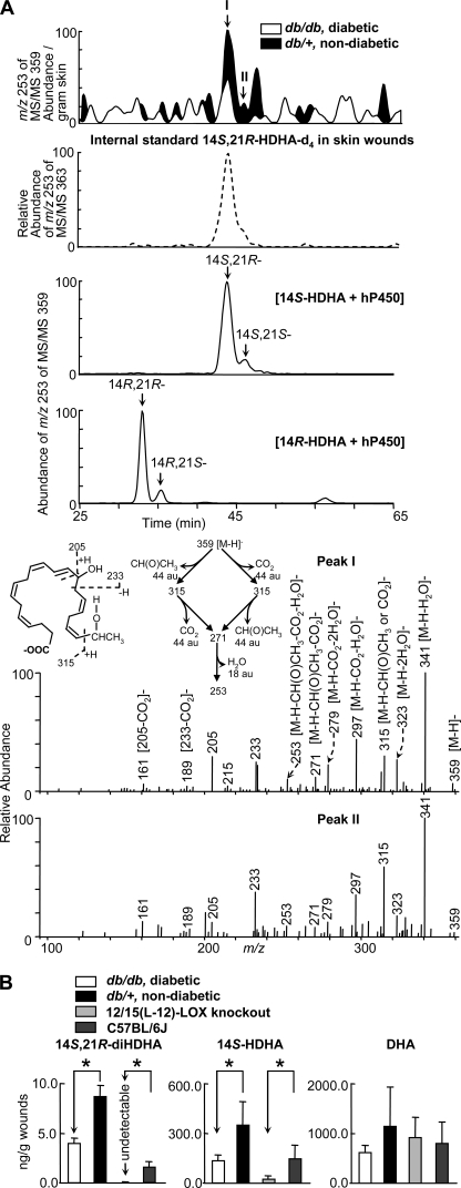
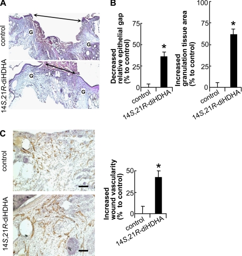
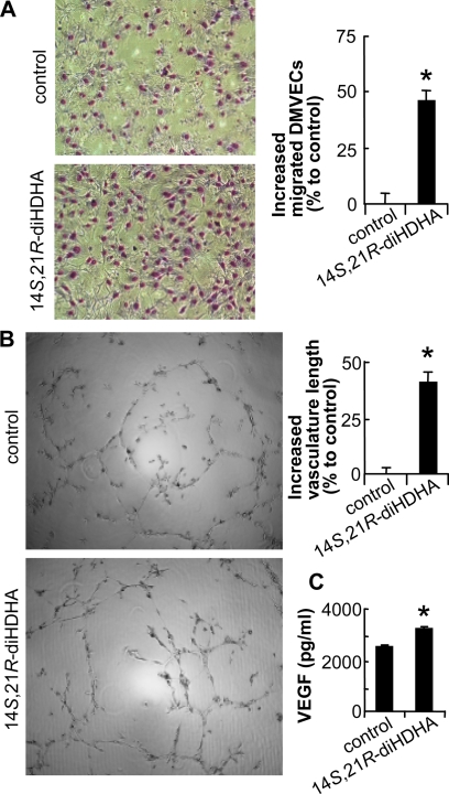
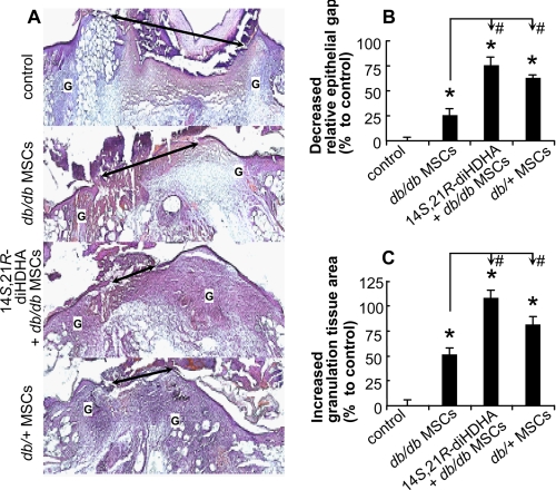
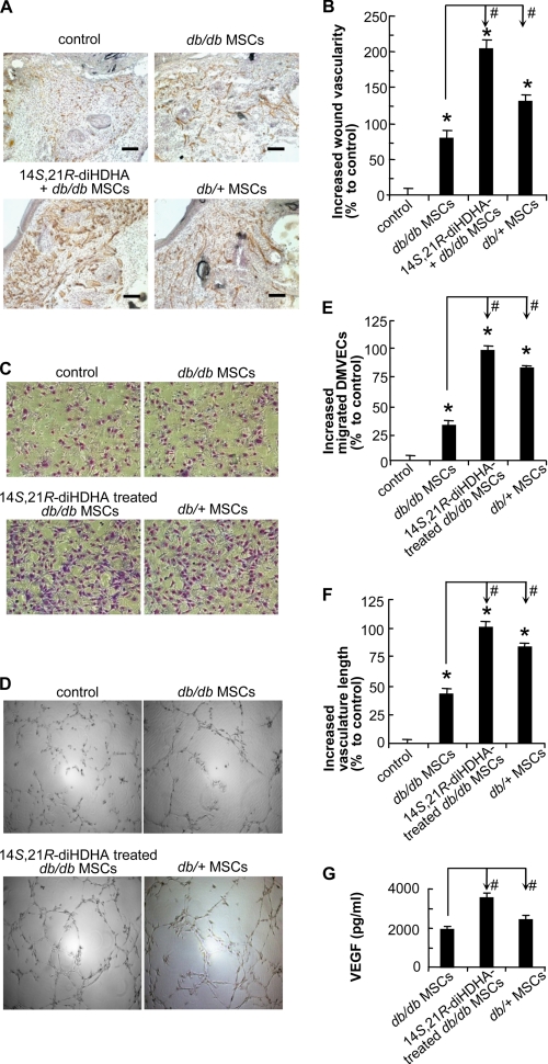
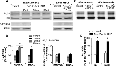
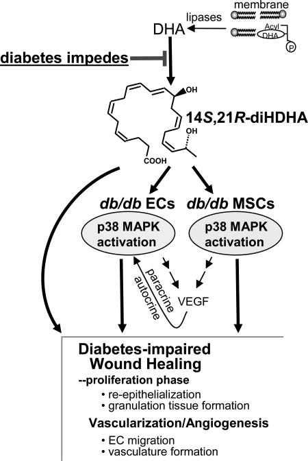
Treatment of diabetes-impaired wound healing remains a major unresolved medical challenge. Here, we identified suppressed formation of a novel reparative lipid mediator 14S,21R-dihydroxydocosa-4Z,7Z,10Z,12E,16Z,19Z-hexaenoic acid (14S,21R-diHDHA) in cutaneous wounds of diabetic db/db mice. These results indicate that diabetes impedes the biosynthetic pathways of 14S,21R-diHDHA in skin wounds. Administration of exogenous 14S,21R-diHDHA to wounds in diabetic animals rescued healing and angiogenesis. When db/db mesenchymal stem cells (MSCs) were administered together with 14S,21R-diHDHA to wounds in diabetic animals, they coacted to accelerate wound re-epithelialization, granulation tissue formation, and synergistically improved vascularization. In the pivotal cellular processes of angiogenesis, 14S,21R-diHDHA enhanced VEGF release, vasculature formation, and migration of db/db dermal microvascular endothelial cells (DMVECs), as well as remedied paracrine angiogenic functions of db/db MSCs, including VEGF secretion and the promotion of DMVEC migration and vasculature formation. Our results show that 14S,21R-diHDHA activates the p38 MAPK pathway in wounds, db/db MSCs, and DMVECs. Overall, the impeded formation of 14S,21R-diHDHA described in this study suggests that diabetes could affect the generation of pro-healing lipid mediators in wound healing. By restoring wound healing and MSC functions, 14S,21R-diHDHA is a new lead for the development of better therapeutics used in treating wounds of diabetics.


Figures

FIGURE 1.
FIGURE 1.
Formation of 14S,21R-diHDHA was suppressed in skin wounds in diabetic db/db mice compared with nondiabetic db/+ mice. Wounded skin was harvested from mice immediately following sacrifice at day 1 after the splinted excisional wounding was performed. A, overlay ChiralPak-IA-based chiral LC-MS/MS chromatograms (top panels) of wounds of db/+ (solid black) and db/db (open line) mice, and spectra (nonwide band) (bottom two panels) of peak I and peak II found in wounds of db/+ mice. For comparison, the ion abundance of each selected MS/MS ion chromatogram was divided by the extraction recovery of the stable deuterated internal standard 14S,21R-diHDHA-d4 added to the sample and then normalized per g of skin. The 2nd panel is a selected ion chromatogram at m/z 253 of MS/MS 363 for internal standard 14S,21R-diHDHA-d4 added to and extracted from skin wounds. This deuterated compound was added to skin wounds collected from mice and used as the internal standard for the quantification of 14S,21R-diHDHA (see details under “Experimental Procedures”). Chromatograms of 14S,21R-diHDHA and 14S,21S-diHDHA (generated from 14S-HDHA by h-P450) as well as 14R,21S-diHDHA and 14R,21R-diHDHA (generated from 14R-HDHA by h-P450) are shown in the 3rd and 4th panels, respectively. B, quantification of 14S,21R-diHDHA, 14S-HDHA, and DHA in wounds of db/+ (solid black), db/db (open bar), 12/15-LOX-knock-out mice (light gray), and C57BL/6J mice (dark gray) via ChiralPak-IA-based chiral LC-MS/MS using stable deuterated internal standards. Results are mean ± S.E. (n = 3). *, p < 0.05 compared with control.
FIGURE 2.
FIGURE 2.
14S,21R-diHDHA remedied impaired wound healing and vascularization in diabetic mice. Splinted excisional wounding was conducted in db/db mice as in Fig. 1, followed by administration of 14S,21R-diHDHA to wounds (50 ng/wound). A, micrographs; B, quantitative analysis of H&E-stained cryosections of wounds show that 14S,21R-diHDHA treatment enhanced re-epithelialization and granulation tissue formation compared with the DMEM control, a decreased percentage of relative epithelial gap (left panel) and increased percentage of granulation tissue area (right panel) were observed (n = 6). G, granulation tissue; double arrow, epithelial gap. The control had a relative epithelial gap of 64.9 ± 2.0% and total granulation tissue area of 4250.0 ± 176.9 pixels/cryosection. C, 14S,21R-diHDHA increased wound vascularity. Micrographs (left panel) showing CD31+ vessels and hematoxylin-stained nuclei in wound cryosections. The increased percentage of wound vascularity is presented on the right (n = 12). The wound vascularity of control animals was 4.4 ± 0.4%. Scale bar, 100 μm. Results are means ± S.E. *, p < 0.05 compared with control.
FIGURE 3.
FIGURE 3.
14S,21R-diHDHA improved the cellular processes of angiogenesis in db/db DMVECs. The cellular angiogenic processes of migration (A), vasculature formation (B), and VEGF release (C) were assayed in vitro using db/db DMVECs cultured under simulated hyperglycemic conditions (25 mm glucose) in the presence and absence (control) of 14S,21R-diHDHA (100 nm). Migration and vasculature formation are presented as increased percentages of migrated DMVECs per field (n = 15) and of vasculature length per field (n = 4), respectively, compared with control (migrated DMVECs, 118.0 ± 2.8 cells/field; vasculature length, 7.2 ± 0.3 mm/field). Representative images (left panel) and the quantification (right panel) are presented. VEGF secreted from DMVECs was measured using the Bio-Plex protein array (n = 3). Results are means ± S.E. *, p < 0.05 compared with control.
FIGURE 4.
FIGURE 4.
14S,21R-diHDHA acted together with db/db MSCs to promote wound healing. Splinted excisional wounding was conducted in db/db mice as in Fig. 1, followed by treatment of the wounds with vehicle, db/db MSCs, db/db MSCs plus 14S,21R-diHDHA, or db/+ MSCs alone (14S,21R-diHDHA, 50 ng/wound; MSCs, 106 cells/wound). Wounds were collected from db/db mice at day 8 post-wounding. A, representative micrographs of H&E-stained cryosections of wounds. Double arrow, epithelial gap; G, granulation tissue. B, decreased percentage of the relative epithelial gap; C, increased percentage of the granulation tissue area as compared with controls that had a relative epithelial gap of 64.9 ± 2.0% and total granulation tissue area of 4250.0 ± 176.9 pixels/cryosection. Results are means ± S.E. (n = 6–8). *, p < 0.05 compared with control; #, p < 0.05 compared with db/db MSC treatment.
FIGURE 5.
FIGURE 5.
14S,21R-diHDHA remedied impaired functions of db/db MSCs and acted synergistically with them in promoting angiogenesis under hyperglycemia. The wound healing model and treatment were conducted in db/db mice as in Fig. 4. A, representative microphotographs show the CD31+ vessels in wounds treated with vehicle (control), db/db MSCs, db/db MSCs plus 14S,21R-diHDHA, or db/+ MSCs (14S,21R-diHDHA, 50 ng/wound; MSCs, 106 cells/wound). B, increased percentages of wound vascularity compared with the control that possessed 4.4 ± 0.4% vascularity (n = 12). Scale bar, 100 μm. Cellular processes of angiogenesis were studied in vitro as in Fig. 3. Representative images of db/db DMVEC migration (C) and vasculature formation (D) are shown. The db/db DMVECs were studied under simulated hyperglycemia (25 mm glucose) in DMEM (control), DMEM conditioned by db/db MSCs with (14S,21R-diHDHA-treated db/db MSCs) or without (db/db MSCs) 14S,21R-diHDHA treatment, as well as DMEM conditioned by db/+ MSCs (db/+ MSCs). Migration (E) and vasculature formation (F) were quantified as increased percentages of migrated DMVECs per field (n = 15) and of vasculature length per field (n = 4), respectively, compared with the controls (migrated DMVECs, 118.0 ± 2.8 cells/field; vasculature length, 7.2 ± 0.3 mm/field). G, VEGF secretion was assayed by Bio-Plex protein array (n = 3). The medium was conditioned with 3 × 10⁵ db/db MSCs, 14S,21R-diHDHA-treated db/db MSCs, or db/+ MSCs. Results are means ± S.E. *, p < 0.05 compared with control; #, p < 0.05 compared with db/db MSC treatment.
FIGURE 6.
FIGURE 6.
14S,21R-diHDHA activated p38 MAPK but not ERK1/2 signaling pathway. Quiescent subconfluent db/db DMVECs and MSCs were treated with 100 nm 14S,21R-diHDHA for 10–120 min, and wounds of db/+ and db/db mice were treated with 14S,21R-diHDHA (50 ng/wound) for 15 min. Whole tissue and cell lysates were analyzed by Western blot with antibodies specific to phospho-p38 (P-p38) and phospho-ERK1/2 (P-ERK1/2) as well as total p38 and ERK1. Densitometric ratios of P-p38 to p38 and P-ERK1/2 to ERK1 are presented as a percentage of control (i.e. without 14S,21R-diHDHA treatment). Representative Western blot images of P-p38, p38, P-ERK1/2, and ERK1 as well as densitometric ratios of P-p38 to p38 and P-ERK1/2 to ERK1 in db/db DMVECs and MSCs (A and B), and in wounds of db/+ and db/db mice (C and D) are shown. Results are mean ± S.E. (n = 3). *, p < 0.05 compared with control.
SCHEME 1.
SCHEME 1.
Diabetes reduces formation of 14S,21R-diHDHA in cutaneous wounds and application of exogenous 14S,21R-diHDHA counteracts the diabetic impairment on healing, angiogenesis, and associated functions of mesenchymal stem cells. Treatment with 14S,21R-diHDHA rescues healing, angiogenesis, and associated MSC functions that are impaired in diabetes. 14S,21R-diHDHA restores the diabetes-impaired cellular processes of angiogenesis and paracrine functions of MSCs, including endothelial cell (EC) migration, vasculature formation, and production of VEGF (autocrine and paracrine). This lipid mediator activates the p38 MAPK pathway in endothelial cells and MSCs. 14S,21R-diHDHA may also act directly to enhance re-epithelialization and granulation tissue formation in wound healing.

Similar articles

Cited by

References

    1. Falanga V. (2005) Lancet 366, 1736–1743 - PubMed
    1. Javazon E. H., Keswani S. G., Badillo A. T., Crombleholme T. M., Zoltick P. W., Radu A. P., Kozin E. D., Beggs K., Malik A. A., Flake A. W. (2007) Wound Repair Regen. 15, 350–359 - PubMed
    1. Montori V. M., Farmer A., Wollan P. C., Dinneen S. F. (2000) Diabetes Care 23, 1407–1415 - PubMed
    1. Shingel K. I., Faure M. P., Azoulay L., Roberge C., Deckelbaum R. J. (2008) J. Tissue Eng. Regen. Med. 2, 383–393 - PubMed
    1. Serhan C. N., Yang R., Martinod K., Kasuga K., Pillai P. S., Porter T. F., Oh S. F., Spite M. (2009) J. Exp. Med. 206, 15–23 - PMC - PubMed

Publication types

MeSH terms

Substances