. 2011 Feb 25;286(8):6627-40.
doi: 10.1074/jbc.M110.167247. Epub 2010 Nov 23.

Intraphagosomal peroxynitrite as a macrophage-derived cytotoxin against internalized Trypanosoma cruzi: consequences for oxidative killing and role of microbial peroxiredoxins in infectivity

Affiliations

Intraphagosomal peroxynitrite as a macrophage-derived cytotoxin against internalized Trypanosoma cruzi: consequences for oxidative killing and role of microbial peroxiredoxins in infectivity

María Noel Alvarez et al. J Biol Chem. .

Abstract

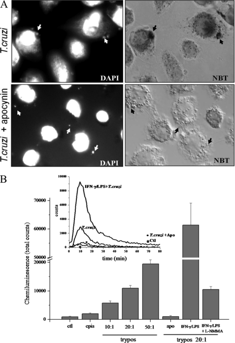
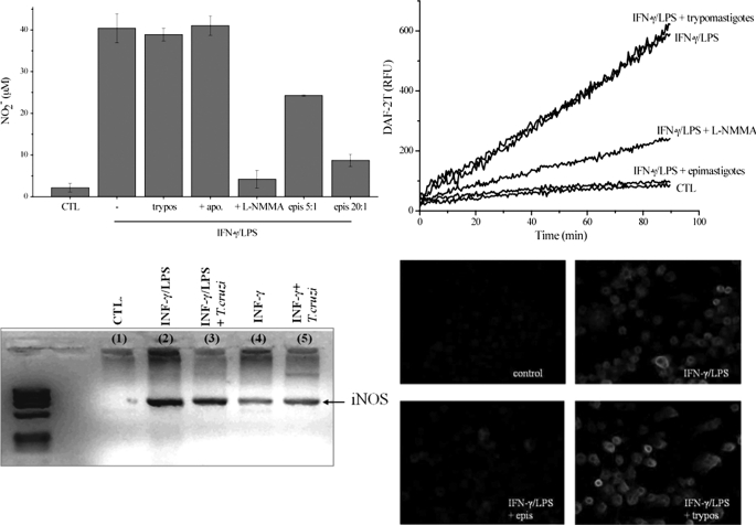
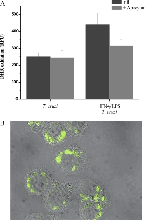
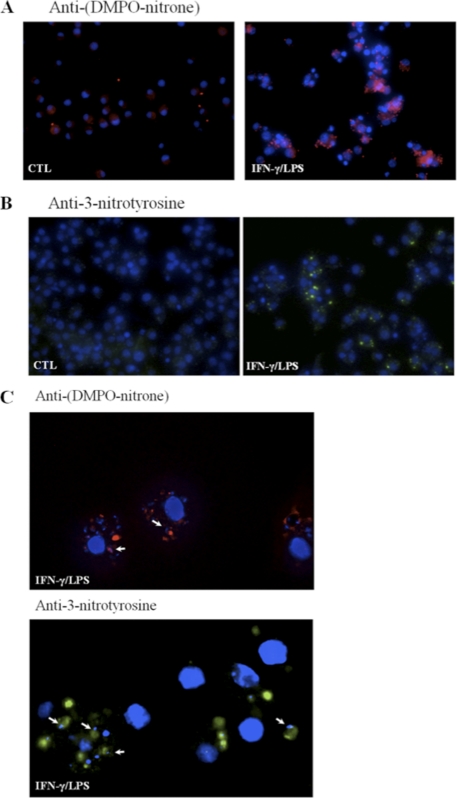
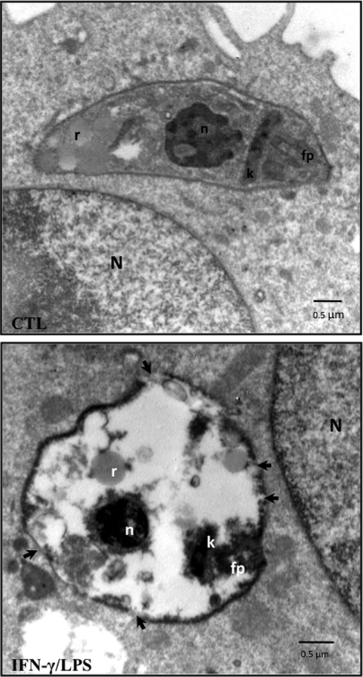
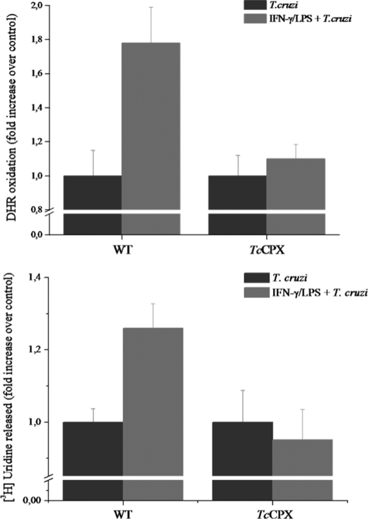
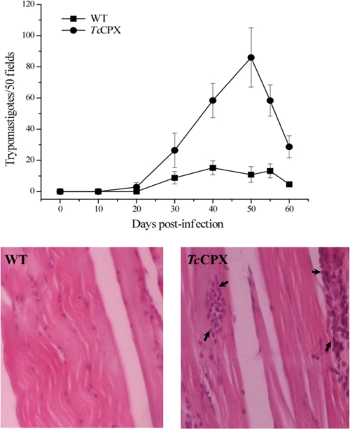
Macrophage-derived radicals generated by the NADPH oxidase complex and inducible nitric-oxide synthase (iNOS) participate in cytotoxic mechanisms against microorganisms. Nitric oxide ((•)NO) plays a central role in the control of acute infection by Trypanosoma cruzi, the causative agent of Chagas disease, and we have proposed that much of its action relies on macrophage-derived peroxynitrite (ONOO(-) + ONOOH) formation, a strong oxidant arising from the reaction of (•)NO with superoxide radical (O(2)(-)). Herein, we have shown that internalization of T. cruzi trypomastigotes by macrophages triggers the assembly of the NADPH oxidase complex to yield O(2)(-) during a 60-90-min period. This does not interfere with IFN-γ-dependent iNOS induction and a sustained (•)NO production (∼24 h). The major mechanism for infection control via reactive species formation occurred when (•)NO and O(2)() were produced simultaneously, generating intraphagosomal peroxynitrite levels compatible with microbial killing. Moreover, biochemical and ultrastructural analysis confirmed cellular oxidative damage and morphological disruption in internalized parasites. Overexpression of cytosolic tryparedoxin peroxidase in T. cruzi neutralized macrophage-derived peroxynitrite-dependent cytotoxicity to parasites and favored the infection in an animal model. Collectively, the data provide, for the first time, direct support for the action of peroxynitrite as an intraphagosomal cytotoxin against pathogens and the premise that microbial peroxiredoxins facilitate infectivity via decomposition of macrophage-derived peroxynitrite.


Figures

FIGURE 1.
FIGURE 1.
Phagocytosis of T. cruzi trypomastigotes is accompanied by intraphagosomal- and NADPH oxidase-dependent O2˙̄ formation. A, superoxide-dependent intraphagosomal NBT reduction. Macrophages were incubated with T. cruzi trypomastigotes (5:1 ratio) in the presence of NBT (1 mg/ml, 30 min, 37 °C) and apocynin (50 μm) as indicated, stained with DAPI as described under “Experimental Procedures,” and observed by fluorescence/Nomarski DIC microscopy (magnification, ×1000). Arrows indicate internalized parasites visualized by DAPI-stained nuclei (upper and lower left) within phagosomes as evidenced by formazan deposits (upper right). Apocynin completely abolished O2˙̄-dependent formazan formation (lower right). Data shown are representative of at least three independent experiments performed on separate days. B, luminol chemiluminescence. Macrophages (1 × 10⁶) incubated in dPBS with 200 μm luminol were exposed to: T. cruzi epimastigotes (2 × 10⁷) and trypomastigotes (1, 2, and 5 × 10⁷) corresponding to a trypomastigotes:macrophage ratio of 10:1, 20:1, and 50:1. The effect on luminol chemiluminescence of apocynin (apo), iNOS induction (IFN-γ/LPS), and inhibition by l-NMMA (IFN-γ/LPS + l-NMMA) was explored at a 20:1 ratio. Chemiluminescence was continuously measured immediately after T. cruzi addition to the macrophage monolayer in a luminescence plate reader at 37 °C. Data represent mean ± S.E. of total counts in 100 min. Inset, shows representative records obtained by the addition of trypomastigotes (20:1) to unstimulated (T. cruzi), apocynin-treated (T. cruzi + Apo), or preactivated macrophages (IFN-γ/LPS + T. cruzi). ctl, control (correspond to macrophages not challenged with T. cruzi.); epis, epimastigotes; trypos, trypomastigotes.
FIGURE 2.
FIGURE 2.
Nitric oxide production and iNOS levels in T. cruzi-infected macrophages. A, nitrite production. Macrophages were exposed to IFN-γ/LPS for 24 h in the absence and presence of epimastigotes (epimastigotes:macrophages, 5:1 and 20:1) or trypomastigotes (20:1). The effect of apocynin (apo) and l-NMMA (2 mm) was also evaluated. Nitrite accumulation was determined using the Griess reaction. epis, epimastigotes; CTL, control; trypos, trypomastigotes. B, DAF-2DA oxidation. Macrophages were exposed to IFN-γ/LPS in the presence of epimastigotes (20:1), trypomastigotes (20:1), or l-NMMA (2 mm) in DMEM at 37 °C. Control experiments were performed in the absence of cytokine stimulation. After 5 h, the medium was replaced with dPBS containing 5 μm DAF-2DA, and probe oxidation was followed in a fluorescence plate reader at 37 °C. RFU, relative fluorescence units. C, iNOS mRNA levels of macrophages exposed to IFN-γ/LPS, IFN-γ/LPS + trypomastigotes (10:1), IFN-γ, and IFN-γ + trypomastigotes (10:1) were evaluated by RT-PCR. Total RNA from 5 × 10⁶ macrophages under all conditions was extracted after 3 h of infection. Host iNOS and GAPDH cDNAs were amplified using primers defined under “Experimental Procedures.” D, iNOS expression was evaluated by immunocytochemistry. Macrophages plated in slides were stimulated for 5 h with IFN-γ/LPS in the presence or absence of T. cruzi epimastigotes or trypomastigotes. Immunocytochemical analysis of iNOS expression was performed using polyclonal anti-NOS antibody and visualized with Alexa-594-conjugated anti-rabbit antibody (magnification, ×400). Data shown are representative of at least three independent experiments performed on separate days.
FIGURE 3.
FIGURE 3.
DHR oxidation inside phagocytosed trypomastigotes. T. cruzi trypomastigotes preloaded with DHR were exposed to macrophages, and cells were washed 30 min after incubation. A, RH 123 accumulation after 2 h of infection in unstimulated (T. cruzi) or preactivated macrophages (IFN-γ/LPS + T. cruzi) was determined in a fluorescence plate reader. The effect of apocynin was evaluated under both conditions (+ Apocynin). RFU, relative fluorescence units. Data are mean ± S.E. of three independent experiments. B, activated macrophages plated in slides were infected with DHR-loaded trypomastigotes (5:1, parasite:macrophage ratio). Merged DIC and fluorescence images were obtained 2 h after infection; the green fluorescence corresponds to oxidized DHR (magnification, ×400).
FIGURE 4.
FIGURE 4.
Detection of peroxynitrite-dependent protein oxidation in phagocytosed trypomastigotes. A, DMPO protein adducts. Unstimulated (CTL) or activated (IFN-γ/LPS) macrophages were infected with DMPO-loaded trypomastigotes (20:1). After 2 h, cells were fixed, permeabilized, incubated with anti-DMPO-nitrone polyclonal antibody, and visualized with Alexa-594-conjugated anti-rabbit antibody. DMPO protein adducts are shown in red and nuclei in blue (magnification, ×400). B, protein 3-nitrotyrosine. Unstimulated or activated macrophages were infected with T. cruzi trypomastigotes (20:1). Following phagocytosis (2 h), macrophages were incubated with anti-3-nitrotyrosine antibody and visualized with Alexa-488-conjugated anti-rabbit antibody. 3-Nitrotyrosine staining is shown in green, and the nucleus is stained in blue (magnification, ×400). C, magnified field (×1000) of anti-DMPO-nitrone (red) or anti- 3-nitrotyrosine (green) in activated macrophages. Arrows indicate the distinctive parasite kinetoplast or nuclei revealed by the blue DAPI staining in close proximity to the oxidized proteins. Results are representative of at least three independent experiments performed on separate days.
FIGURE 5.
FIGURE 5.
Electron microscopy studies of T. cruzi infection. Micrographs showing unstimulated (CTL) and activated (IFN-γ/LPS) infected macrophages at 1 h post-infection. The arrows in the lower panel indicate disruptions of membrane integrity. N, macrophage nucleus; T. cruzi n, T. cruzi nucleus; k, kinetoplast; fp, flagellar pocket; r, reservosomes. The electron micrographs are representative of at least three independent experiments.
FIGURE 6.
FIGURE 6.
T. cruzi killing by intraphagosomal peroxynitrite and protective effect of parasite cytosolic peroxiredoxin. A, wild type (WT) or TcCPX-overexpressing trypomastigotes preloaded with DHR were exposed to unstimulated (T. cruzi) or activated macrophages (IFN-γ/LPS + T. cruzi). RH 123 accumulation after 2 h of infection was determined in a fluorescence plate reader. B, macrophages were infected with wild type or TcCPX-overexpressing trypomastigotes preloaded with [5,6-3H]uridine. After incubation (for 2 h) supernatants were collected, and radioactivity was measured in a liquid scintillation counter as described under “Experimental Procedures.” Data are the mean ± S.E. of three independent experiments.
FIGURE 7.
FIGURE 7.
Parasitemia time course and tissue inflammation of mice infected with wild type trypomastigotes or TcCPX overexpressers. Behavior of wild type (WT) and TcCPX-overexpressing-trypomastigotes in experimental mice infections. Two month-old BALB/c mice were infected by intraperitoneal inoculation with 1 × 10⁶ metacyclic trypomastigote forms. Parasitemia and histopathology were analyzed as described under “Experimental Procedures.” A, parasitemia (trypomastigotes/50 fields) levels elicited by the wild type and TcCPX overexpresser strains. Values are given as means ± S.E. B, microphotographs of skeletal muscle tissue sections from mice inoculated with wild type (left) and TcCPX overexpressers (right) (magnification, ×400). Arrows in the right panel (TcCPX) indicate inflammatory infiltrates.

Similar articles

Cited by

References

    1. Babior B. M. (1984) J. Clin. Invest. 73, 599–601 - PMC - PubMed
    1. Groemping Y., Rittinger K. (2005) Biochem. J. 386, 401–416 - PMC - PubMed
    1. Alvarez M. N., Trujillo M., Radi R. (2002) Methods Enzymol. 359, 353–366 - PubMed
    1. De Groote M. A., Ochsner U. A., Shiloh M. U., Nathan C., McCord J. M., Dinauer M. C., Libby S. J., Vazquez-Torres A., Xu Y., Fang F. C. (1997) Proc. Natl. Acad. Sci. U.S.A. 94, 13997–14001 - PMC - PubMed
    1. Fridovich I. (1997) J. Biol. Chem. 272, 18515–18517 - PubMed

Publication types

LinkOut - more resources