. 2011 Jan;85(2):705-14.
doi: 10.1128/JVI.01557-10. Epub 2010 Nov 17.

Inhibition of calmodulin-dependent kinase kinase blocks human cytomegalovirus-induced glycolytic activation and severely attenuates production of viral progeny

Affiliations

Inhibition of calmodulin-dependent kinase kinase blocks human cytomegalovirus-induced glycolytic activation and severely attenuates production of viral progeny

Jessica McArdle et al. J Virol. 2011 Jan.

Erratum in

  • J Virol. 2013 Jun;87(12):7197

Abstract

Viruses depend on the host cell to provide the energy and biomolecular subunits necessary for production of viral progeny. We have previously reported that human cytomegalovirus (HCMV) infection induces dramatic changes to central carbon metabolism, including glycolysis, the tricarboxylic acid (TCA) cycle, fatty acid biosynthesis, and nucleotide biosynthesis. Here, we explore the mechanisms involved in HCMV-mediated glycolytic activation. We find that HCMV virion binding and tegument protein delivery are insufficient for HCMV-mediated activation of glycolysis. Viral DNA replication and late-gene expression, however, are not required. To narrow down the list of cellular pathways important for HCMV-mediated [corrected] activation of glycolysis, we utilized pharmaceutical inhibitors to block pathways reported to be both involved in metabolic control and activated by HCMV infection. We find that inhibition of calmodulin-dependent kinase kinase (CaMKK), but not calmodulin-dependent kinase II (CaMKII) or protein kinase A (PKA), blocks HCMV-mediated activation of glycolysis. HCMV infection was also found to target calmodulin-dependent kinase kinase 1 (CaMKK1) expression, increasing the levels of CaMKK1 mRNA and protein. Our results indicate that inhibition of CaMKK has a negligible impact on immediate-early-protein accumulation yet severely attenuates production of HCMV viral progeny, reduces expression of at least one early gene, and blocks viral DNA replication. Inhibition of CaMKK did not affect the glycolytic activation induced by another herpes virus, herpes simplex virus type 1 (HSV-1). Furthermore, inhibition of CaMKK had a much smaller impact on HSV-1 replication than on that of HCMV. These data suggest that the role of CaMKK during the viral life cycle is, in this regard, HCMV specific. Taken together, our results suggest that CaMKK is an important factor for HCMV replication and HCMV-mediated glycolytic activation.


Figures

FIG. 1.
FIG. 1.
Impact of HCMV infection on glycolysis. (A) Representation of the major intermediates of the glycolytic pathway. Through a series of enzymatic reactions, glucose is converted into the final product of glycolysis, pyruvate. Both the conversion of glucose to hexose phosphate and the conversion of hexose phosphate to fructose-1,6-bisphosphate (FBP) are ATP-dependent and are rate-limiting steps of glycolysis. Hexose-P, glucose-6 phosphate and its isomers; G3P/DHAP, glyceraldehyde 3-phosphate and dihydroxyacetone phosphate; PG, phosphorylated glycerate isoforms. (B) Time course of glycolytic flux activation. Serum-starved MRC-5 human fibroblast cells were mock infected (M) or infected with HCMV (H) (MOI = 3). Cells were labeled with [13C]glucose containing medium at 24, 48, and 72 h postinfection for 1 min. After the labeling, metabolism was quenched by the addition of cold 80% methanol. Accumulation of [13C]fructose-1,6-bisphosphate was measured upon [13C]glucose pulse via measurement by LC-MS-MS. Values are means ± standard errors (SE) (n = 4). (C) Serum-starved MRC-5 fibroblasts were mock infected or infected with HCMV (MOI = 3). After adsorption, cells were treated with DMSO, an inhibitor of protein synthesis, cycloheximide (10 μg/ml), or an inhibitor of viral DNA replication, PAA (50 μg/ml). At 48 hpi, cells were labeled with [13C]glucose containing DMEM for 1 min, quenched with cold methanol, and processed for LC-MS-MS analysis. Values are means ± SE (n = 4). (D) Serum-starved MRC-5 fibroblasts were mock infected, HCMV infected, or infected with UV-irradiated HCMV (MOI = 3.0). At 48 hpi, cells were labeled with [13C]glucose containing DMEM for 1 min, quenched with cold methanol, and processed for LC-MS-MS analysis. Values are means ± SE (n = 2).
FIG. 2.
FIG. 2.
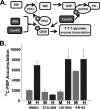
(A) Regulation of glycolysis by selected kinase cascades. PKA, CaMKK, and CaMKII can activate glycolysis transcriptionally through CREB activation or through direct/indirect phosphorylation/activation of glycolytic regulatory enzymes, e.g., PFK-2 or GAPDH. (B) Pharmaceutical inhibition of CaMKK blocks HCMV-mediated glycolytic activation. Serum-starved MRC-5 human fibroblasts were mock infected or infected with HCMV (MOI = 3). After adsorption, cells were treated with the CaMKK inhibitor STO-609 (10 μg/ml), the PKA inhibitor HA1004 (10 μg/ml), the CaMKII inhibitor KN-62 (7 μg/ml), or DMSO alone. At 48 h postinfection, cells were labeled for 1 min with [13C]glucose containing DMEM, quenched with cold methanol, and processed for LC-MS-MS to measure the accumulation of [13C]fructose-1,6-bisphosphate. Values are means ± SE (n = 4).
FIG. 3.
FIG. 3.
Pharmaceutical inhibition of CaMKK attenuates HCMV viral replication. (A) Serum-starved MRC-5 human fibroblasts were infected with HCMV (MOI = 3). After adsorption, cells were treated with the CaMKK-specific inhibitor STO-609 at concentrations of 5 μg/ml or 10 μg/ml or with DMSO alone. Cells were harvested at 24, 72, and 120 h postinfection, and the production of infectious progeny was measured by a plaque assay. Values are means ± SE (n = 2). Points marked with an asterisk were below the limit of detection (∼20 PFU/ml). (B) Analysis of the potential toxicity of STO-609 treatment. Confluent MRC-5 fibroblasts were mock infected or infected with HCMV (MOI = 3). After adsorption, cells were treated with 10 μg/ml STO-609. At 72 h postinfection, cell viability was measured via a Live/Dead cell viability assay. Green indicates the presence of esterase activity associated with viable cells, while red indicates loss of cellular membrane integrity associated with cell death. As a positive control for staining of a breakdown in membrane integrity, cells were treated with ethanol. The images were obtained by inverted fluorescence microscopy.
FIG. 4.
FIG. 4.
Impact of CaMKK pharmaceutical inhibition on viral protein and DNA accumulation. (A) Analysis of the impact of CaMKK inhibition on viral protein accumulation. Serum-starved MRC-5 human fibroblasts were mock infected or infected with HCMV (MOI = 3). After adsorption, cells were treated with the CaMKK-specific inhibitor STO-609 (5 μg/ml or 10 μg/ml) or DMSO alone. Cells were harvested at 24, 48, and 72 h postinfection and analyzed by Western blotting with antisera specific for IE1, UL26, pp28, and tubulin. Mw, molecular mass. (B) Analysis of the impact of CaMKK inhibition on viral DNA accumulation. Serum-starved MRC-5 cells were mock infected or infected with HCMV (MOI = 3). After adsorption, cells were treated with the CaMKK-specific inhibitor STO-609 (10 μg/ml) or DMSO. Viral DNA was extracted from cells that were harvested at 48, 72, and 96 h postinfection and processed for qPCR analysis of viral DNA accumulation. Values are means ± SE (n = 6). (C) Serum-starved MRC-5 human fibroblasts were HCMV infected (MOI = 3). After adsorption, two sets of cells were treated with STO-609 (10 μg/ml) or DMSO, as indicated on the graph. The last set of MRC-5s was treated with STO-609 (10 μg/ml) at 24 hpi and harvested at 96 hpi with the rest of the treatments. Viral replication was measured by a plaque assay. Values are means ± SE (n = 2). An asterisk indicates that the number of infectious virions were below the limit of detection (∼20 PFU/ml). (D) Serum-starved MRC-5 human fibroblasts were mock or HCMV infected (MOI = 3). After adsorption, fresh serum-free medium was placed on the cells. At 24 h postinfection, cells were treated with either DMSO or STO-609. Cells were pulsed at 48 or 72 h postinfection for 1 min with [13C]glucose containing DMEM, quenched with cold methanol, and processed for LC-MS-MS to measure the accumulation of [13C]fructose-1,6-bisphosphate. Values are means ± SE (n = 2).
FIG. 5.
FIG. 5.
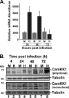
Impact of HCMV infection on CaMKK1 mRNA and protein levels. (A) Analysis of CaMKK1 mRNA during HCMV infection by qPCR. Serum-starved MRC-5 human fibroblasts were mock infected or infected with HCMV (MOI = 3). Cellular mRNA isolated from cells harvested at 24, 48, and 72 h postinfection was processed for qPCR using CaMKK1-specific primers. Values are plotted as mean relative abundances ± SE observed after normalization to GAPDH (n = 3). (B) Analysis of CaMKK1 protein accumulation over a time course of HCMV infection. Serum-starved MRC-5 human fibroblasts were mock infected or infected with HCMV (MOI = 3). Cells were harvested at 4, 24, 48, and 72 h postinfection and analyzed by Western blotting with two independent CaMKK1-specific antibodies. Tubulin was also blotted for as a loading control.
FIG. 6.
FIG. 6.
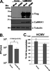
Impact of expression of a CaMKK kinase-dead allele on HCMV replication. (A) Analysis of Flag-CaMKK-KD protein expression in fibroblasts. MRC-5 fibroblasts were transduced with an empty vector or a vector expressing a Flag-tagged kinase-dead allele of CaMKK (CaMKK-KD). Fibroblasts containing the CaMKK-KD variant or an empty-vector control were mock infected or infected with HCMV (MOI = 0.1). Cells were harvested 10 days postinfection (α-Flag blot) or 48 h postinfection (mouse α-CaMKKα and tubulin blots). The presence of the CaMKK-KD or tubulin was monitored by Western blotting with the indicated antibodies. (B) Impact of CaMKK-KD expression on HCMV viral replication. Serum-starved MRC-5 fibroblasts expressing either CaMKK-KD or an empty vector were infected with HCMV (MOI = 0.1). Cells were harvested at 10 days postinfection, and viral replication was measured by a viral plaque assay. Values are plotted as means ± SE (n = 6). (C) Impact of CaMKK-KD expression on glycolysis in HCMV-infected cells. Serum-starved MRC-5 fibroblasts expressing either CaMKK-KD or an empty vector were infected with HCMV (MOI = 3.0). At 48 h postinfection, cells were labeled for 1 min with [13C]glucose containing DMEM, quenched with cold methanol, and processed for LC-MS-MS to measure the accumulation of [13C]fructose-1,6-bisphosphate. Values are means ± SE (n = 2).
FIG. 7.
FIG. 7.
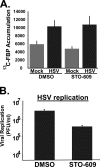
Impact of CaMKK inhibition on HSV viral replication and glycolytic activation. (A) Impact of CaMKK inhibition on HSV viral replication. Serum-starved MRC-5 human fibroblasts were infected with HSV (MOI = 10). After adsorption, cells were treated with DMSO or STO-609 (10 μg/ml). Cells were harvested at 24 h postinfection, and viral replication was measured by a plaque assay. Values are means ± SE (n = 4). (B) Impact of CaMKK inhibition on HSV-mediated activation of glycolysis. MRC-5 fibroblasts were mock infected or infected with HSV (MOI = 10). After adsorption, cells were treated with DMSO or STO-609 (10 μg/ml). Cells were harvested at 12 h postinfection, and flux analysis was performed using LC-MS-MS. Values are means ± SE (n = 4).
FIG. 8.
FIG. 8.
Impact of glycolytic inhibition on HCMV and HSV viral replication. MRC-5 human fibroblasts were infected with HCMV (MOI = 3) or HSV (MOI = 10). After adsorption, cells were treated with DMSO or 2-deoxyglucose (20 mM), a glycolytic inhibitor. HSV-infected cells were harvested at 24 h postinfection, while HCMV-infected cells were harvested at 96 h postinfection. Viral replication was measured by a viral plaque assay. Values are means ± SE (n = 2).

Similar articles

Cited by

References

    1. Andrei, G., E. De Clercq, and R. Snoeck. 2008. Novel inhibitors of human CMV. Curr. Opin. Invest. Drugs 9:132-145. - PubMed
    1. Beisser, P. S., H. Lavreysen, C. A. Bruggeman, and C. Vink. 2008. Chemokines and chemokine receptors encoded by cytomegaloviruses. Curr. Top. Microbiol. Immunol. 325:221-242. - PubMed
    1. Bito, H., and S. Takemoto-Kimura. 2003. Ca(2+)/CREB/CBP-dependent gene regulation: a shared mechanism critical in long-term synaptic plasticity and neuronal survival. Cell Calcium 34:425-430. - PubMed
    1. Bradley, P. L. 1957. Metabolism of pyruvate and alpha-ketoglutarate in virus-infected mouse brain. Nature 180:1418-1419. - PubMed
    1. Burny, W., C. Liesnard, C. Donner, and A. Marchant. 2004. Epidemiology, pathogenesis and prevention of congenital cytomegalovirus infection. Expert Rev. Anti Infect. Ther. 2:881-894. - PubMed

Publication types

MeSH terms

Substances

LinkOut - more resources