. 2010 Dec 15;185(12):7681-90.
doi: 10.4049/jimmunol.1002803. Epub 2010 Nov 12.

The inflammatory response after an epidermal burn depends on the activities of mouse mast cell proteases 4 and 5

Affiliations

The inflammatory response after an epidermal burn depends on the activities of mouse mast cell proteases 4 and 5

George Younan et al. J Immunol. .

Abstract

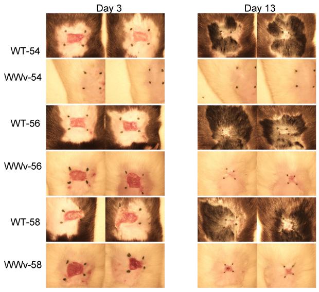
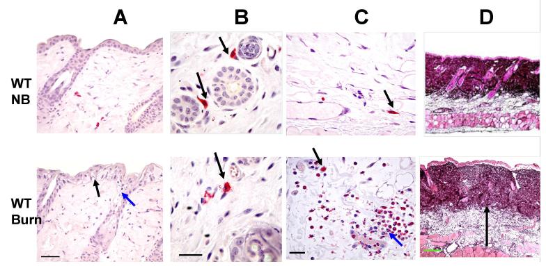
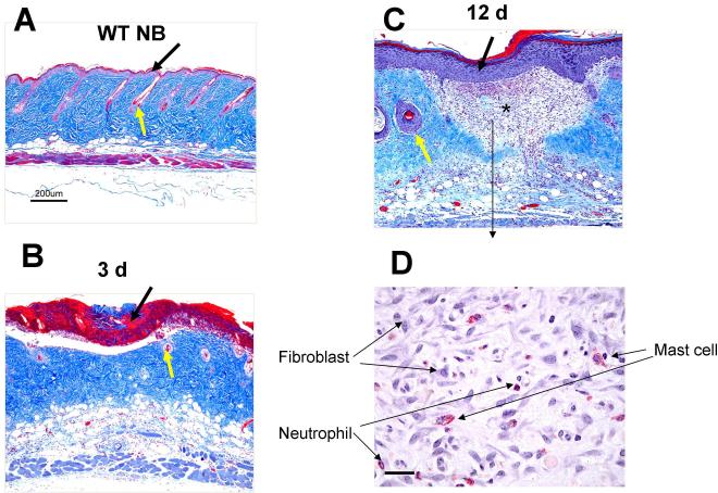
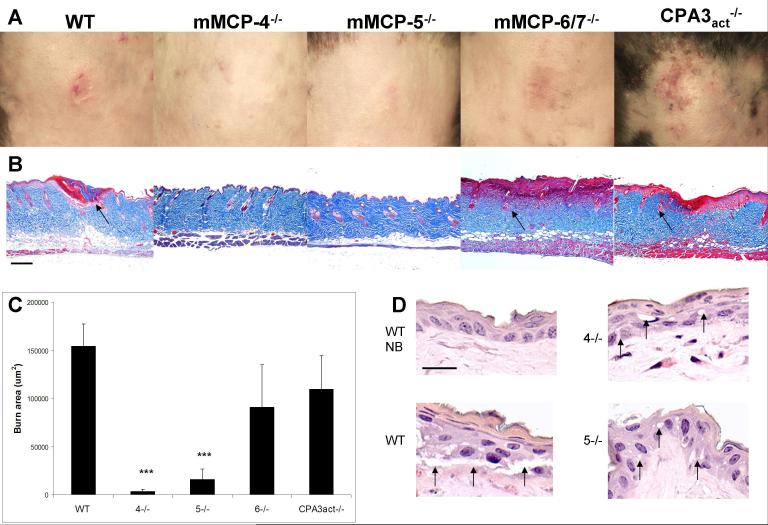
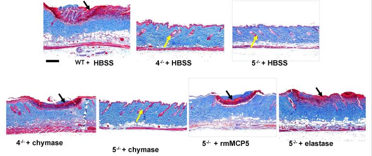
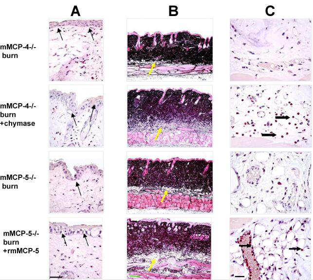
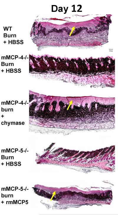
A second-degree epidermal scald burn in mice elicits an inflammatory response mediated by natural IgM directed to nonmuscle myosin with complement activation that results in ulceration and scarring. We find that such burn injury is associated with early mast cell (MC) degranulation and is absent in WBB6F1-Kit(W)/Kit(Wv) mice, which lack MCs in a context of other defects due to a mutation of the Kit receptor. To address further an MC role, we used transgenic strains with normal lineage development and a deficiency in a specific secretory granule component. Mouse strains lacking the MC-restricted chymase, mouse MC protease (mMCP)-4, or elastase, mMCP-5, show decreased injury after a second-degree scald burn, whereas mice lacking the MC-restricted tryptases, mMCP-6 and mMCP-7, or MC-specific carboxypeptidase A3 activity are not protected. Histologic sections showed some disruption of the epidermis at the scald site in the protected strains suggesting the possibility of topical reconstitution of full injury. Topical application of recombinant mMCP-5 or human neutrophil elastase to the scalded area increases epidermal injury with subsequent ulceration and scarring, both clinically and morphologically, in mMCP-5-deficient mice. Restoration of injury requires that topical administration of recombinant mMCP-5 occurs within the first hour postburn. Importantly, topical application of human MC chymase restores burn injury to scalded mMCP-4-deficient mice but not to mMCP-5-deficient mice revealing nonredundant actions for these two MC proteases in a model of innate inflammatory injury with remodeling.


Figures

FIGURE 1
FIGURE 1
MC-deficient mice are protected against epidermal scald injury at 54 °C. The clinical response to an epidermal scald of 25 seconds duration in MC-sufficient WBB6F1-+/+ (WT) mice and MC-deficient WWv mice was compared at 3 different temperatures at 3 and 13 d after injury. Pictures show the 2 mice in each group. Hair regrowth at the scald site of the 54 °C treated WWv mice is not seen due to its lack of pigment. The black marks were replaced each day to define the residual injury and do not represent the area of the original scald site. Protection against a visible burn in MC-deficient mice exposed to a 54 °C scald for 25 s was confirmed in 2 further experiments with sufficient and deficient strains with 5 mice per group.
FIGURE 2
FIGURE 2
The histological changes occurring 2 h after a scald burn in the skin of WT C57BL/6 mice. The histologic changes in the shaved skin in WT mice without (top panels, NB, no burn) and with a scald burn of 25 s duration at 54 °C (bottom panels) are compared at 2 h post burn. Sections in A-C are stained for CAE reactivity. A, Epidermis at the burn site shows disruption of the tight junctions between the basal cells of the epithelium (black arrow) and cytoplasmic vacuolization in epithelial cells of the epidermis and the hair follicles (red arrow). B, MCs in the dermis at the shaved site are intact (arrows) whereas MCs are degranulating with extracellular granules 2 h after the scald burn (arrow). C, Similarly, MCs in the hypodermis at the shaved site are intact (arrow) while 2 h after the scald burn they are degranulating (black arrow). There is also neutrophil margination in a blood vessel and influx into the hypodermis (red arrow). D, Jones’ stain shows lighter staining of the collagen matrix of the dermis and expansion of the hypodermis at the scald site due to edema (arrow in bottom panel denotes depth of hypodermis). Scale bars: A=50 μm, B, C=25um, D=200um).
FIGURE 3
FIGURE 3
The histologic changes occurring at 3 d and 12 d after a scald burn in WT mice. A-C are stained with Masson’s trichrome and D is stained for CAE reactivity. A, WT mice with no burn (NB) show intact epithelium (black arrow) and hair follicles (yellow arrow). B, The epidermis 3 d post burn is denuded leaving an ulceration demarcated by denatured collagen (stained red, black arrow), The denatured collagen extends through about one half the dermis reflecting the breadth and depth of the burn and loss of hair follicles (yellow arrow). C, By 12 d post burn, the morphological changes include a thickened epithelium covering the wound (black arrow), granulation tissue without any hair follicles in the dermis (star) and damaged hair follicles at the edge of the scar (yellow arrow). D. High power photograph of the granulation tissue shows accumulation of fibroblasts, MCs and neutrophils. Scale bar: A-C = 200 μm, D=25um).
FIGURE 4
FIGURE 4
The absence of MC-specific secretory granule proteases, mMCP-4 or mMCP-5, protects against epidermal scald injury. A, The progression of clinical injury to ulceration at d 3 occurs in the WT, mMCP-6/7- and CPA3act-deficient mice, but not in mMCP-4- or mMCP-5-deficient mice. B, The corresponding histological sections after Masson’s trichrome staining to assess the breadth and depth of injury by the appearance of denatured collagen (red), injured hair follicles (arrows), and edema of the dermis revealed by thickness and lighter staining. Each picture is from a single mouse representative of the injury characteristic of each strain. C, Burn injury area (red area of denatured collagen in histological sections) in the same groups as in A. Values for the burn area are the mean (± SE) um2. The value for the WT mice is derived from 3 experiments with 20 mice while the other values are from 1 experiment with 4, 4, 6, and 5 mice, respectively. Statistical values are from a t-test relative to the WT value. D, A high magnification of the epidermis in unburned WT (NB) and in burned WT , mMCP-4−/− (4−/−), and mMCP-5−/− (5−/−) mice at 2 h post burn shows disruption of epithelial tight junctions and epithelial vacuolization (arrows) in the burned WT mice but only minimal changes in these parameters mMCP-4−/− and mMCP-5−/− strains. Additional histologic data showing protection in the mMCP-4- and mMCP-5-deficient strains are shown in Fig. 5 and quantitated in Fig. 6. Scale bar: B=200 um, D= 20 um.
FIGURE 5
FIGURE 5
Topical application of human chymase and of recombinant mMCP-5 or human elastase reconstitutes the histologic changes of scald burn injury in mMCP-4−/− and mMCP-5−/− strains, respectively. WT, mMCP-4−/− (4−/−), and mMCP-5−/− (5−/−) mice were burned for 35 s at 54 °C and treated topically with a dressing providing HBSS, human chymase (chymase), human elastase (elastase) or rmMCP-5 for 1 h post burn. Masson’s trichrome staining of scald site is depicted at d 3. The injury is indicated by the red staining denatured collagen (black arrows) while protection is indicated by absence of denatured collagen and the presence of intact hair follicles (yellow arrows). Sections are representative of the aggregate findings for each group presented in Fig 6. Scale bar: 200 um.
FIGURE 6
FIGURE 6
The quantitative assessment of burn injury after the topical application of human chymase to mMCP4−/− mice and of rmMCP-5 or human elastase to mMCP-5−/− mice. Mice were burned for 35 s at 54 °C, ,then treated with topically applied HBSS, chymase (chym), rmMCP-5 or elastase (elast) as indicated for 1 h and the injury evaluated on d 3 post scald burn. A, The mean area (±SE) of burn injury area was assessed by Masson’s trichrome staining. The value for the WT mice is derived from 3 experiments with 16 mice. The value for the HBSS-treated mMCP-5−/− is derived from 2 experiments while all others are from 1 experiment. Numbers of mice are 16, 4, 5, 7, 5, 4, and 5 in each group, respectively. B, The mean number ( ±SE) of injured hair follicles (per 4 HPF) in the same animals as in A. C, The mean number (± SE) of neutrophils (per 4 HPF) identified by CAE reactivity in the hypodermis of the same animals as in A. D, The mean depth (± SE) of the skin from the same mice as in A with the depth of the various individual tissues indicated by burned dermis (red), unburned dermis (green) and hypodermis (blue). The mean (± ½ range) values from 2 non burned WT (WT NB) mice are shown for comparison. For statistical analysis, the mMCP-4- and mMCP-5-deficient mice treated with HBSS were compared to the WT controls, while the enzyme reconstituted groups were compared to the respective HBSS-treated strain. *, p<0.05; **, p<0.01; ***, p<0.001.
FIGURE 7
FIGURE 7
The histological changes 2 h after an epidermal scald in the skin of mMCP-4- and mMCP-5-deficient mice without and with topical application of human MC chymase or rmMCP-5, respectively. All mice were treated with topical HBSS without or without a protease for the first h post burn. A, Cytoplasmic vacuolization and disruption of the tight junctions between the basal cells of the epithelium at the scald site is apparent after CAE in the protease-deficient strains and is accentuated with topical application of chymase to the mMCP-4-deficient and of rmMCP-5 to the mMCP-5-deficient strain (arrows). B, Jones’ staining shows increased edema as indicated by the lighter color of the dermis and greater depth of the hypodermis (yellow arrows) in mMCP-4−/− mice treated with chymase, and of mMCP-5−/− mice treated with rmMCP-5 relative to their HBBS-treated protease-deficient controls. C, The CAE reactivity in the hypodermis shows neutrophils (arrows) in the blood vessels and tissue of mMCP-4−/− mice treated with chymase, and in mMCP-5−/− mice treated with rmMCP-5. Scale bar: A=50 um, B=200 um, C= 25 um.
FIGURE 8
FIGURE 8
The topical application of chymase and rmMCP-5 to the burn site on mMCP-4−/− and mMCP-5−/− mice, respectively, leads to remodeling at d 12. Jones’ stain of the skin reveals thickened epithelium, granulation tissue (light pink, yellow arrows) and loss of hair follicles in WT mice, in mMCP-4−/− mice treated with chymase and in mMCP-5−/− mice treated with rmMCP-5, whereas the protease deficient mice treated with HBSS alone had normal epithelium, hair follicles, and sebaceous glands. Scale bar; 200um.

Similar articles

Cited by

References

    1. Horton JW, Mileski WJ, White DJ, Lipsky P. Monoclonal Antibody to Intercellular Adhesion Molecule-1 Reduces Cardiac Contractile Dysfunction after Burn Injury in Rabbits. J. Surg. Res. 1996;64:49–56. - PubMed
    1. Nwariaku FEM, Mileski WJM, Lightfoot EJ, Sikes PJB, Lipsky PEM. Alterations in Leukocyte Adhesion Molecule Expression after Burn Injury. J. Trauma. 1995;39:285–288. - PubMed
    1. Weiser MR, Williams JP, Moore FD, Jr., Kobzik L, Ma M, Hechtman HB, Carroll MC. Reperfusion injury of ischemic skeletal muscle is mediated by natural antibody and complement. J. Exp. Med. 1996;183:2343–2348. - PMC - PubMed
    1. Chan RK, Ibrahim SI, Takahashi K, Kwon E, McCormack M, Ezekowitz A, Carroll MC, Moore FD, Jr., Austen WG., Jr. The differing roles of the classical and mannose-binding lectin complement pathways in the events following skeletal muscle ischemia-reperfusion. J. Immunol. 2006;177:8080–8085. - PubMed
    1. Chan RK, Verna N, Afnan J, Zhang M, Ibrahim S, Carroll MC, Moore FD., Jr. Attenuation of skeletal muscle reperfusion injury with intravenous 12 amino acid peptides that bind to pathogenic IgM. Surgery. 2006;139:236–243. - PubMed

Publication types

MeSH terms