. 2011 Jan 21;286(3):2308-19.
doi: 10.1074/jbc.M110.169839. Epub 2010 Nov 11.

Fractalkine attenuates excito-neurotoxicity via microglial clearance of damaged neurons and antioxidant enzyme heme oxygenase-1 expression

Affiliations

Fractalkine attenuates excito-neurotoxicity via microglial clearance of damaged neurons and antioxidant enzyme heme oxygenase-1 expression

Mariko Noda et al. J Biol Chem. .

Erratum in

Abstract

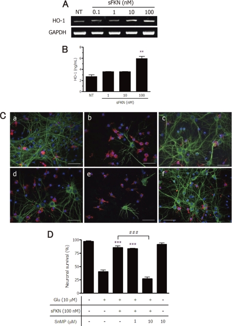
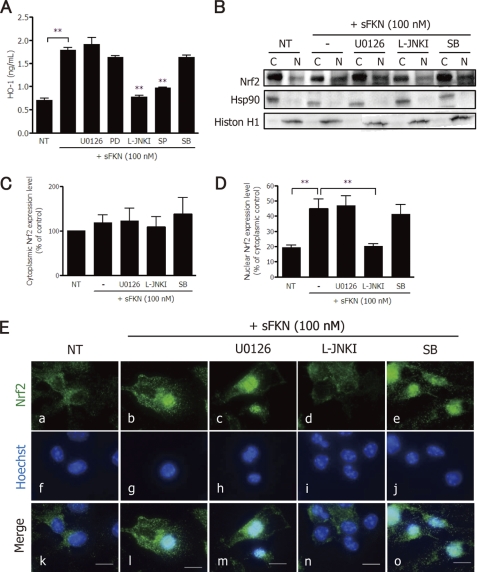
Glutamate-induced excito-neurotoxicity likely contributes to non-cell autonomous neuronal death in neurodegenerative diseases. Microglial clearance of dying neurons and associated debris is essential to maintain healthy neural networks in the central nervous system. In fact, the functions of microglia are regulated by various signaling molecules that are produced as neurons degenerate. Here, we show that the soluble CX3C chemokine fractalkine (sFKN), which is secreted from neurons that have been damaged by glutamate, promotes microglial phagocytosis of neuronal debris through release of milk fat globule-EGF factor 8, a mediator of apoptotic cell clearance. In addition, sFKN induces the expression of the antioxidant enzyme heme oxygenase-1 (HO-1) in microglia in the absence of neurotoxic molecule production, including NO, TNF, and glutamate. sFKN treatment of primary neuron-microglia co-cultures significantly attenuated glutamate-induced neuronal cell death. Using several specific MAPK inhibitors, we found that sFKN-induced heme oxygenase-1 expression was primarily mediated by activation of JNK and nuclear factor erythroid 2-related factor 2. These results suggest that sFKN secreted from glutamate-damaged neurons provides both phagocytotic and neuroprotective signals.


Figures

FIGURE 1.
FIGURE 1.
Excitotoxically damaged neurons release sFKN to promote microglial clearance of neuronal debris. A, expression of FKN and CX3CR1 mRNA in microglia (Mi) and neurons (Neu) was assessed using RT-PCRs. GAPDH expression was used as a control. B, sFKN in the culture medium of microglia and neuronal cultures was measured using ELISAs. C, neurons were treated with glutamate at the indicated concentrations. sFKN concentrations in the neuronal culture supernatants were measured. Results show the mean ± S.E. (n = 3). Glutamate treatment significantly induced sFKN expression in 6 h compared with untreated control samples. *, p < 0.05; **, p < 0.01 (one-way ANOVA with Dunnett's post hoc test). D, microglial phagocytosis assay. Primary mouse cortical neurons were labeled with CM-DiI, and treated with or without glutamate (Glu) for 24 h. The culture medium was changed and microglia were added to the culture with or without sFKN for 24 h. A few DiI-incorporated microgila were detected in co-cultures of untreated neurons (NT) and microglia (a). sFKN (10 nm) did not increase the number of phagocytosed cells (b). Neurons that were pretreated with 10 μm glutamate (pre-Glu neurons) showed an increase in microglial phagocytosis (c); 10 nm sFKN increase the number of DiI-incorporated microglia (d). The arrows in the fluorescence micrograph denote microglial phagocytosis of DiI-stained neuronal debris. Scale bar = 20 μm. E, quantification of the phagocytosis index, which is defined as the percentage of total microglia staining (green) that overlaps with DiI staining (red) (relevant areas are shown in yellow). The columns indicate the mean ± S.E. obtained from three independent experiments, each of which included analysis of 10 randomly selected fields. Significant differences compared with untreated samples (*) or samples without sFKN treatment (#) are noted. ***, p < 0.001; ###, p < 0.001; n.s., not significant (one-way ANOVA with Tukey's post hoc test). F–H, microglia treated with the indicated concentrations of sFKN did not induce the production of glutamate (G), nitrite and iNOS (H), or TNF-α (I) as measured using the assay kit or ELISAs for protein levels, and mRNA expression levels of iNOS (H) or TNF-α (I) as measured using RT-PCR.
FIGURE 2.
FIGURE 2.
sFKN-induced microglial phagocytosis of excitotoxically damaged neurons is mediated through expression of MFG-E8. A, microglia were treated with the indicated concentrations of sFKN for 24 h, and MFG-E8 mRNA and protein expression levels were measured using ELISAs and RT-PCRs, respectively. The columns indicate the mean ± S.E. (n = 3). Treatment with 10 nm sFKN significantly increased MFG-E8 protein levels compared with untreated (NT) samples. *, p < 0.05 (one-way ANOVA with Dunnett's post hoc test). B, microglial phagocytosis of primary cortical neurons exposed to glutamate was examined by addition of anti-MFG-E8 antibody or isotype-matched hamster IgG control: (a) without sFKN, (b) 100 nm sFKN, (c) 100 nm sFKN and 10 μg/ml anti-MFG-E8 antibody, and (d) 100 nm sFKN and 20 μg/ml anti-MFG-E8 antibody, (e) 100 nm sFKN and 20 μg/ml of hamster IgG control. The arrows denote phagocytosis of neuronal debris (red) by microglia (green). Scale bar = 20 μm. C, phagocytosis index. The columns indicate mean ± S.E. from three independent experiments. In each experiment, 10 randomly selected fields were analyzed. sFKN significantly increased phagocytosis of neural debris, which was dose-dependently suppressed by anti-MFG-E8 antibody. ***, p < 0.001 compared with cultures without antibody (one-way ANOVA with Tukey's post hoc test).
FIGURE 3.
FIGURE 3.
sFKN exerts neuroprotective effects in the presence of microglia. A, untreated neuronal cultures (NT; a) and neuron-microglia co-cultures (f; 1:2 neurons to microglia). Glutamate (Glu) induced neuronal loss in both neuronal (b) and neuron-microglia co-cultures (g). Addition of 100 nm sFKN did not significantly increase the survival rate (c), whereas the same treatment significantly increased survival in the presence of microglia (h). Anti-FKN antibody reduced survival of neurons in the neuron-microglia co-cultures (i), but this antibody had no significant effect in neuronal cultures (d). Addition of goat IgG (isotype-matched control for anti-FKN antibody) had no effect on the survival rate (e and j). Neurons were stained with anti-MAP-2 antibody (green), and microglia were stained with a Cy5-conjugated anti-CD11b antibody (red). Scale bar = 50 μm. B, neuronal survival was estimated as the percentage of intact neurons in the sample relative to the untreated sample. The columns indicate the mean ± S.E. from three independent experiments. In each experiment, 10 randomly selected fields were analyzed. *, indicates significant differences compared with Glu treatment (*, p < 0.05; ***, p < 0.001; n.s., not significant).
FIGURE 4.
FIGURE 4.
Inhibition of endogenous MFG-E8 expression abrogates sFKN-induced neuroprotection. A, untreated neuron-microglia co-cultures (NT; a). The cultures were pretreated with (c–e) or without (b and f) sFKN for 3 h in the presence of 20 μg/ml of anti-MFG-E8 antibody (d and f) or 20 μg/ml of isotype-matched hamster IgG control (e). The cultures were then treated with glutamate for 24 h (b–e). Neurons were stained with anti-MAP-2 antibody (green), and microglia were stained with Cy5-conjugated anti-CD11b antibody (red). Scale bar = 50 μm. B, the neuronal survival rate in the presence of sFKN and anti-MFG-E8 antibody or control IgG was estimated. Columns indicate the mean ± S.E. from three independent experiments, each of which included analysis of 10 randomly selected fields. * indicates significant differences compared with sFKN treatment (***, p < 0.001).
FIGURE 5.
FIGURE 5.
sFKN exerts neuroprotective effects via expression of HO-1 in microglia. Microglia were treated with the indicated concentrations of sFKN for 24 h, and HO-1 mRNA (A) and protein (B) expression levels were measured using RT-PCRs and ELISAs, respectively. The columns indicate the mean ± S.E. (n = 3). **, p < 0.01 compared with untreated control (NT) samples. C, effects of an HO-1 inhibitor on the neuroprotective function of sFKN. Untreated neuron-microglia co-cultures (NT; a). The cultures without pretreatment (b and f) or pretreated with sFKN (c–e) for 3 h in the presence of the HO-1 inhibitor SnMP at 1 μm (c) or 10 μm (d and f). The cultures were treated with glutamate for 24 h. Neurons were stained with anti-MAP-2 antibody (green), and microglia were stained with Cy5-conjugated anti-CD11b antibody (red). Scale bar, 50 μm. D, the neuronal survival rate in the presence of sFKN and SnMP was estimated. Columns indicate the mean ± S.E. from three independent experiments, each of which included analysis of 10 randomly selected fields. ***, p < 0.001 compared with neuronal survival in the presence of only glutamate. ###, p < 0.001 compared with cultures without SnMP.
FIGURE 6.
FIGURE 6.
sFKN exerts neuroprotective effects via ERK and JNK MAPK signaling. A, protein extracts from BV-2 cells were analyzed by immunoblotting with antibodies specific for phosphorylated and total MAPKs (ERK1/2, JNK, and p38). Cells were treated with 100 nm sFKN for the indicated time periods or 1 μg/ml of LPS for 60 min. *, p < 0.05; **, p < 0.01 compared with untreated control (NT) samples. B, neuron-microglia co-cultures were pretreated with (a–d) or without (e and f) 100 nm sFKN for 3 h in the presence of MAPK inhibitors (MEK1/2 (b and e), 1 μm U0126; JNK (c and f), 10 μm JNK peptide inhibitor L-JNKI; p38 (d), 10 μm SB203580). The cultures were then treated with 10 μm glutamate for 24 h. Staining for neurons (MAP2; green) and microglia (CD11b; red) was then performed. Scale bar, 50 μm. C, neuronal survival rate against glutamate excitotoxicity in the presence of 100 nm sFKN and each MAPK inhibitor (MEK1/2, 1 μm U0126; MEK1, 10 μm PD98059 (PD); JNK, 10 μm L-JNKI and 10 μm SP600125 (SP); p38, 10 μm SB203580 (SB)). The columns indicate the mean ± S.E. from three independent experiments, each of which included analysis of 10 randomly selected fields. ***, p < 0.001 compared with the cultures without MAPK inhibitors.
FIGURE 7.
FIGURE 7.
sFKN induces microglial HO-1 expression via JNK-Nrf2 signaling. A, after microglia were treated with sFKN and the indicated MAPK inhibitors for 24 h, HO-1 protein expression levels were measured using ELISAs. **, p < 0.01 compared with sFKN-treated samples. B, BV-2 cells were treated with various MAPK inhibitors (MEK1/2, 1 μm U0126; JNK, 10 μm L-JNKI; or p38, 10 μm SB203580 (SB)) in the presence or absence of 100 nm sFKN. Protein extracts from cytoplasmic (c) or nuclear (n) fractions were analyzed by immunoblotting with the antibodies specific for Nrf2, Hsp90, and histone H1. Hsp90 expression was used as a cytoplasmic protein control. Histon H1 was used as nuclear protein expression control. C, relative amount of Nrf2 per cytoplasmic protein Hsp90 in BV-2 cells. The columns show the mean ± S.E. from three independent experiments. D, relative amount of Nrf2 per nuclear protein histone H1 in BV-2 cells. The columns show the mean ± S.E. from three independent experiments. * indicates significant differences compared with sFKN-treated nuclear extracts (**, p < 0.01). E, immunofluorescence images of Nrf2 (green; a–e) and nuclei (Hoechst blue; f–j) in microglia treated with 100 nm sFKN in the presence of various MAPK inhibitors (U0126 (c, h, and m), L-JNKI (d, i, and n), SB (e, j, and o)). The merged images (k–o) show decreased levels of nuclear Nrf2 in response to L-JNKI (n). Scale bar, 10 μm.
FIGURE 8.
FIGURE 8.
Model of the role of sFKN in microglial phagocytosis and neuroprotection. sFKN, which is secreted from neurons that are damaged by glutamate, promotes microglial phagocytosis of neuronal debris through the release of MFG-E8. sFKN also induces the expression of the antioxidant enzyme HO-1 in microglia via Nrf2 recruitment and activation of the JNK MAPK signaling pathway. The neuroprotective effects of sFKN are also mediated in part by activation of ERK MAPK, although the downstream signaling pathway has not yet been elucidated. Therefore, sFKN may be an intrinsic neuroprotectant for damaged yet surviving neurons.

Similar articles

Cited by

References

    1. Farfara D., Lifshitz V., Frenkel D. (2008) J. Cell Mol. Med. 12, 762–780 - PMC - PubMed
    1. Takeuchi H., Jin S., Wang J., Zhang G., Kawanokuchi J., Kuno R., Sonobe Y., Mizuno T., Suzumura A. (2006) J. Biol. Chem. 281, 21362–21368 - PubMed
    1. Takeuchi H. (2010) Clin. Exp. Neuroimmun. 1, 12–21
    1. Doi Y., Mizuno T., Maki Y., Jin S., Mizoguchi H., Ikeyama M., Doi M., Michikawa M., Takeuchi H., Suzumura A. (2009) Am. J. Pathol. 175, 2121–2132 - PMC - PubMed
    1. Elbirt K. K., Bonkovsky H. L. (1999) Proc. Assoc. Am. Physicians 111, 438–447 - PubMed

Publication types

MeSH terms