. 2011 Jan;152(1):313-25.
doi: 10.1210/en.2010-0874. Epub 2010 Nov 3.

The involvement of specific PKC isoenzymes in phorbol ester-mediated regulation of steroidogenic acute regulatory protein expression and steroid synthesis in mouse Leydig cells

Affiliations

The involvement of specific PKC isoenzymes in phorbol ester-mediated regulation of steroidogenic acute regulatory protein expression and steroid synthesis in mouse Leydig cells

Pulak R Manna et al. Endocrinology. 2011 Jan.

Abstract

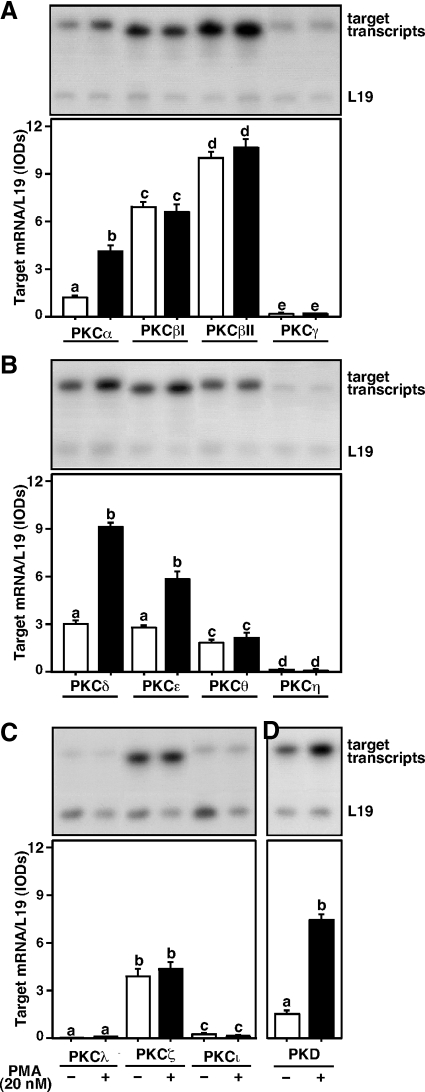
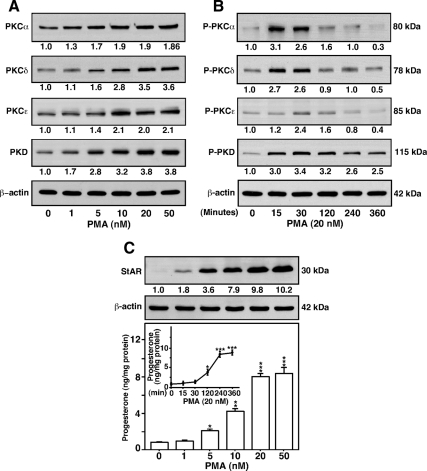
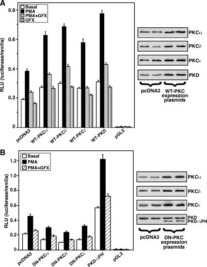
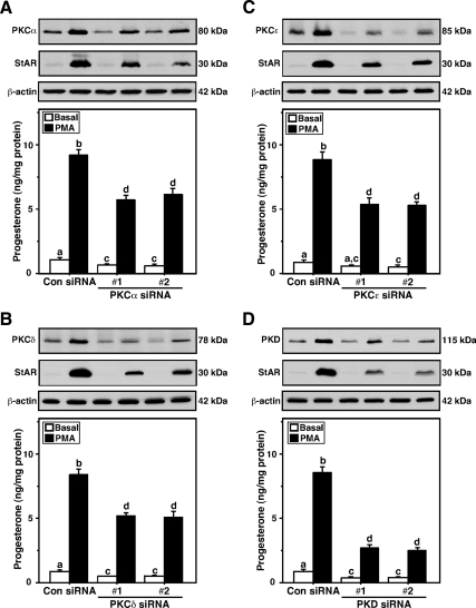
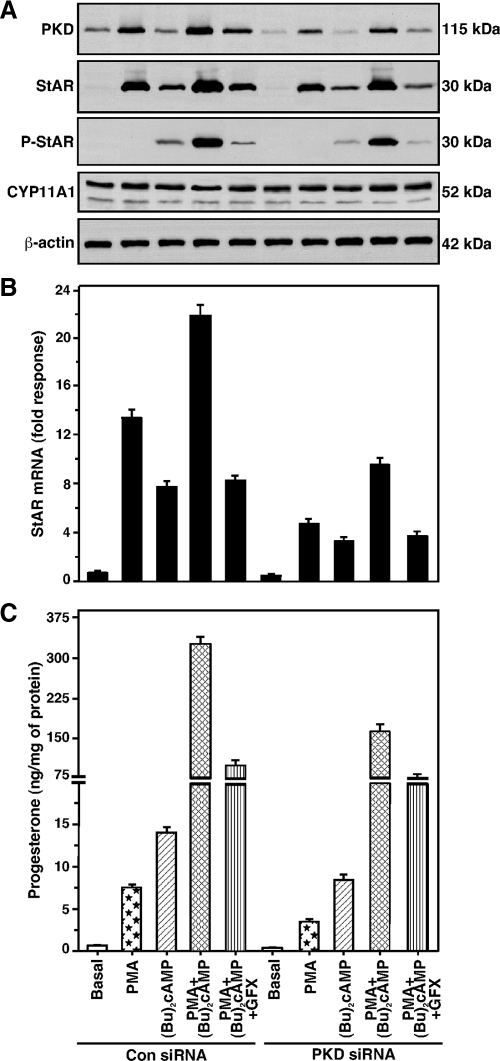
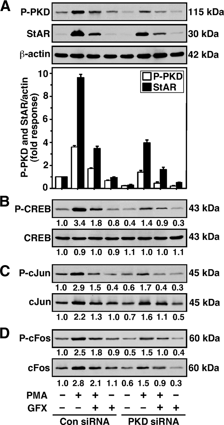
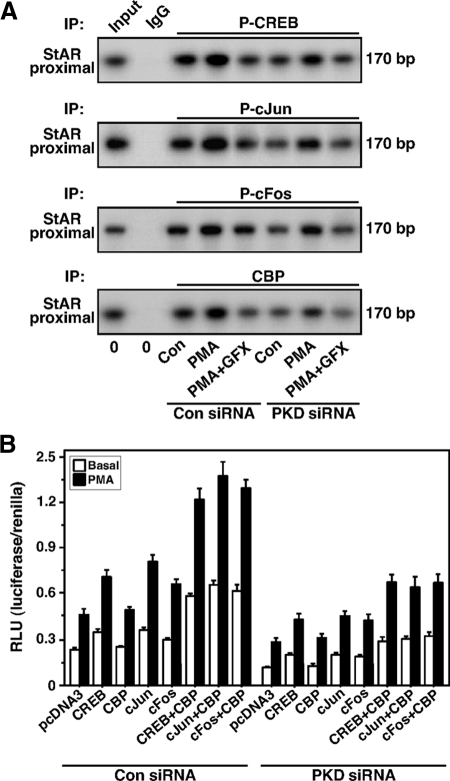
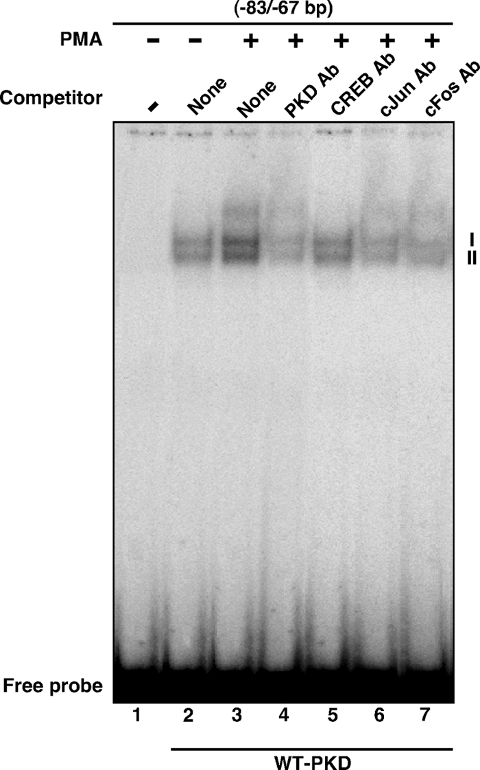
Protein kinase C (PKC) is a multigene family of serine/threonine kinases. PKC is involved in regulating adrenal and gonadal steroidogenesis; however, the functional relevance of the different PKC isoenzymes remains obscure. In this study, we demonstrate that MA-10 mouse Leydig tumor cells express several PKC isoforms to varying levels and that the activation of PKC signaling, by phorbol 12-myristate 13-acetate (PMA) elevated the expression and phosphorylation of PKCα, -δ, -ε, and -μ/protein kinase D (PKD). These responses coincided with the expression of the steroidogenic acute regulatory (StAR) protein and progesterone synthesis. Targeted silencing of PKCα, δ, and ε and PKD, using small interfering RNAs, resulted in deceases in basal and PMA-mediated StAR and steroid levels and demonstrated the importance of PKD in steroidogenesis. PKD was capable of controlling PMA and cAMP/PKA-mediated synergism involved in the steroidogenic response. Further studies pointed out that the regulatory events effected by PKD are associated with cAMP response element-binding protein (CREB) and c-Jun/c-Fos-mediated transcription of the StAR gene. Chromatin immunoprecipitation studies revealed that the activation of phosphorylated CREB, c-Jun, and c-Fos by PMA was correlated with in vivo protein-DNA interactions and the recruitment of CREB-binding protein, whereas knockdown of PKD suppressed the association of these factors with the StAR promoter. Ectopic expression of CREB-binding protein enhanced the trans-activation potential of CREB and c-Jun/c-Fos in StAR gene expression. Using EMSA, a -83/-67-bp region of the StAR promoter was shown to bind PKD-transfected MA-10 nuclear extract in a PMA-responsive manner, targeting CREB and c-Jun/c-Fos proteins. These findings provide evidence for the presence of multiple PKC isoforms and demonstrate the molecular events by which selective isozymes, especially PKD, influence PMA/PKC signaling involved in the regulation of the steroidogenic machinery in mouse Leydig cells.


Figures

Figure 1
Figure 1
Detection of various PKC isoforms in MA-10 cells. Cells were treated without (white bar) or with (black bar) 20 nm PMA for 6 h and subjected to isolation of total RNA for determining relative expression of different PKC isoforms by RT-PCR analysis. Representative autoradiograms illustrate expression of PKCα, -βI, -βII, and -γ (A); PKCδ, -ε, -θ, and -η (B); PKCλ, -ζ, and -ι (C); and PKD (D) in control and stimulated groups. L19 expression was assessed as loading controls. Integrated OD (IOD) values of each band were quantified, normalized with the corresponding L19 bands, and presented as target mRNA/L19. Data represent the mean ± se of four independent experiments. Letters above the bars indicate that these groups differ significantly from each other at least at P < 0.05.
Figure 2
Figure 2
Effect of PMA on expression and phosphorylation of PKCα, -δ, and -ε, PKD, StAR, and progesterone synthesis. MA-10 cells were treated either with increasing (0–50 nm, 6 h) or with a fixed (20 nm, 0–360 min) dose of PMA and subjected to preparation of cellular protein for immunoblotting. Representative immunoblots show expression (A) and phosphorylation (B) of PKCα, -δ, and -ε, PKD, and StAR (C) in different groups using 25–30 μg of total protein. The average integrated ODs for different PKC isoforms and StAR were normalized to those of β-actin for each treatment. The fold change of these values is presented below the blots relative to unstimulated cells. Results are representative of four to seven independent experiments. C, The levels of progesterone in media, obtained with either varying doses or different time points (inset) in response to PMA, were determined and expressed as nanograms per milligram protein (n = 4, ±se). *, P < 0.05; **, P < 0.01; ***, P < 0.001 vs. control.
Figure 3
Figure 3
Roles of PKCα, -δ, and -ε and PKD isoforms in PMA mediated StAR promoter responsiveness. MA-10 cells were transfected with empty vector (pcDNA3), WT (A), and DN (B) mutants of PKCα, -δ, or -ε or a PKD mutant lacking the PH domain (PKD-ΔPH), within the context of the −151/−1 StAR luciferase reporter segment, in the presence of pRL-SV40, as described in Materials and Methods. pGL3 basic (pGL3) was used as a control. After 36 h of transfection, cells were pretreated without or with PKC inhibitor GFX (20 μm) for 30 min and then treated for 6 h in the absence (basal) or presence of PMA (20 nm), PMA plus GFX, and GFX, as indicated. After 36 h of transfection, cells were also determined for protein levels by Western blotting. Representative immunoblots (n = 3) illustrate expression (in duplicates) of different WT and DN PKC isoforms in control (pcDNA3) and PKC-overexpressing cells (A and B, right panels). Luciferase activity in the cell lysates was determined and expressed as relative light units (RLU, luciferase/renilla). Data represent the mean ± se of five independent experiments.
Figure 4
Figure 4
Silencing of PKCα, -δ, and -ε and PKD isoenzymes in MA-10 cells and its consequences on StAR expression and steroid synthesis. Cells were transfected with either a negative control siRNA (Con siRNA) or two different (no. 1 and no. 2) PKCα (A), -δ (B), and -ε (C) and PKD (D) specific siRNAs at 100 nm concentration each, as described in Materials and Methods. After 48 h of transfection, cells were treated without (basal) or with PMA (20 nm) for an additional 6 h, and cells were subjected to cellular protein preparation for immunoblotting. Representative immunoblots illustrate expression of PKCα, -δ, and -ε, PKD, and StAR in different treatment groups using 20–30 μg of total protein. β-Actin expression was assessed as loading controls. Immunoblots are representative of five to seven independent experiments. C, Accumulation of progesterone in media of different treatment groups was determined (n = 5, ±se) and expressed as nanograms per milligram protein. Letters above the bars indicate that these groups differ significantly from each other at least at P < 0.05.
Figure 5
Figure 5
Effect of PKD knockdown on PMA- and (Bu)2cAMP-mediated StAR, P-StAR, StAR mRNA, CYP11A1, and progesterone levels. MA-10 cells were transfected with either a negative control siRNA (Con siRNA) at 100 nm or a mixture of two PKD-specific siRNAs at 50 nm each (100 nm total, PKD siRNA). After 48 h of transfection, cells were pretreated without or with GFX (20 μm) for 30 min and then treated without or with PMA (20 nm), (Bu)2cAMP (0.1 mm), or a combination of them for 6 h, as indicated. Cells were then processed for either immunoblotting or real-time RT-PCR, as described in Materials and Methods. A, Representative immunoblots illustrate PKD, StAR, P-StAR, and CYP11A1 levels in different treatment groups using 25–30 μg of total cellular protein. β-Actin expression was assessed as a loading control in immunoblotting. B, Levels of StAR mRNAs of the same treatment groups were determined by real-time RT-PCR. Immunoblots and RT-PCR analyses are representative of four to six independent experiments. C, Accumulation of progesterone in media of the same groups was determined and expressed as nanograms per milligram protein (n = 4, ±se).
Figure 6
Figure 6
Silencing of PKD on StAR, CREB, c-Jun, and c-Fos levels in MA-10 cells. Cells were transfected with either a negative control (Con siRNA) or a mixture of two PKD siRNAs (PKD siRNA), as described in the legend of Fig. 5. After 48 h of transfection, cells were pretreated without or with GFX (20 μm) for 30 min, then treated in the absence or presence of PMA (20 nm) for 6 h, and then subjected to cellular protein preparation. Representative immunoblots illustrate P-PKD and StAR (A), P-CREB and CREB (B), P-c-Jun and c-Jun (C), and P-c-Fos and c-Fos (D) in different treatment groups, using 20–30 μg of total cellular protein. The average integrated ODs for P-PKD and StAR were normalized to those of β-actin for each treatment. Integrated OD values for P-CREB, CREB, P-c-Jun, c-Jun, P-c-Fos, and c-Fos were quantified. The fold changes of these values are reported for each treatment relative to the unstimulated control siRNA group. Compiled data from four experiments are presented in A (±se, lower panel). Immunoblots shown are representative of three to six independent experiments.
Figure 7
Figure 7
Effect of PKD knockdown on association of P-CREB, P-c-Jun, P-c-Fos, and CBP with the StAR promoter and its relevance to StAR gene expression. MA-10 cells were transfected with either a negative control (Con siRNA) or a mixture of two PKD siRNAs (PKD siRNA), as described in the legend of Fig. 5. After 48 h of transfection, cells were pretreated without or with GFX (20 μm) for 30 min and then treated in the absence or presence of PMA (20 nm) for an additional 30 min as indicated, and ChIP assays were carried out as described in Materials and Methods. Cross-linked sheared chromatin obtained from different treatment groups was immunoprecipitated (IP) either with IgG or anti-P-CREB, anti-P-c-Jun, anti-P-c-Fos, and anti-CBP Abs. Recovered chromatin was subjected to PCR using the −170/−1-bp region of the StAR promoter. A, Representative autoradiograms illustrate the association of P-CREB, P-c-Jun, P-c-Fos, and CBP to the proximal StAR promoter. Data are representative of three to four independent experiments. B, Cells were transfected either with empty vector (pcDNA3), CREB, c-Jun, c-Fos, and CBP expression plasmids, or a combination of them as indicated, within the context of the −151/−1 StAR reporter segment in the presence of pRL-SV40. After 36 h of transfection, cells were treated in the absence (basal) or presence of PMA (20 nm) for an additional 6 h. Luciferase activity in the cell lysates was determined and expressed as relative light units (RLU, luciferase/renilla). Data represent the mean ± se of four independent experiments.
Figure 8
Figure 8
Binding of MA-10 NE to the 83/−67-bp region of the StAR promoter, using EMSA. MA-10 cells were transfected with WT PKD expression plasmid. After 48 h of transfection, cells were treated without (lane 2) or with (lanes 3–7) 10 nm PMA for 6 h and then subjected to NE preparation. NEs (12–15 μg) obtained from different treatment groups were incubated with the 32P-labeled probe specific to the −83/−67-bp region of the StAR promoter. DNA-protein complexes (I and II) were challenged without (lanes 2 and 3) or with PKD (lane 4), CREB (lane 5), c-Jun (lane 6), and c-Fos (lane 7) Abs. Migration of free probes is shown for each lane. Data are representative of three independent experiments.

Similar articles

Cited by

References

    1. Nishizuka Y 1995 Protein kinase C and lipid signaling for sustained cellular responses. FASEB J 9:484–496 - PubMed
    1. Peters CA, Maizels ET, Hunzicker-Dunn M 1999 Activation of PKCδ in the rat corpus luteum during pregnancy. Potential role of prolactin signaling. J Biol Chem 274:37499–37505 - PubMed
    1. Parker PJ, Murray-Rust J 2004 PKC at a glance. J Cell Sci 117:131–132 - PubMed
    1. Feng C, Zhang J, Gasana V, Fu W, Liu Y, Zong Z, Yu B 2005 Differential expression of protein kinase C alpha and delta in testes of mouse at various stages of development. Cell Biochem Funct 23:415–420 - PubMed
    1. Valverde AM, Sinnett-Smith J, Van Lint J, Rozengurt E 1994 Molecular cloning and characterization of protein kinase D: a target for diacylglycerol and phorbol esters with a distinctive catalytic domain. Proc Natl Acad Sci USA 91:8572–8576 - PMC - PubMed

Publication types

MeSH terms