. 2010 Nov;120(11):3969-78.
doi: 10.1172/JCI42563. Epub 2010 Oct 11.

The cytomegalovirus-encoded chemokine receptor US28 promotes intestinal neoplasia in transgenic mice

Affiliations

The cytomegalovirus-encoded chemokine receptor US28 promotes intestinal neoplasia in transgenic mice

Gerold Bongers et al. J Clin Invest. 2010 Nov.

Abstract

US28 is a constitutively active chemokine receptor encoded by CMV (also referred to as human herpesvirus 5), a highly prevalent human virus that infects a broad spectrum of cells, including intestinal epithelial cells (IECs). To study the role of US28 in vivo, we created transgenic mice (VS28 mice) in which US28 expression was targeted to IECs. Expression of US28 was detected in all IECs of the small and large intestine, including in cells expressing leucine rich repeat containing GPCR5 (Lgr5), a marker gene of intestinal epithelial stem cells. US28 expression in IECs inhibited glycogen synthase 3β (GSK-3β) function, promoted accumulation of β-catenin protein, and increased expression of Wnt target genes involved in the control of the cell proliferation. VS28 mice showed a hyperplastic intestinal epithelium and, strikingly, developed adenomas and adenocarcinomas by 40 weeks of age. When exposed to an inflammation-driven tumor model (azoxymethane/dextran sodium sulfate), VS28 mice developed a significantly higher tumor burden than control littermates. Transgenic coexpression of the US28 ligand CCL2 (an inflammatory chemokine) increased IEC proliferation as well as tumor burden, suggesting that the oncogenic activity of US28 can be modulated by inflammatory factors. Together, these results indicate that expression of US28 promotes development of intestinal dysplasia and cancer in transgenic mice and suggest that CMV infection may facilitate development of intestinal neoplasia in humans.


Figures
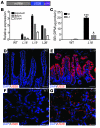
Figure 1
Figure 1. Expression of the US28 transgene in VS28 mice.
(A) Diagram of the VS28 transgene. Expression of US28 is driven by the 9-kb mouse villin promoter (mVillin). (B) US28 mRNA expression in jejunum, ileum, and colon of WT, VS28L18, VS28L19, and VS28L36 mice. Values were standardized to ubiquitin. Results represent the mean ± SEM (n = 3–4). (C) Radioligand competition assay using [125I]-CCL5 on membranes of IECs isolated from of the small intestine (SI) or large intestine (LI) of WT or VS28L18 mice. Results are expressed as specific binding (SB) in disintegrations per minute (DPM) divided by total protein. Results represent the mean ± SEM (n = 3). *P < 0.05, ***P < 0.001 vs. WT. (DG) Representative images of the jejunum (D and E) and colon (F and G) of WT (D and F) and VS28L19 (E and G) mice stained with US28 (red) and DAPI (blue). Scale bar: 50 μm (D and E); 25 μm (F and G).
Figure 2
Figure 2. Histological changes in VS28 mice.
(A and B) H&E staining of the jejunum of WT (A) and VS28L19 mice (B). Scale bar: 100 μm. (CE) Decreased villus length (C) and increased crypt size (D) in VS28L19 mice compared with those of WT mice. (E) Correspondingly, the villus/crypt ratio is decreased in VS28L19 mice compared with that of WT. On average, 80 crypts were measured for each mouse. Results represent the mean ± SEM (n = 12 mice per group). ***P < 0.001 vs. WT.
Figure 3
Figure 3. US28 expression induces IEC proliferation.
(A and B) FACS analysis showing a relative (A) and absolute (B) increase of BrdU-positive CD45 IECs from the jejunum of VS28L19 mice compared with those of WT littermates. Numbers indicate the percentage of BrdU+ cells. Results represent the mean ± SEM (n = 3 mice per group). **P < 0.01 vs. WT mice. (C and D) Increased Ki67-positive cells in VS28L19 mice (D) compared with those in WT mice (C). The inset shows a high magnification of intestinal crypts and reveals expression of US28 in Ki67+ cells. Staining of the jejunum with DAPI (blue), US28 (red), and Ki67 (green). Scale bar: 100 μm (C and D); 25 μm (D, inset).
Figure 4
Figure 4. Expression of US28 in IECs leads to activation of Wnt and MAPK pathways.
(A) Increased mRNA levels of the Wnt target genes c-myc, Birc5, and Ccnd1 in IECs from VS28L19 mice. Notice that the mRNA levels of Ctnnb1 were not changed. The dashed line indicates WT values. Values were standardized to ubiquitin Results represent the mean ± SEM (n = 3–4 mice per group). **P < 0.01, ***P < 0.001 vs. WT. (B) Western blot analysis of total β-catenin, phosphorylated GSK-3β/Ser9, active non-phosphorylated β-catenin (αABC), cyclin-D1, survivin, and phosphorylated Erk1/2 in IECs from VS28L19 and WT mice. A representative blot is show of from 3 independent experiments (n = 4).
Figure 5
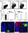
Figure 5. US28 is expressed in Lgr5+ intestinal stem cells.
(A) GFPhi and GFPneg IECs were sorted from LGR5 and LGR5S28 mice. Numbers indicate the percentage of GFPhi IECs. (B and C) GFP (B) and US28 (C) mRNA expression in sorted cells from the small intestine of LGR5 and LGR5S28 mice. Values were standardized to ubiquitin. Results represent the mean ± SEM (n = 3). (D and E) US28 is expressed in Lgr5+ (GFP) cells. Staining of the jejunum of LGR5S28 mice with DAPI (blue), US28 (red), and GFP (green). The inset shows colocalization of US28 in Lgr5+ cells in white. Scale bar: 25 μm (D and E).
Figure 6
Figure 6. Colonic and jejunal tumors in VS28 mice.
(A and B) H&E-stained sections of a poorly differentiated colon carcinoma found in a VS28L18 mouse at 42 weeks of age. (C) H&E-stained section of jejunal tubular adenoma with high-grade dysplasia found in a VS28L19 mouse. (D and E) Tumors found in VS28 mice are positive for pan-keratin, Ki67 (D), and US28 (E). Representative sections stained with DAPI (blue), pan-keratin (D, green), Ki67 (D, red), or US28 (E, red) are shown. (F) β-catenin is present in the nucleus (white) of an adenoma (right side of the dashed line) of a VS28L19 mouse. No nuclear translocation is seen in normal crypts (left side of the dashed line). Representative sections stained with DAPI (red) and β-catenin (green) are shown. Nuclear translocation (double staining) is indicated in white. (G) Jejunal adenomas express cyclin-D1. A representative section stained with DAPI (blue) and cyclin-D1 (red) is shown. (H) Survivin was found in the cytosol of jejunal tubular adenomas (right side of the dashed line), in contrast to its normal nuclear localization (left side of the dashed line). Scale bar: 200 μm (A); 100 μm (BD); 50 μm (E and G); 20 μm (F and H).
Figure 7
Figure 7. Response of VS28 and WT mice to AOM/DSS.
(A) Schematic of AOM/DSS administration. Mice were injected with 12 mg/kg AOM and subjected to three 7-day cycles of 2.5% DSS. (B and C) Representative H&E-stained sections of adenomas found after AOM/DSS treatment in WT (B) and VS28L18 mice (C). Scale bar: 50 μm. (D and E) The number of polyps (E) and the polyp area (D) were significantly increased in the VS28L18 mice (n = 11) compared with those of the WT littermates (n = 11). Results represent the mean ± SEM. *P < 0.05, ***P < 0.001 vs. WT.
Figure 8
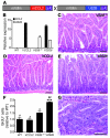
Figure 8. Transgenic coexpression of the inflammatory chemokine CCL2 exacerbates the phenotype of VS28L18 mice.
The inflammatory chemokine CCL2 is an agonist for US28. To test whether CCL2 can activate US28 in vivo, we generated mice expressing both CCL2 and US28 in IECs. (A) Generation of mice expressing both CCL2 and US28 in IECs (V2S28 mice). A 9-kb fragment of the villin promoter was used to drive expression of mouse CCL2 (mCCL2) to IECs (VCCL2 mice). Subsequently, the VCCL2 transgenic mice were crossed with VS28L18 mice to generate V2S28 mice. (B) CCL2 and US28 mRNA expression in the jejunum of WT, VCCL2, VS28L18, and V2S28 mice. Values were standardized to ubiquitin (n = 3). (CE) H&E staining of the jejunum of VS28L18 (C), VCCL2 (D), and V2S28 mice (E). (F) Analysis of BrdU+ incorporation in CD45 IECs from the jejunum of WT, VCCL2, VS28L18, and V2S28 mice was determined using FACS analysis (n = 4 mice per group). *P < 0.05, ***P < 0.001 vs. WT. #P < 0.05, ##P < 0.01 vs. VS28L18. (G) H&E-stained section of jejunal tubular adenoma found in a V2S28 mouse. Scale bar: 100 μm (CE and G). Results represent the mean ± SEM.

Similar articles

Cited by

References

    1. Soderberg-Naucler C. Does cytomegalovirus play a causative role in the development of various inflammatory diseases and cancer? J Intern Med. 2006;259(3):219–246. doi: 10.1111/j.1365-2796.2006.01618.x. - DOI - PubMed
    1. Mocarski ES, Shenk T, Pass RF. Cytomegaloviruses. In: Knipe DM, ed.Fields Virology . Philadelphia, Pennsylvania, USA: Lippincott, Williams, and Wilkins; 2006: 2701–2772.
    1. Maussang D, Vischer HF, Leurs R, Smit MJ. Herpesvirus-encoded G protein-coupled receptors as modulators of cellular function. Mol Pharmacol. 2009;76(4):692–701. doi: 10.1124/mol.109.057091. - DOI - PubMed
    1. Neote K, DiGregorio D, Mak JY, Horuk R, Schall TJ. Molecular cloning, functional expression, and signaling characteristics of a C-C chemokine receptor. Cell. 1993;72(3):415–425. doi: 10.1016/0092-8674(93)90118-A. - DOI - PubMed
    1. Kledal TN, Rosenkilde M, Schwartz TW. Selective recognition of the membrane-bound CX3C chemokine, fractalkine, by the human cytomegalovirus-encoded broad-spectrum receptor US28. FEBS Lett. 1998;441(2):209–214. doi: 10.1016/S0014-5793(98)01551-8. - DOI - PubMed

Publication types

Substances