. 2011 Feb;21(2):350-64.
doi: 10.1038/cr.2010.144. Epub 2010 Oct 19.

Oncostatin M inhibits myoblast differentiation and regulates muscle regeneration

Affiliations

Oncostatin M inhibits myoblast differentiation and regulates muscle regeneration

Fang Xiao et al. Cell Res. 2011 Feb.

Abstract

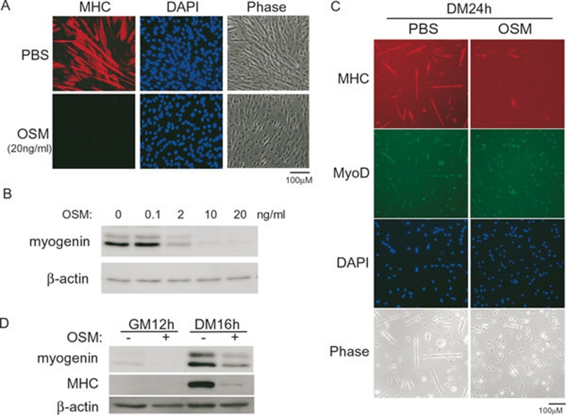
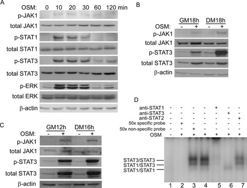
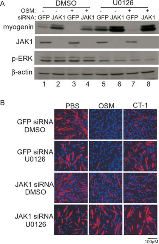
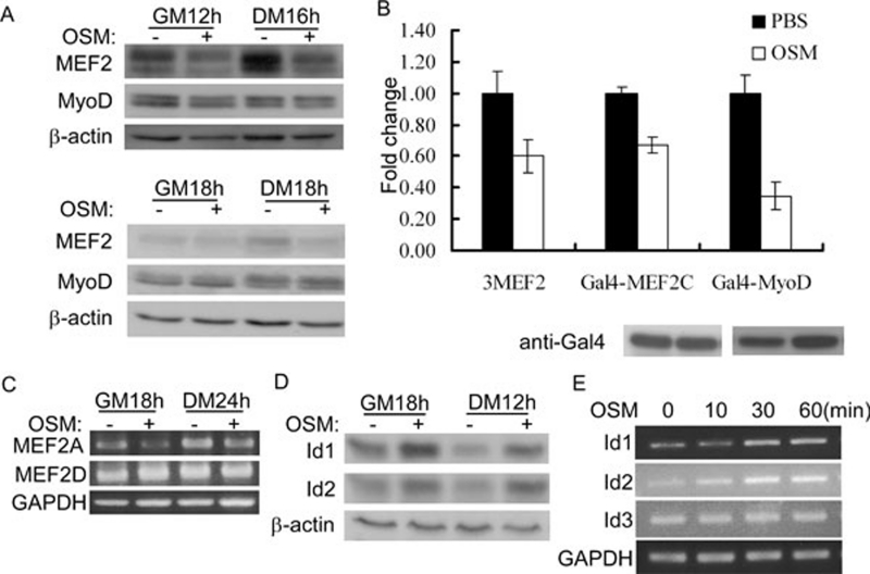
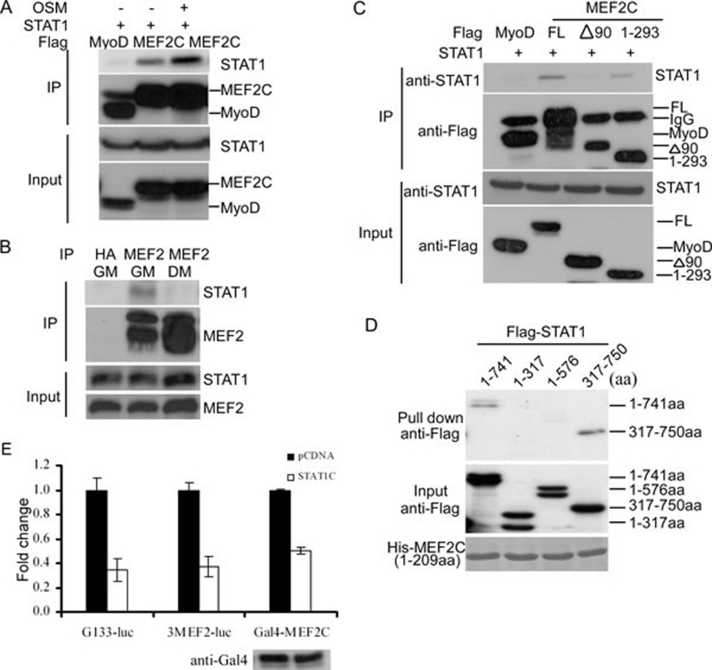
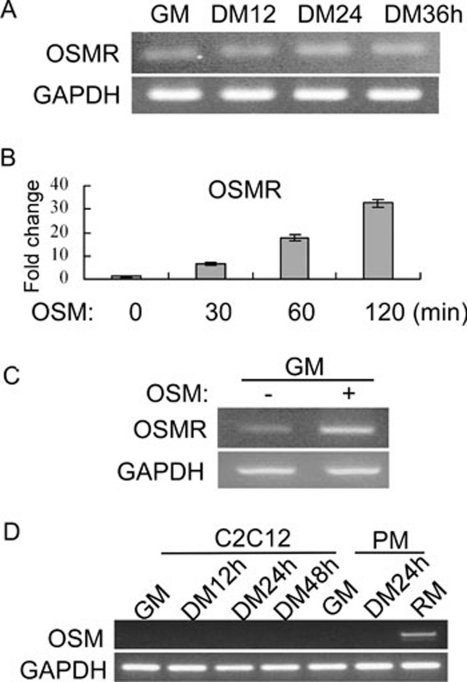
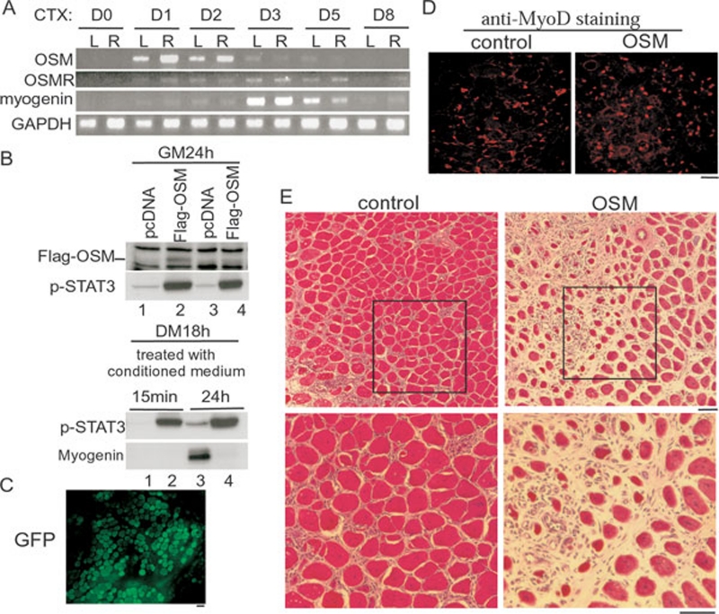
Oncostatin M (OSM) is a cytokine of the interleukin-6 family and plays important roles during inflammation. However, its roles in myoblast differentiation and muscle regeneration remain unexplored. We show here that OSM potently inhibited myoblast differentiation mainly by activating the JAK1/STAT1/STAT3 pathway. OSM downregulated myocyte enhancer-binding factor 2A (MEF2A), upregulated the expression of Id1 and Id2, and inhibited the transcriptional activity of MyoD and MEF2. In addition, OSM also enhanced the expression of STAT3 and OSM receptor, which constituted a positive feedback loop to further amplify OSM-induced signaling. Moreover, we found that STAT1 physically associated with MEF2 and repressed its transcriptional activity, which could account for the OSM-mediated repression of MEF2. Although undetectable in normal muscles in vivo, OSM was rapidly induced on muscle injury and then promptly downregulated just before the majority of myoblasts differentiate. Prolonged expression of OSM in muscles compromised the regeneration process without affecting myoblast proliferation, suggesting that OSM functions to prevent proliferating myoblasts from premature differentiation during the early phase of muscle regeneration.


Figures

Figure 1
Figure 1
OSM inhibited myoblast differentiation. (A, B) Near-confluent C2C12 cells were induced to differentiate for either 72 h (A) or 24 h (B), with or without different doses of OSM. (C, D) Primary myoblasts grown in either GM or DM were treated with or without 20 ng/ml of OSM for various time periods as indicated. In A and C, cells were fixed and subjected to immunostaining for MHC (red) or MyoD (green). Nuclei were counter-stained with DAPI (blue). Phase: phase-contrast images. PBS: phosphate-buffered saline. In B and D, 50 μg of whole cell extracts (WCE) were subjected to SDS-PAGE and western blot analysis for myogenin, MHC, and β-actin (loading control). GM: growth medium. DM: differentiation medium.
Figure 2
Figure 2
OSM activated the JAK1/STAT1/STAT3 pathway in myoblasts. (A) C2C12 cells were treated with 20 ng/ml of OSM for various time periods as indicated. min: minutes. (B, C) C2C12 cells (B) or primary myoblasts (C) grown in GM or DM were treated with or without 20 ng/ml of OSM for 12-18 h. WCE (50 μg), from A to C, were subjected to SDS-PAGE and western blot analysis for various proteins as indicated. (D) C2C12 cells were treated with PBS or 20 ng/ml of OSM for 15 min before harvest. A total of 20 μg of WCE was subjected to EMSA using SIE as a probe.
Figure 3
Figure 3
The JAK1-mediated pathway was mainly responsible for the inhibitory effect of OSM on myoblast differentiation. (A, B) C2C12 cells were firstly transfected with either GFP-siRNA or JAK1 siRNA and then grown in GM for 24 h. Cells were induced to differentiate for 18 h (A) or 48 h (B), with or without 20 ng/ml of OSM or 20 ng/ml of CT-1 in the presence of either DMSO or U0126 (10 μM). In A, cells were harvested, and 50 μg of WCE were subjected to SDS-PAGE and western blot analysis for myogenin, JAK1, p-ERK, and β-actin. In B, cells were fixed and subjected to immunostaining for MHC (red). Nuclei were counter-stained with DAPI (blue).
Figure 4
Figure 4
OSM suppressed the expression of MEF2A, upregulated the expression of Id1/Id2, and inhibited the transcriptional activity of MyoD and MEF2. (A) Primary myoblasts (top three panels) and C2C12 cells (bottom three panels) grown in GM or DM were treated with or without 20 ng/ml of OSM for 12-18 h. Cells were harvested and 50 μg of WCE were subjected to SDS-PAGE and western blot analysis for MEF2, MyoD, or β-actin. (B) C2C12 cells were transfected in triplicate with 3xMEF2-luc or gal4-luc, together with constructs encoding Gal4-MyoD or Gal4-MEF2. At 24 h after transfection, cells were induced to differentiate with or without 20 ng/ml of OSM for another 24 h before harvest. WCE were subjected to luciferase assays and SDS-PAGE/western blot analysis for Gal4-MyoD and Gal4-MEF2C. Fold change was calculated as the ratio of the luciferase activity of the OSM-treated cells over that of PBS-treated cells. The results were presented as mean±s.d. (C) C2C12 cells grown in GM or DM were treated with or without 20 ng/ml of OSM for 18 or 24 h before harvest. Total RNA was prepared and subjected to RT-PCR analysis for MEF2A and MEF2D genes. GAPDH: glyceraldehyde 3-phosphate dehydrogenase (loading control). (D) C2C12 cells grown in GM or DM were treated with or without 20 ng/ml of OSM for 18 or 12 h. Cells were harvested and 50 μg of WCE were subjected to SDS-PAGE and western blot analysis for Id1, Id2, and β-actin. (E) C2C12 cells grown in GM were treated with 20 ng/ml of OSM for 0, 10, 30, and 60 min. Total RNA was prepared and subjected to RT-PCR analysis for Id1, Id2, Id3, and GAPDH.
Figure 5
Figure 5
STAT1 interacted with MEF2 and repressed its transcriptional activity. (A) C2C12 cells were cotransfected with STAT1 together with Flag-MyoD or Flag-MEF2C, with or without OSM (20 ng/ml) treatment. Flag-MyoD/or Flag-MEF2C was immunoprecipitated from WCE. (B) WCE were prepared from nontransfected C2C12 cells grown in GM or DM. The endogenous MEF2 was immunoprecipitated. HA: anti-HA antibody. (C) C2C12 cells were cotransfected with STAT1 together with Flag-MyoD, full-length Flag-MEF2C, or two truncated Flag-MEF2C constructs. Flag-tagged proteins were immunoprecipitated from WCE. The immunoprecipitates from A to C were subjected to SDS-PAGE and western blot analysis for either the transfected (A, C) or the endogenous STAT1 (B). (D) Equal amount of purified His-MEF2C (1-209 aa) was incubated with WCE prepared from 293T cells expressing either the full-length Flag-STAT1 or three-truncated Flag-STAT1 together with Talon beads. After extensive washing, the retained proteins were subjected to SDS-PAGE and western blot analysis. (E) C2C12 cells were transfected in triplicate with different reporter constructs together with either an empty vector or STAT1c in the absence or presence of Gal4-MEF2C. At 24 h after transfection, cells were further induced to differentiate in DM for another 24 h. WCE was subjected to luciferase assays and SDS-PAGE/western blots for Gal4-MEF2C. (F) Fold change was calculated as the ratio of the luciferase activity in cells transfected with STAT1c over that in cells transfected with the empty vector. The results were presented as mean±s.d.
Figure 6
Figure 6
OSMR mRNA was expressed in myoblasts and induced by OSM. (A) C2C12 cells were grown in GM or induced to differentiate in DM for various time periods. (B) C2C12 cells were treated with 20 ng/ml of OSM for various time periods as indicated. (C) Proliferating primary myoblasts in GM were treated with 20 ng/ml of OSM for 3 h before harvest. (D) C2C12 cells or primary myoblasts (PM) grown in GM or induced to differentiate in DM for various time periods as indicated. Total RNA was prepared from cells in A to D. For samples from A, C, and D, semiquantitative RT-PCR was performed for OSMR or OSM genes. GAPDH gene was used as a loading control. For samples from B, SYBR Green-based quantitative RT-PCR was performed for OSMR gene, with GAPDH serving as an internal control. Total RNA from regenerating mouse tibialis anterior muscles (RM) in D was used as a positive control.
Figure 7
Figure 7
Prolonged expression of OSM delayed muscle regeneration. (A) 6- to 8-week-old mice were either left untreated (D0) or injected with 20 μl of 10 μM cardiotoxin (CTX) into tibialis anterior (TA) muscles of both legs. TA muscles were surgically isolated at different time points as indicated. Total RNA was prepared and subjected to semiquantitative RT-PCR analysis for OSM, OSMR, and myogenin genes. GAPDH gene was used as a loading control. The experiment was repeated three times with similar results and one set of the representative data was shown. Dx, x day after injury; L, left leg; R, right leg. (B) Upper two panels: C2C12 cells were transfected with either an empty vector or Flag-OSM. At 24 h after transfection, cells were either left in GM for another 24 h or induced to differentiate in DM for 18 h before harvest. The conditioned media were collected for the following experiment. Bottom two panels: near-confluent C2C12 cells were treated with the conditioned media prepared above (in the same order) for 15 min or 24 h before harvest. WCE were subjected to SDS-PAGE and western blot analysis for Flag-OSM, p-STAT3, and myogenin. (C-E) TA muscles of 6-week-old mice were electroporated with either a GFP-expressing vector (C, and left panel in D) or an OSM-expressing vector (right panel in D). At 3 (C, D) or 10 (E) days after electroporation, TA muscles were dissected and embedded for cryostat sectioning. Muscle sections were then subjected to direct microscopic examination for autofluorescence (C), immunostaining for MyoD (D), or hematoxylin/eosin staining (E). The experiments in C to E were repeated three times with similar results. Representative images were shown here. Bars: 100 μm.

Similar articles

Cited by

References

    1. Buckingham M. Skeletal muscle formation in vertebrates. Curr Opin Genet Dev. 2001;11:440–448. - PubMed
    1. Puri PL, Sartorelli V. Regulation of muscle regulatory factors by DNA-binding, interacting proteins, and post-transcriptional modifications. J Cell Physiol. 2000;185:155–173. - PubMed
    1. Sabourin LA, Rudnicki MA. The molecular regulation of myogenesis. Clin Genet. 2000;57:16–25. - PubMed
    1. Arnold HH, Braun T. Targeted inactivation of myogenic factor genes reveals their role during mouse myogenesis: a review. Int J Dev Biol. 1996;40:345–353. - PubMed
    1. Tapscott SJ. The circuitry of a master switch: Myod and the regulation of skeletal muscle gene transcription. Development. 2005;132:2685–2695. - PubMed

Publication types

MeSH terms