. 2011 Jan;31(1):226-36.
doi: 10.1128/MCB.00821-10. Epub 2010 Oct 18.

Genomic collaboration of estrogen receptor alpha and extracellular signal-regulated kinase 2 in regulating gene and proliferation programs

Affiliations

Genomic collaboration of estrogen receptor alpha and extracellular signal-regulated kinase 2 in regulating gene and proliferation programs

Zeynep Madak-Erdogan et al. Mol Cell Biol. 2011 Jan.

Abstract

The nuclear hormone receptor, estrogen receptor α (ERα), and mitogen-activated protein kinases (MAPKs) play key roles in hormone-dependent cancers, and yet their interplay and the integration of their signaling inputs remain poorly understood. In these studies, we document that estrogen-occupied ERα activates and interacts with extracellular signal-regulated kinase 2 (ERK2), a downstream effector in the MAPK pathway, resulting in ERK2 and ERα colocalization at chromatin binding sites across the genome of breast cancer cells. This genomic colocalization, predominantly at conserved distal enhancer sites, requires the activation of both ERα and ERK2 and enables ERK2 modulation of estrogen-dependent gene expression and proliferation programs. The ERK2 substrate CREB1 was also activated and recruited to ERK2-bound chromatin following estrogen treatment and found to cooperate with ERα/ERK2 in regulating gene transcription and cell cycle progression. Our study reveals a novel paradigm with convergence of ERK2 and ERα at the chromatin level that positions this kinase to support nuclear receptor activities in crucial and direct ways, a mode of collaboration likely to underlie MAPK regulation of gene expression by other nuclear receptors as well.


Figures
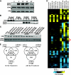
FIG. 1.
FIG. 1.
cDNA microarray gene expression analysis after ERK1 or ERK2 knockdown in MCF-7 cells and effects of kinase depletion on estradiol (E2)-mediated gene regulation. (A) Time course of MAPK activation by E2. MCF-7 cells were treated with 10 nM E2 for the indicated times. Protein was harvested in RIPA buffer and subjected to SDS-PAGE analysis. pMAPK, ERK2, and ERα antibodies were used for Western blot analysis. (B) ERα and ERK2 interact upon E2 treatment of cells and can be immunoprecipitated from MCF-7 cells. Cells were treated with 10 nM E2 for the indicated times and then harvested with RIPA buffer. ERK2- or ERK1-containing complexes were immunoprecipitated from whole-cell extracts and immunoprecipitated proteins were subjected to SDS-PAGE and Western blot analysis for ERα and total MAPK. (C) Validation of selective ERK1 or ERK2 knockdowns in MCF-7 cells. Cells were transfected with siCtrl or single siRNA from each siGENOME or with siGENOME pool reagents for 60 h. ERK2, ERK1, and ERα protein levels after knockdowns were verified by Western blotting. (D) Cluster diagram of genes impacted by ERK2 or ERK1 knockdown. MCF-7 cells were treated with siCtrl, siERK2, or siERK1 for 60 h prior to treatment with 0.1% ethanol vehicle or 10 nM E2 for 4 or 24 h. Affymetrix gene expression microarrays were analyzed by LIMMA and Tightcluster software. The cluster map is visualized using Treeview Java. Fold expression is indicated below. Vertical red bar with star at right indicates gene cluster associated with cell proliferation. (E) Venn diagram depicting numbers of E2 regulated genes in each cell background (siCtrl, siERK2, and siERK1) at 4 and 24 h.
FIG. 2.
FIG. 2.
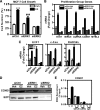
ERK2 controls E2-regulated cell proliferation and the expression of proliferation associated genes. (A) ERK2 is critical for E2-stimulated cell proliferation. MCF-7 cells were plated at 1,000 cells/well in 96-well plates. Cells were transfected with 20 nM siGENOME for Ctrl, ERK1, or ERK2 and the following day (day 0) were treated with 0.1% ethanol vehicle (Veh) or 10 nM E2. Treatment was repeated on day 2 and cell numbers were examined using the MTS assay at day 4. ***, P < 0.001; ###, P < 0.01 (versus vehicle). (B) ERK2 is essential for E2 stimulation of proliferation group genes. MCF-7 cells were transfected with 20 nM siGENOME reagent for Ctrl or ERK2 for 60 h and were then treated with 0.1% ethanol vehicle or 10 nM E2 for 24 h. Total RNA was isolated and reverse transcribed, and expression of the proliferation group genes from the 21 gene signature, which predicts tamoxifen responsiveness of ERα positive breast tumors, was examined by using quantitative PCR (Q-PCR). (C) ERK2 is essential for E2 stimulation of S-phase genes. MCF-7 cells were transfected with 20 nM siGENOME reagent for Ctrl, ERK1, or ERK2 for 60 h and then treated with control vehicle or 10 nM E2 for 24 h. Total RNA was isolated and reverse transcribed, and the expression of DNA synthesis-associated genes was examined by using Q-PCR. (D) CCND1 (cyclin D1) and E2F1 expression and E2 stimulation are affected by ERK2. MCF-7 cells were transfected with 20 nM siGENOME for Ctrl, ERK1, or ERK2 for 60 h and were then treated with control 0.1% ethanol vehicle or 10 nM E2 for 8 h. CCND1 and E2F1 protein levels were assessed by Western blotting. (E) ERK2 is recruited to the ERα binding site at the 3′ enhancer of CCND1. MCF-7 cells were treated with vehicle or 10 nM E2 for 45 min after exposure to siERα for 60 h or 10 μM MEK1 inhibitor U0126 for 1 h. Chromatin was cross-linked and sonicated. ERK2-DNA or background IgG complexes were immunoprecipitated using ERK2 antibody or normal mouse IgG antibody overnight. Recovered DNA was subjected to Q-PCR analysis. Values are expressed as the percent input and are means ± the standard errors of the mean (SEM) from four independent experiments.
FIG. 3.
FIG. 3.
ChIP-on-Chip analysis of genome-wide ERα and ERK2 binding sites. (A) UCSC Genome Browser view of ERα and ERK2 binding sites identified by our ChIP-chip studies. MCF-7 cells were treated with vehicle or 10 nM E2 for 45 min. After formaldehyde cross-linking and sonication, ERα and ERK2 containing complexes were immunoprecipitated. After amplification of the immunoprecipitated or input DNA, microarray analysis was performed using whole-genome Affymetrix GeneChip Human Tiling 2.0R Array sets. (B) Localization of binding sites relative to annotated genes. The location of binding sites was determined relative to the nearest gene in both upstream and downstream directions on both strands, within a 300-kb window. Distributions shown are percentage values. If the binding region is within a gene, CEAS software indicates whether it is in a 5′ untranslated region (5′UTR), a 3′UTR, a coding exon, or an intron. Proximal promoter is defined as 1 kb upstream from RefSeq 5′ start and immediate downstream is 1 kb downstream from RefSeq 3′ end. If a binding site is more than 1 kb away from the RefSeq TSS, it is considered an enhancer. (C) Conservancy of binding sites. Conservancy plots of binding sites were generated by using CEAS software.
FIG. 4.
FIG. 4.
Characterization of ERK2 recruitment to ERα binding sites upon E2 treatment. (A) Time course of ERK2 recruitment to ERα binding sites in the estrogen-responsive LRRC54 and pS2 genes. MCF-7 cells were treated with 10 nM E2 for the indicated times. Chromatin was cross-linked and sonicated. ERK2-DNA or background IgG complexes were immunoprecipitated using ERK2 antibody or normal mouse IgG antibody overnight. Precipitated DNA was subjected to Q-PCR analysis. Values are the means ± the SEM from three independent experiments. (B) ERα is required for ERK2 recruitment to chromatin. MCF-7 cells were transfected with 20 nM siCtrl or siGENOME ERα for 60 h and were then treated with vehicle or 10 nM E2 for 45 min. ERK2-DNA or background IgG complexes were immunoprecipitated using ERK2 (D-2; Santa Cruz) antibody or normal mouse IgG antibody overnight. Precipitated DNA was subjected to Q-PCR analysis. Values are the means ± the SEM from four independent experiments. (C) ERα and ERK2 are present together at the ER binding sites. ChIP/reChIP experiments were performed using antibodies for ERK2 (D-2, sc-1647) and ERα (HC-20, sc-543). Recovered DNA was analyzed by Q-PCR. Values are the means ± the SEM from four independent experiments. (D) ERK2 is not required for ERα recruitment. MCF-7 cells were transfected with 20 nM siCtrl or siGENOME ERK2 for 60 h and then treated with vehicle or 10 nM E2 for 45 min. ERα-DNA or background IgG complexes were immunoprecipitated using ERα (F-10) or normal mouse IgG (Santa Cruz) antibodies overnight. Precipitated DNA was subjected to Q-PCR analysis. Values are the means ± the SEM from four independent experiments. (E) ERK2 activation by MEK1 is required for ERK2 but not ERα recruitment to ER binding sites. MCF-7 cells were pretreated with vehicle or 10 μM U0126 for 1 h and then treated with vehicle or 10 nM E2 for 45 min in the presence or absence of inhibitor. ERK2-DNA, ERα-DNA, or background IgG complexes were immunoprecipitated and precipitated DNA was subjected to Q-PCR analysis. Values are the means ± the SEM from four independent experiments.
FIG. 5.
FIG. 5.
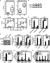
CREB1 is a cooperating transcription factor in ERK2 recruitment to chromatin binding sites and in E2 regulation of cell proliferation. (A) Box plots showing that E2 treatment stimulates recruitment of ERK2 and CREB1 to ERα binding sites (n = 7 genes evaluated). MCF-7 cells were treated with vehicle or 10 nM E2 for 45 min. ChIP was performed using specific antibodies for ERK2 or CREB1, and DNA was analyzed by Q-PCR. Values are the means ± the SEM of at least three independent experiments. (B) Time course of CREB1 phosphorylation after E2 treatment. Total CREB1 is also shown as a control for loading. (C) Time course of CREB1 recruitment. MCF-7 cells were treated with 10 nM E2 for the indicated times. Chromatin was cross-linked and sonicated, and CREB1-DNA or background IgG complexes were immunoprecipitated overnight using antibody for CREB1 or IgG as control. Precipitated DNA was subjected to Q-PCR analysis. Values are the means ± the SD of two experiments. (D) ChIP-reChIP for CREB1 and ERK2 shows that they are present together at ERα binding sites of the LRRC54 and pS2 genes. (E) CREB1 is required for full ERK2 recruitment to binding sites in the LRRC54 and pS2 genes. The left panel shows a Western blot for CREB1, ERα, and ERK2 after control siGL3 or siCREB1 transfection into MCF-7 cells for 72 h prior to vehicle or E2 treatment. ChIP was performed with specific antibodies as described in panel A. (F) MCF-7 cells were transfected with 20 nM siCtrl or siCREB1 and the following day (day 0) were treated with 0.1% ethanol vehicle or 10 nM E2. The treatment was repeated on day 2, and cell numbers were examined using the MTS assay at day 4. (G) CREB1 is required for the E2 stimulation of S-phase genes. MCF-7 cells were transfected with 20 nM siCtrl or siCREB1 for 60 h and then treated with vehicle or 10 nM E2 for 24 h. Total RNA was isolated and reverse transcribed, and expression of DNA synthesis-associated genes was examined by using Q-PCR. (H) CREB1 is required for the E2 stimulation of proliferation group genes. MCF-7 cells were transfected with 20 nM siCtrl or siCREB1 for 60 h and then treated with vehicle or 10 nM E2 for 24 h. Total RNA was isolated and reverse transcribed, and expression of proliferation group genes was examined by using Q-PCR.
FIG. 6.
FIG. 6.
Model depicting the interrelationships elucidated in the present study between ERα, ERK2, and CREB1 in the hormonal regulation of gene expression and cell proliferation. Our findings reveal rapid activation of ERK2 and CREB1 (red stars) in response to estrogen and their colocalization with ERα at enhancer binding sites. ERK2 and CREB1 collaborate with ERα in regulating hormone stimulation of proliferation and of cell cycle-related genes. The findings indicate that ERK2 has not only a signaling function but also a nuclear role at chromatin in integrating with and supporting the actions of this nuclear hormone receptor. See the text for details. CoA, coactivator.

Similar articles

Cited by

References

    1. Agoulnik, I. U., W. E. Bingman III, M. Nakka, W. Li, Q. Wang, X. S. Liu, M. Brown, and N. L. Weigel. 2008. Target gene-specific regulation of androgen receptor activity by p42/p44 mitogen-activated protein kinase. Mol. Endocrinol. 22:2420-2432. - PMC - PubMed
    1. Amazit, L., L. Pasini, A. T. Szafran, V. Berno, R. C. Wu, M. Mielke, E. D. Jones, M. G. Mancini, C. A. Hinojos, B. W. O'Malley, and M. A. Mancini. 2007. Regulation of SRC-3 intercompartmental dynamics by estrogen receptor and phosphorylation. Mol. Cell. Biol. 27:6913-6932. - PMC - PubMed
    1. Avruch, J. 2007. MAP kinase pathways: the first twenty years. Biochim. Biophys. Acta 1773:1150-1160. - PMC - PubMed
    1. Belakavadi, M., P. K. Pandey, R. Vijayvargia, and J. D. Fondell. 2008. MED1 phosphorylation promotes its association with mediator: implications for nuclear receptor signaling. Mol. Cell. Biol. 28:3932-3942. - PMC - PubMed
    1. Britton, D. J., I. R. Hutcheson, J. M. Knowlden, D. Barrow, M. Giles, R. A. McClelland, J. M. Gee, and R. I. Nicholson. 2006. Bidirectional cross talk between ERα and EGFR signaling pathways regulates tamoxifen-resistant growth. Breast Cancer Res. Treat 96:131-146. - PubMed

Publication types

MeSH terms

LinkOut - more resources