. 2010 Dec;21(24):4400-8.
doi: 10.1091/mbc.E10-06-0507. Epub 2010 Oct 13.

AS160 associates with the Na+,K+-ATPase and mediates the adenosine monophosphate-stimulated protein kinase-dependent regulation of sodium pump surface expression

Affiliations

AS160 associates with the Na+,K+-ATPase and mediates the adenosine monophosphate-stimulated protein kinase-dependent regulation of sodium pump surface expression

Daiane S Alves et al. Mol Biol Cell. 2010 Dec.

Abstract

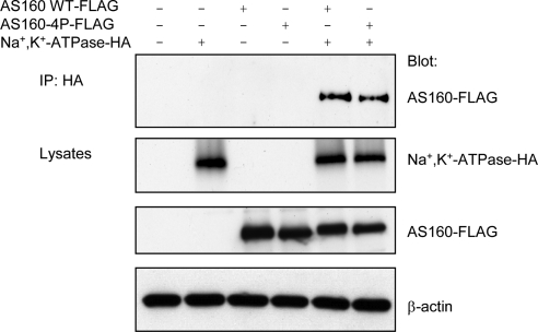
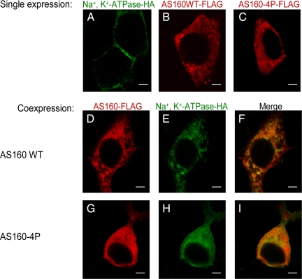
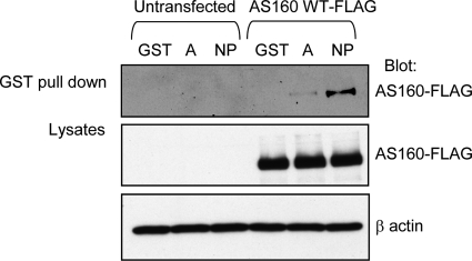
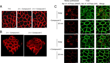
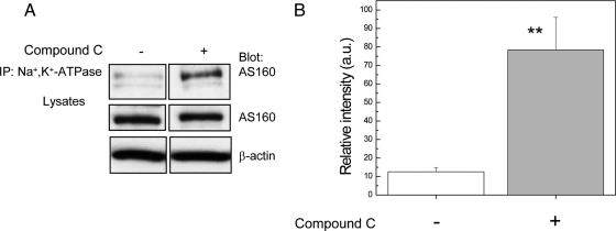
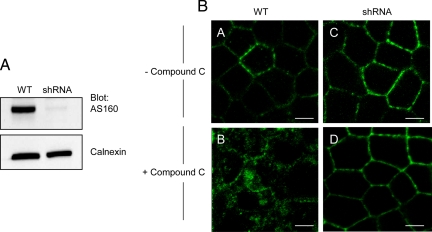
The Na(+),K(+)-ATPase is the major active transport protein found in the plasma membranes of most epithelial cell types. The regulation of Na(+),K(+)-ATPase activity involves a variety of mechanisms, including regulated endocytosis and recycling. Our efforts to identify novel Na(+),K(+)-ATPase binding partners revealed a direct association between the Na(+),K(+)-ATPase and AS160, a Rab-GTPase-activating protein. In COS cells, coexpression of AS160 and Na(+),K(+)-ATPase led to the intracellular retention of the sodium pump. We find that AS160 interacts with the large cytoplasmic NP domain of the α-subunit of the Na(+),K(+)-ATPase. Inhibition of the activity of the adenosine monophosphate-stimulated protein kinase (AMPK) in Madin-Darby canine kidney cells through treatment with Compound C induces Na(+),K(+)-ATPase endocytosis. This effect of Compound C is prevented through the short hairpin RNA-mediated knockdown of AS160, demonstrating that AMPK and AS160 participate in a common pathway to modulate the cell surface expression of the Na(+),K(+)-ATPase.


Figures

Figure 1.
Figure 1.
Endogenous AS160 coimmunoprecipitates with Na+,K+-ATPase-SNAP-HA expressed by transfection in MDCK cells. Coimmunoprecipitation was performed using Sepharose beads conjugated to mouse anti-HA immunoglobulin G to pull down Na+,K+-ATPase-HA, and AS160 was detected by Western blotting with anti-AS160 antibody. Endogenous AS160 coimmunoprecipitates with Na+,K+-ATPase in lysates prepared from cells transfected with Na+,K+-ATPase-HA but not from untransfected cell lysates. As a control, the immunoprecipitates were blotted with rabbit anti-HA antibody to confirm that Na+,K+-ATPase-SNAP-HA was indeed recovered in the immunoprecipitates. Total membrane lysates from untransfected (WT) and transfected MDCK cells were blotted with antibodies directed against AS160 and HA to assess the levels of AS160 and Na+,K+-ATPase-HA expression. Typical results from one of three experiments are shown.
Figure 2.
Figure 2.
Elimination of four potential phosphorylation sites does not affect the interaction between Na+,K+-ATPase and AS160. COS cells were untransfected, singly transfected with Na+,K+-ATPase-HA, AS160WT-FLAG, or AS160-4P-FLAG or cotransfected with Na+,K+-ATPase-HA and AS160WT-FLAG or AS160-4P-FLAG. The immunoprecipitation was performed using mouse anti-HA immunoglobulin G-Sepharose beads (to capture the Na+,K+-ATPase-HA), and immunoprecipitates were probed with a rabbit anti-FLAG antibody to detect exogenous AS160 coimmunoprecipitating with Na+,K+-ATPase-HA. The immunoblot indicates that AS160 WT and AS160-4P mutant coimmunoprecipitated with Na+,K+-ATPase. As a control for protein loading and for the expression levels of AS160 and Na+,K+-ATPase, the lysates were blotted with mouse anti-β-actin, anti-FLAG, and anti-HA, respectively. Typical results from one of four experiments are shown.
Figure 3.
Figure 3.
Coexpression of Na+,K+-ATPase and AS160 in COS cells produces intracellular accumulation of the Na+,K+-ATPase. Immunofluorescence analysis was performed to localize exogenous Na+,K+-ATPase-HA, AS160WT-FLAG, and AS160-4P-FLAG. Cells were stained with anti-HA for Na+,K+-ATPase (A, E, and H) and anti-FLAG for AS160WT (B and D) or AS160-4P (C and G). Overlay patterns are shown in F and I. COS cells were singly transfected with Na+,K+-ATPase-HA (A), AS160WT-FLAG (B), and AS160-4P-FLAG (C) or cotransfected with Na+,K+-ATPase-HA and AS160WT-FLAG (D and E) and Na+,K+-ATPase-HA and AS1604P-FLAG (G–I). Na+,K+-ATPase is localized predominantly at the plasma membrane when it is singly expressed in COS cells. However, the Na+,K+-ATPase is located in intracellular structures when it is coexpressed with AS160WT or AS160-4P. Typical results from one of five experiments are presented. Bar, 5 μm.
Figure 4.
Figure 4.
AS160 directly associates with the Na+,K+-ATPase α-subunit. In vitro fusion protein pull-down assay was performed to map the site on the Na+,K+-ATPase that interacts with AS160. COS cells lysates were incubated with glutathione-Sepharose 4B beads carrying GST alone (GST), GST fused to the A-domain (A), or the NP domain of the Na+,K+-ATPase (NP) α-subunit. Precipitated AS160 was detected by immunoblotting with anti-FLAG. As a control for nonspecific binding, lysates from untransfected COS cells also were incubated with the GST-fusion proteins (lanes 1–3). When GST-fusion proteins were incubated with lysates from COS cells overexpressing AS160WT-FLAG, AS160 was precipitated with both A domain and NP domain of Na+,K+-ATPase α-subunit, although the extent of the interaction with the NP domain was considerably greater than that with the A domain. Lysates were blotted with anti-FLAG to detect the total expression of AS160WT-FLAG and with anti-β-actin to control for the total amount of protein loaded. The total GST-fusion protein input is shown in Supplemental Figure 2. Typical results of one of three experiments are shown.
Figure 5.
Figure 5.
AMPK inhibition by Compound C induces Na+,K+-ATPase internalization in MDCK cells. MDCK cells were treated with vehicle (0) or with Compound C for 2 h at different concentrations (1, 10, 20, 30, or 40 μM). (A) Lysates of MDCK cells were blotted with phosphorylated (p)-AMPK Thr172 and p-ACC Ser79 to assess the levels of AMPK activity, and the β-actin antibody immunoblot reports on the total amount of protein loaded. Typical results of one of three experiments are shown. (B) Immunofluorescence analysis of endogenous Na+,K+-ATPase. MDCK cells were stained with an antibody directed against Na+,K+-ATPase (α5). An increase of the amount of Na+,K+-ATPase internalization was observed with increasing concentrations of Compound C (0 (vehicle), 1 (1 μM), 10 (10 μM), 20 (20 μM), 30 (30 μM), and 40 (40 μM)). Typical results of one of three experiments are shown. Bar, 5 μm.
Figure 6.
Figure 6.
Compound C leads to Na+,K+-ATPase internalization and does not affect Na+,K+-ATPase trafficking to the plasma membrane. (A) Cells were incubated with 0.5 μM TMR-STAR to “live label” the sodium pump (red). Essentially all of the Na+,K+-ATPase labeled in this manner is detected at the cell surface. Next, cells were washed and incubated for a further 30 min at 37°C to remove excess TMR-STAR (T = 0). Finally, cultures were incubated in the presence or absence of 40 μM Compound C for 2 h at 37°C. After the incubation, cells were fixed and prepared for imaging. Bar, 5 μm. Compound C treatment causes a fraction of the TMR-STAR–labeled pumps that were present at the cell surface immediately after labeling to be redistributed to intracellular compartments. (B) 3D image reconstructions of confocal z-stacks generated in A were performed with Volocity software 4.3 (Improvision, Coventry, United Kingdom). 1 U = 5.73 μm. (C) SNAP cells were incubated with BTP to block “old” pump, incubated at 37°C for 30 min to permit synthesis of new “unblocked” cohort of Na+,K+-ATPase and placed at 19°C for 2 h to accumulate the newly synthesized pump proteins in the Golgi. The 19°C incubation was performed in the presence or absence of 40 μM Compound C. Samples were then fixed immediately or warmed to 37°C to allow trafficking to the cell surface. TMR-STAR is depicted in red and anti-HA, which stains the total pool of Na+,K+-ATPase, is shown in green. Bar, 5 μm. Compound C treatment did not delay or reduce the delivery of the Golgi-accumulated newly synthesized Na+,K+-ATPase to the plasma membrane.
Figure 7.
Figure 7.
AMPK inhibition by Compound C enhances the interaction between the Na+,K+-ATPase and AS160 proteins endogenously expressed in MDCK cells. Endogenous Na+,K+-ATPase was immunoprecipitated using a mouse antibody directed against the α-subunit (α5), and the resultant precipitates were blotted with anti-AS160 antibody. MDCK cells were incubated with vehicle (−) or with 40 μM Compound C (+), for 2 h. (A) Immunoblot of the coimmunoprecipitation. Lysates were blotted with anti-AS160 to detect the endogenous levels of AS160 and with anti-β-actin to control for the total amount of protein loaded. (B) Coimmunoprecipitation quantification (**p < 0.001, n = 3). The results demonstrated that the interaction between AS160 and Na+,K+-ATPase increases by a factor 7 after Compound C treatment.
Figure 8.
Figure 8.
shRNA-mediated knockdown of AS160 inhibits the internalization of Na+,K+-ATPase induced by Compound C treatment in MDCK cells. (A) Lysates from MDCK cells: wild type cells (WT) and a clonal AS160 shRNA-expressing line (shRNA) were immunoblotted with an anti-AS160 antibody and with an anti-calnexin antibody to report on the total amount of protein loaded. The levels of AS160 expression were robustly decreased in the AS160 shRNA clone. Typical results of one of four experiments are shown. (B) Immunofluorescence analysis of endogenous Na+,K+-ATPase. MDCK cells were stained with antibody directed against Na+,K+- ATPase (α5). Wild-type (WT) and AS160 knockdown (shRNA) MDCK cells were treated with vehicle (A and C) or treated with 40 μM of Compound C (B and D) for 2 h. Compound C treatment induces Na+,K+-ATPase internalization in wild-type MDCK cells. This phenomenon is not observed in the AS160 shRNA knockdown cells. Typical results of one of three experiments are shown. Bar, 5 μm.

Similar articles

Cited by

References

    1. Al-Khalili L., Kotova O., Tsuchida H., Ehren I., Feraille E., Krook A., Chibalin A. V. ERK1/2 mediates insulin stimulation of Na(+),K(+)-ATPase by phosphorylation of the alpha-subunit in human skeletal muscle cells. J. Biol. Chem. 2004;279:25211–25218. - PubMed
    1. Al-Khalili L., Yu M., Chibalin A. V. Na+,K+-ATPase trafficking in skeletal muscle: insulin stimulates translocation of both alpha 1- and alpha 2-subunit isoforms. FEBS Lett. 2003;536:198–202. - PubMed
    1. Babbey C. M., Ahktar N., Wang E., Chen C. C., Grant B. D., Dunn K. W. Rab10 regulates membrane transport through early endosomes of polarized Madin-Darby canine kidney cells. Mol. Biol. Cell. 2006;17:3156–3175. - PMC - PubMed
    1. Barlet-Bas C., Khadouri C., Marsy S., Doucet A. Enhanced intracellular sodium concentration in kidney cells recruits a latent pool of Na-K-ATPase whose size is modulated by corticosteroids. J. Biol. Chem. 1990;265:7799–7803. - PubMed
    1. Bertorello A. M., Komarova Y., Smith K., Leibiger I. B., Efendiev R., Pedemonte C. H., Borisy G., Sznajder J. I. Analysis of Na+,K+-ATPase motion and incorporation into the plasma membrane in response to G protein-coupled receptor signals in living cells. Mol. Biol. Cell. 2003;14:1149–1157. - PMC - PubMed

Publication types

MeSH terms

LinkOut - more resources