. 2010 Oct 13;30(41):13707-17.
doi: 10.1523/JNEUROSCI.4040-10.2010.

Apolipoprotein E4 causes age- and Tau-dependent impairment of GABAergic interneurons, leading to learning and memory deficits in mice

Affiliations

Apolipoprotein E4 causes age- and Tau-dependent impairment of GABAergic interneurons, leading to learning and memory deficits in mice

Yaisa Andrews-Zwilling et al. J Neurosci. .

Abstract

Apolipoprotein E4 (apoE4) is the major genetic risk factor for Alzheimer's disease. However, the underlying mechanisms are unclear. We found that female apoE4 knock-in (KI) mice had an age-dependent decrease in hilar GABAergic interneurons that correlated with the extent of learning and memory deficits, as determined in the Morris water maze, in aged mice. Treating apoE4-KI mice with daily peritoneal injections of the GABA(A) receptor potentiator pentobarbital at 20 mg/kg for 4 weeks rescued the learning and memory deficits. In neurotoxic apoE4 fragment transgenic mice, hilar GABAergic interneuron loss was even more pronounced and also correlated with the extent of learning and memory deficits. Neurodegeneration and tauopathy occurred earliest in hilar interneurons in apoE4 fragment transgenic mice; eliminating endogenous Tau prevented hilar GABAergic interneuron loss and the learning and memory deficits. The GABA(A) receptor antagonist picrotoxin abolished this rescue, while pentobarbital rescued learning deficits in the presence of endogenous Tau. Thus, apoE4 causes age- and Tau-dependent impairment of hilar GABAergic interneurons, leading to learning and memory deficits in mice. Consequently, reducing Tau and enhancing GABA signaling are potential strategies to treat or prevent apoE4-related Alzheimer's disease.


Figures

Figure 1.
Figure 1.
Age-dependent significant decrease in numbers of GABAergic interneurons in the hilus of the dentate gyrus of female apoE4-KI mice. A–D, Representative photomicrographs (200×) from female apoE3-KI and apoE4-KI mice at 16 months of age show GABAergic interneurons in the hilus after staining with anti-GAD67 (A, B) and anti-somatostatin (C, D). E, F, Hilar GABAergic interneurons positive for GAD67 (E) or somatostatin (F) in female apoE3-KI and apoE4-KI mice at 1, 3, 6, 12, 16, and 21 months of age (n = 4–12 mice per group) were quantified as described in Materials and Methods. Values are mean ± SEM. *p < 0.05; **p < 0.01 versus apoE3-KI mice at the same age (t test). G, H, Representative photomicrographs (200×) show GABAergic interneurons in CA1 region of the hippocampus after staining with anti-GAD67. I, Quantification of GAD67-positive GABAergic interneurons in CA1 region of the hippocampus in 16-month-old female apoE3-KI (n = 10) and apoE4-KI (n = 12) mice. Values are mean ± SEM. J, K, Representative photomicrographs (200×) from female wild-type mice at 16 months of age show GABAergic interneurons in the hilus after staining with anti-GAD67 (J) and anti-somatostatin (K). L, M, Hilar GABAergic interneurons positive for GAD67 (L) or somatostatin (M) in female wild-type, apoE3-KI, and apoE4-KI mice at 3, 12, and 16 months of age (n = 6–12 mice per group) were quantified as described in Materials and Methods. Values are mean ± SEM. *p < 0.05; **p < 0.01 versus wild-type and apoE3-KI mice at the same age (t test).
Figure 2.
Figure 2.
Presynaptic GABAergic input onto granule cells is reduced in female apoE4-KI mice. A–I, Representative confocal images of the granule cell layer of the dentate gyrus of female wild-type (A–C), apoE3-KI (D–F), and apoE4-KI (G–I) mice at 16 months of age stained with anti-GAD67 (A, D, G) and anti-synaptophysin (B, E, H). Merged images are shown in C, F, and I. J, GAD67 immunoreactivity (IR) of sections from different mice was quantified and normalized by area. Values are mean ± SEM (four images per mouse and 4–5 mice per genotype). ***p < 0.005 versus wild-type and apoE3-KI mice (t test). K, The ratio of GAD67-IR to synaptophysin-IR (a general presynaptic marker) in sections from different mice. Values are mean ± SEM (four images per mouse and 4–5 mice per genotype). ***p < 0.005 versus wild-type and apoE3-KI mice (t test). L–N, Traces of miniature IPSCs (mIPSCs) in granule cells from wild-type (L), apoE3-KI (M), or apoE4-KI (N) mice during whole-cell voltage-clamp recording in the presence of DNQX (20 μm), d-AP5 (50 μm), and TTX (1 μm). Calibration: 20 pA, 700 ms. O, Average mIPSC frequency in granule cells was lower in apoE4-KI mice than in wild-type and apoE3-KI mice. Values are mean ± SEM (n = 9–11 cells from 3 to 4 mice per genotype). **p < 0.01 versus wild-type and apoE3-KI mice (t test). P, Average mIPSC amplitude in granule cells was similar in wild-type, apoE3-KI, and apoE4-KI mice. Values are mean ± SEM (n = 9–11 cells from 3 to 4 mice per genotype). Q, Average membrane resistance of granule cells was similar in wild-type, apoE3-KI, and apoE4-KI mice. Values are mean ± SEM (n = 8–11 cells from 3 to 4 mice per genotype).
Figure 3.
Figure 3.
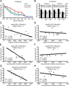
Correlation of hilar GABAergic interneuron impairment with spatial learning deficits in apoE4-KI mice. A, Nine wild-type, ten apoE3-KI, and twelve apoE4-KI female mice were tested at 16 months of age in the Morris water maze. Points represent averages of daily trials. HD, hidden platform day (2 trials/session, 2 sessions/d); HD0, first trial on HD1; VD, visible platform day (2 trials/session, 2 sessions/d). y-Axis indicates time to reach the target platform (escape latency, mean ± SEM). In the hidden platform days, learning curves differed significantly by genotypes (repeated-measures ANOVA, p < 0.01). In post hoc comparisons, apoE4-KI mice learned poorly (p < 0.01 versus other groups). Wild-type and apoE4-KI mice performed at a similar level. B, The probe trials of female wild-type, apoE3-KI, and apoE4-KI mice at 16 months of age were performed 72 h (probe 2) and 120 h (probe 3) after the last hidden platform training. Percentage time spent in the target quadrant versus the average time spent in other quadrants differed by genotype in probe 3 (p < 0.05). Values are mean ± SEM. *p < 0.05, **p < 0.01, ***p < 0.005 (t test). C, D, Escape latency in hidden platform days 1–5 correlated inversely with the number of GAD67-positive hilar GABAergic interneurons in apoE4-KI mice (C, n = 12) but not apoE3-KI mice (D, n = 10) at 16 months of age. E, F, Escape latency in hidden platform days 1–5 correlated inversely with the number of somatostatin-positive hilar GABAergic interneurons in apoE4-KI mice (E, n = 12) but not apoE3-KI mice (F, n = 10) at 16 months of age. G, H, Eight apoE3-KI and eight apoE4-KI female mice were tested at 21 months of age in the Morris water maze. Escape latency in hidden platform days 1–5 correlated inversely with the number of somatostatin-positive hilar GABAergic interneurons in apoE4-KI mice (G, n = 8) but not apoE3-KI mice (H, n = 8) at 21 months of age.
Figure 4.
Figure 4.
GABAA receptor potentiator pentobarbital rescues spatial learning and memory deficits in apoE4-KI mice. A, Female 16-month-old apoE4-KI mice were treated with pentobarbital (PB, 20 mg/kg i.p.) or saline (n = 6–13 mice per group) for 21 d before and daily during the Morris water maze test. Untreated wild-type (n = 9) and apoE3-KI (n = 10) mice served as controls. The learning curve of pentobarbital-treated apoE4-KI mice differed from that of saline-treated apoE4-KI mice (repeated-measures ANOVA, p < 0.05) but resembled that of untreated wild-type and apoE3-KI mice. Values are mean ± SEM. HD, Hidden day; VD, visible day. B, In the third probe trial 120 h after the last hidden session, pentobarbital treatment rescued memory deficits in 16-month-old apoE4-KI mice in the target quadrant and target cross tests (n = 6–13 mice per group). Values are mean ± SEM. ***p < 0.005 (t test). C, Total number of GAD67-positive GABAergic interneurons in the hilus of wild-type mice, apoE3-KI mice, apoE4-KI mice, and apoE4-KI mice treated with pentobarbital. Values are mean ± SEM. **p < 0.01, ***p < 0.005 versus wild-type and apoE3-KI mice (t test). D, Female 21-month-old apoE4-KI mice were treated with pentobarbital (PB, 20 mg/kg) or saline (n = 8 per group) for 21 d before and daily during the Morris water maze test. Saline-treated apoE3-KI mice (n = 8) served as controls. The learning curve of pentobarbital-treated apoE4-KI mice differed from that of saline-treated apoE4-KI mice (repeated-measures ANOVA, p < 0.05) but resembled that of saline-treated apoE3-KI mice. Values are mean ± SEM. HD, Hidden day; VD, visible day.
Figure 5.
Figure 5.
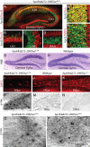
Localization of apoE4(Δ272–299) in the hippocampus and its effects on neurodegeneration and Tau pathology in the presence and absence of Tau. A–D, Double immunofluorescence staining for apoE (green) and NeuN (red) in the hippocampus of apoE4(Δ272–299)mE−/−Tau+/+ mice (magnification: A, 100×; B–D, 400×). E, Double immunofluorescence staining for apoE (green) and synaptophysin (Syn, red) in the CA3 region of apoE4(Δ272–299)mE−/−Tau+/+ mice (600×). F, Double immunofluorescence staining for apoE (green) and MAP2 (red) in the CA3 region of apoE4(Δ272–299)mE−/−Tau+/+ mice (magnification, 600×). G, H, Hematoxylin-eosin staining of the dentate gyrus of apoE4(Δ272–299)mE−/−Tau+/+ (G) and wild-type (H) mice (magnification, 200×). I–K, Immunofluorescence staining for MAP2 in the hilus of the dentate gyrus of apoE4(Δ272–299)mE−/−Tau+/+ (I), wild-type (J), and apoE4(Δ272–299)mE−/−Tau−/− (K) mice (magnification, 200×). L–N, Anti-p-Tau (AT8 monoclonal antibody) immunostaining of the hilus of apoE4(Δ272–299)mE−/−Tau+/+ (L), mE−/−Tau+/+ (M), and apoE4(Δ272–299)mE−/−Tau−/− (N) mice (magnification, 400×). O, P, Anti-p-Tau (AT8 monoclonal antibody) immunostaining of the CA3 region (O) of the hippocampus and the subiculum (P) of apoE4(Δ272–299)mE−/−Tau+/+ mice (magnification, 400×). All mice were 11–13 months of age.
Figure 6.
Figure 6.
Loss of GABAergic interneurons in the hilus of the dentate gyrus of apoE4(Δ272–299)mE−/−Tau+/+ mice and rescue by Tau removal. The brains of 14 mE−/−Tau+/+, 10 apoE4(Δ272–299)mE−/−Tau+/+, 12 apoE4(Δ272–299)mE−/−Tau−/−, 8 mE−/−Tau−/−, and 16 wild-type mice (all females) were collected at 12 months of age after behavioral assessment, sectioned, and immunostained with antibodies against GAD67, neuropeptide Y (NPY), or somatostatin. A–O, Photomicrographs (200×) of GABAergic interneurons in the hilus after staining with anti-GAD67 (A–E), anti-NPY (F–J), or anti-somatostatin (K–O). P–R, Total number of GAD67-positive (P), NPY-positive (Q), and somatostatin-positive (R) GABAergic interneurons in the hilus. Values are mean ± SEM. ***p < 0.005 (t test).
Figure 7.
Figure 7.
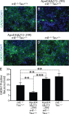
Eliminating Tau prevents the neurotoxic effect of apoE4 fragments on primary hippocampal GABAergic neurons. A–D, Primary hippocampal neurons from individual P0 pups (mE−/−Tau+/+, apoE4(Δ272–299)mE−/−Tau+/+, apoE4(Δ272–299)mE−/−Tau−/−, and mE−/−Tau−/−) were cultured for 14 d in vitro (DIV14) and double stained with anti-GAD67 (green) and DAPI (blue). Shown are representative images collected from three mice of each genotype and five fields per coverslip (magnification, 200×). E, Numbers of GAD67-positive neurons were quantified as described in Materials and Methods. Values are mean ± SEM. **p < 0.01, ***p < 0.005 (t test).
Figure 8.
Figure 8.
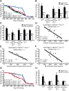
Spatial learning and memory deficits in apoE4(Δ272–299)mE−/−Tau+/+ mice and rescue by Tau removal. A, Fourteen mE−/−Tau+/+, ten apoE4(Δ272–299)mE−/−Tau+/+, twelve apoE4(Δ272–299)mE−/−Tau−/−, eight mE−/−Tau−/−, and sixteen wild-type mice (all females) were tested at 12 months of age in the Morris water maze. Values are mean ± SEM. In the hidden platform days, learning curves differed significantly by genotype (repeated-measures ANOVA, p < 0.001). In post hoc comparisons, apoE4(Δ272–299)mE−/−Tau+/+ mice learned poorly (p < 0.01 vs other groups). ApoE4(Δ272–299)mE−/−Tau−/−, mE−/−Tau+/+, and wild-type mice performed at a similar level. B, In the probe trial 24 h after the last hidden platform training, the number of target platform crossings versus crossings of the equivalent area in the other quadrants differed by genotype (p < 0.05). In post hoc comparisons, apoE4(Δ272–299)mE−/−Tau−/− mice performed better than apoE4(Δ272–299)mE−/−Tau+/+ mice (p < 0.01) in the target crossing test. Only apoE4(Δ272–299)mE−/−Tau+/+ mice showed impaired memory in the probe trail, and the deficit was rescued by Tau removal. Values are mean ± SEM. ***p < 0.005. C, In the probe trial 24 h after the last hidden platform training, the time spent in the target quadrant versus the other quadrants differed by genotypes (p < 0.01). In post hoc comparisons, only apoE4(Δ272–299)mE−/−Tau+/+ mice showed impaired memory in the probe test, and the deficit was rescued by Tau removal. Values are mean ± SEM. ***p < 0.005. D–F, Latency on hidden days 1–5 correlated inversely with the number of GAD67-positive (D), somatostatin-positive (E), and NPY-positive (F) GABAergic interneurons in the hilus in apoE4(Δ272–299)mE−/−Tau+/+ mice. n = 10 per analysis. G, ApoE4(Δ272–299)mE−/−Tau−/− mice were treated with picrotoxin (Picro, 1 mg/kg i.p.) or saline (n = 6–8 per group) for 3 d before and daily during the Morris water maze test. Saline-treated apoE4(Δ272–299)mE−/−Tau+/+ and mE−/−Tau+/+ mice (n = 6–8 per group) served as controls. The learning curve of picrotoxin-treated apoE4(Δ272–299)mE−/−Tau−/− mice resembled that of saline-treated apoE4(Δ272–299)mE−/−Tau+/+ mice, which differed significantly from those of saline-treated controls (p < 0.01). Values are mean ± SEM. H, In the probe trial 24 h after the last hidden session, picrotoxin-treated apoE4(Δ272–299)mE−/−Tau−/− mice performed significantly worse than saline-treated apoE4(Δ272–299)mE−/−Tau−/− (p < 0.05) or mE−/−Tau+/+ mice (p < 0.01) in the target crossing test. n = 6–8 mice per group. Values are mean ± SEM. *p < 0.05, **p < 0.01.

Similar articles

Cited by

References

    1. Bareggi SR, Franceschi M, Bonini L, Zecca L, Smirne S. Decreased CSF concentrations of homovanillic acid and γ-aminobutyric acid in Alzheimer's disease. Age- or disease-related modifications? Arch Neurol. 1982;39:709–712. - PubMed
    1. Bell RD, Zlokovic BV. Neurovascular mechanisms and blood-brain barrier disorder in Alzheimer's disease. Acta Neuropathol. 2009;118:103–113. - PMC - PubMed
    1. Bour A, Grootendorst J, Vogel E, Kelche C, Dodart J-C, Bales K, Moreau P-H, Sullivan PM, Mathis C. Middle-aged human apoE4 targeted-replacement mice show retention deficits on a wide range of spatial memory tasks. Behav Brain Res. 2008;193:174–182. - PubMed
    1. Brecht WJ, Harris FM, Chang S, Tesseur I, Yu G-Q, Xu Q, Fish JD, Wyss-Coray T, Buttini M, Mucke L, Mahley RW, Huang Y. Neuron-specific apolipoprotein E4 proteolysis is associated with increased tau phosphorylation in brains of transgenic mice. J Neurosci. 2004;24:2527–2534. - PMC - PubMed
    1. Brosh I, Barkai E. Learning-induced enhancement of feedback inhibitory synaptic transmission. Learn Mem. 2009;16:413–416. - PubMed

Publication types

MeSH terms

LinkOut - more resources