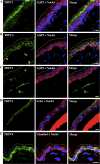
Expression and distribution of transient receptor potential (TRP) channels in bladder epithelium
- PMID: 20943764
- PMCID: PMC3023226
- DOI: 10.1152/ajprenal.00349.2010
Expression and distribution of transient receptor potential (TRP) channels in bladder epithelium
Abstract
The urothelium is proposed to be a sensory tissue that responds to mechanical stress by undergoing dynamic membrane trafficking and neurotransmitter release; however, the molecular basis of this function is poorly understood. Transient receptor potential (TRP) channels are ideal candidates to fulfill such a role as they can sense changes in temperature, osmolarity, and mechanical stimuli, and several are reported to be expressed in the bladder epithelium. However, their complete expression profile is unknown and their cellular localization is largely undefined. We analyzed expression of all 33 TRP family members in mouse bladder and urothelium by RT-PCR and found 22 specifically expressed in the urothelium. Of the latter, 10 were chosen for closer investigation based on their known mechanosensory or membrane trafficking functions in other cell types. Western blots confirmed urothelial expression of TRPC1, TRPC4, TRPV1, TRPV2, TRPV4, TRPM4, TRPM7, TRPML1, and polycystins 1 and 2 (PKD1 and PKD2) proteins. We further defined the cellular and subcellular localization of all 10 TRP channels. TRPV2 and TRPM4 were prominently localized to the umbrella cell apical membrane, while TRPC4 and TRPV4 were identified on their abluminal surfaces. TRPC1, TRPM7, and TRPML1 were localized to the cytoplasm, while PKD1 and PKD2 were expressed on the apical and basolateral membranes of umbrella cells as well as in the cytoplasm. The cellular location of TRPV1 in the bladder has been debated, but colocalization with neuronal marker calcitonin gene-related peptide indicated clearly that it is present on afferent neurons that extend into the urothelium, but may not be expressed by the urothelium itself. These findings are consistent with the hypothesis that the urothelium acts as a sentinel and by expressing multiple TRP channels it is likely it can detect and presumably respond to a diversity of external stimuli and suggest that it plays an important role in urothelial signal transduction.
Figures


Similar articles
-
Functional characterization of transient receptor potential channels in mouse urothelial cells.Am J Physiol Renal Physiol. 2010 Mar;298(3):F692-701. doi: 10.1152/ajprenal.00599.2009. Epub 2009 Dec 16. Am J Physiol Renal Physiol. 2010. PMID: 20015940 Free PMC article.
-
Differential localizations of the transient receptor potential channels TRPV4 and TRPV1 in the mouse urinary bladder.J Histochem Cytochem. 2009 Mar;57(3):277-87. doi: 10.1369/jhc.2008.951962. Epub 2008 Nov 24. J Histochem Cytochem. 2009. PMID: 19029406 Free PMC article.
-
Functional TRP and ASIC-like channels in cultured urothelial cells from the rat.Am J Physiol Renal Physiol. 2009 Apr;296(4):F892-901. doi: 10.1152/ajprenal.90718.2008. Epub 2009 Jan 21. Am J Physiol Renal Physiol. 2009. PMID: 19158342 Free PMC article.
-
The role of the transient receptor potential (TRP) superfamily of cation-selective channels in the management of the overactive bladder.BJU Int. 2010 Oct;106(8):1114-27. doi: 10.1111/j.1464-410X.2010.09650.x. BJU Int. 2010. PMID: 21156013 Review.
-
Receptors, channels, and signalling in the urothelial sensory system in the bladder.Nat Rev Urol. 2016 Apr;13(4):193-204. doi: 10.1038/nrurol.2016.13. Epub 2016 Mar 1. Nat Rev Urol. 2016. PMID: 26926246 Free PMC article. Review.
Cited by
-
Functional expression of purinergic P2 receptors and transient receptor potential channels by the human urothelium.Am J Physiol Renal Physiol. 2013 Aug 1;305(3):F396-406. doi: 10.1152/ajprenal.00127.2013. Epub 2013 May 29. Am J Physiol Renal Physiol. 2013. PMID: 23720349 Free PMC article.
-
Urothelial TRPV1: TRPV1-Reporter Mice, a Way to Clarify the Debate?Front Physiol. 2012 May 7;3:130. doi: 10.3389/fphys.2012.00130. eCollection 2012. Front Physiol. 2012. PMID: 22586410 Free PMC article. No abstract available.
-
Transient receptor potential (TRP) channels: a clinical perspective.Br J Pharmacol. 2014 May;171(10):2474-507. doi: 10.1111/bph.12414. Br J Pharmacol. 2014. PMID: 24102319 Free PMC article. Review.
-
Bladder cancer and urothelial impairment: the role of TRPV1 as potential drug target.Biomed Res Int. 2014;2014:987149. doi: 10.1155/2014/987149. Epub 2014 May 8. Biomed Res Int. 2014. PMID: 24901005 Free PMC article. Review.
-
Endothelin inhibits renin release from juxtaglomerular cells via endothelin receptors A and B via a transient receptor potential canonical-mediated pathway.Physiol Rep. 2014 Dec 18;2(12):e12240. doi: 10.14814/phy2.12240. Print 2014 Dec 1. Physiol Rep. 2014. PMID: 25524278 Free PMC article.
References
-
- Acharya P, Beckel J, Ruiz WG, Wang E, Rojas R, Birder L, Apodaca G. Distribution of the tight junction proteins ZO-1, occludin, and claudin-4, -8, and -12 in bladder epithelium. Am J Physiol Renal Physiol 287: F305–F318, 2004 - PubMed
-
- Apodaca G. The uroepithelium: not just a passive barrier. Traffic 5: 117–128, 2004 - PubMed
-
- Apodaca G, Balestreire E, Birder LA. The uroepithelial-associated sensory web. Kidney Int 72: 1057–1064, 2007 - PubMed
-
- Avelino A, Cruz C, Nagy I, Cruz F. Vanilloid receptor 1 expression in the rat urinary tract. Neuroscience 109: 787–798, 2002 - PubMed
Publication types
MeSH terms
Substances
Grants and funding
LinkOut - more resources
Full Text Sources
Molecular Biology Databases
Miscellaneous

