

Whole brain resting-state analysis reveals decreased functional connectivity in major depression
- PMID: 20941370
- PMCID: PMC2950744
- DOI: 10.3389/fnsys.2010.00041
Whole brain resting-state analysis reveals decreased functional connectivity in major depression
Abstract
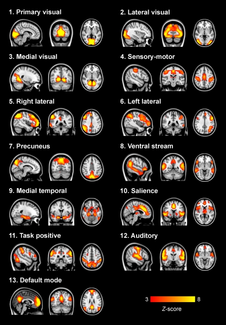
Recently, both increases and decreases in resting-state functional connectivity have been found in major depression. However, these studies only assessed functional connectivity within a specific network or between a few regions of interest, while comorbidity and use of medication was not always controlled for. Therefore, the aim of the current study was to investigate whole-brain functional connectivity, unbiased by a priori definition of regions or networks of interest, in medication-free depressive patients without comorbidity. We analyzed resting-state fMRI data of 19 medication-free patients with a recent diagnosis of major depression (within 6 months before inclusion) and no comorbidity, and 19 age- and gender-matched controls. Independent component analysis was employed on the concatenated data sets of all participants. Thirteen functionally relevant networks were identified, describing the entire study sample. Next, individual representations of the networks were created using a dual regression method. Statistical inference was subsequently done on these spatial maps using voxel-wise permutation tests. Abnormal functional connectivity was found within three resting-state networks in depression: (1) decreased bilateral amygdala and left anterior insula connectivity in an affective network, (2) reduced connectivity of the left frontal pole in a network associated with attention and working memory, and (3) decreased bilateral lingual gyrus connectivity within ventromedial visual regions. None of these effects were associated with symptom severity or gray matter density. We found abnormal resting-state functional connectivity not previously associated with major depression, which might relate to abnormal affect regulation and mild cognitive deficits, both associated with the symptomatology of the disorder.
Keywords: amygdala; functional connectivity; independent component analysis; major depression; resting-state functional magnetic resonance imaging.
Figures

Similar articles
-
Disruption of resting-state functional connectivity of right posterior insula in adolescents and young adults with major depressive disorder.J Affect Disord. 2019 Oct 1;257:23-30. doi: 10.1016/j.jad.2019.06.057. Epub 2019 Jul 2. J Affect Disord. 2019. PMID: 31299401
-
Resting-state functional connectivity abnormalities in limbic and salience networks in social anxiety disorder without comorbidity.Eur Neuropsychopharmacol. 2013 Mar;23(3):186-95. doi: 10.1016/j.euroneuro.2012.04.018. Epub 2012 Jun 30. Eur Neuropsychopharmacol. 2013. PMID: 22749355
-
Large-Scale Network Dysfunction in Major Depressive Disorder: A Meta-analysis of Resting-State Functional Connectivity.JAMA Psychiatry. 2015 Jun;72(6):603-11. doi: 10.1001/jamapsychiatry.2015.0071. JAMA Psychiatry. 2015. PMID: 25785575 Free PMC article.
-
[Resting state fMRI study of emotional network in patients with postconcussion syndrome].Zhonghua Yi Xue Za Zhi. 2017 Jul 4;97(25):1951-1955. doi: 10.3760/cma.j.issn.0376-2491.2017.25.007. Zhonghua Yi Xue Za Zhi. 2017. PMID: 28693073 Chinese.
-
Resting state brain network function in major depression - Depression symptomatology, antidepressant treatment effects, future research.J Psychiatr Res. 2017 Sep;92:147-159. doi: 10.1016/j.jpsychires.2017.04.007. Epub 2017 Apr 24. J Psychiatr Res. 2017. PMID: 28458140 Review.
Cited by
-
Resting-state functional connectivity in late-life depression: higher global connectivity and more long distance connections.Front Psychiatry. 2013 Jan 9;3:116. doi: 10.3389/fpsyt.2012.00116. eCollection 2012. Front Psychiatry. 2013. PMID: 23316175 Free PMC article.
-
Amplitudes of resting-state functional networks - investigation into their correlates and biophysical properties.Neuroimage. 2023 Jan;265:119779. doi: 10.1016/j.neuroimage.2022.119779. Epub 2022 Dec 1. Neuroimage. 2023. PMID: 36462729 Free PMC article.
-
Introduction to research topic - brain connectivity analysis: investigating brain disorders. Part 2: original research articles.Front Syst Neurosci. 2012 Feb 13;6:4. doi: 10.3389/fnsys.2012.00004. eCollection 2012. Front Syst Neurosci. 2012. PMID: 22347850 Free PMC article. No abstract available.
-
Abnormal resting state activity of left middle occipital gyrus and its functional connectivity in female patients with major depressive disorder.BMC Psychiatry. 2018 Nov 26;18(1):370. doi: 10.1186/s12888-018-1955-9. BMC Psychiatry. 2018. PMID: 30477561 Free PMC article.
-
Disrupted interhemispheric coordination of sensory-motor networks and insula in major depressive disorder.Front Neurosci. 2023 Mar 7;17:1135337. doi: 10.3389/fnins.2023.1135337. eCollection 2023. Front Neurosci. 2023. PMID: 36960171 Free PMC article.
References
-
- American Psychiatric Association (1994). Diagnostic and Statistical Manual of Mental Disorders, 4th Edn.Washington, D.C.: American Psychiatric Association
-
- Anand A., Li Y., Wang Y., Wu J., Gao S., Bukhari L., Mathews V. P., Kalnin A., Lowe M. J. (2005b). Antidepressant effect on connectivity of the mood-regulating circuit: an FMRI study. Neuropsychopharmacology 30, 1334–1344 - PubMed
LinkOut - more resources
Full Text Sources
Other Literature Sources