Elevation of high-mobility group protein box-1 in serum correlates with severity of acute intracerebral hemorrhage
- PMID: 20936104
- PMCID: PMC2948906
- DOI: 10.1155/2010/142458
Elevation of high-mobility group protein box-1 in serum correlates with severity of acute intracerebral hemorrhage
Abstract
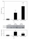
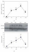
High-mobility group protein box-1 (HMGB1) is a proinflammatory involved in many inflammatory diseases. However, its roles in intracerebral hemorrhage (ICH) remain unknown. The purpose of this study was to examine the correlation between changes in serum levels of HMGB1 following acute ICH and the severity of stroke as well as the underlying mechanism. Changes in serum levels of HMGB1 in 60 consecutive patients with primary hemispheric ICH within 12 hours of onset of symptoms were determined. The correlation of HMGB1 with disease severity, IL-6, and TNF-α was analyzed. Changes in HMGB1 levels were detected with ELISA and Western blot. Compared with normal controls, patients with ICH had markedly elevated levels of HMGB1, which was significantly correlated with the levels of IL-6 and TNF-α, NIHSS score at the 10th day, and mRS score at 3 months. In comparison with the control group, the levels of HMGB1 in the perihematomal tissue in mice with ICH increased dramatically, peaked at 72 hours, and decreased at 5 days. Meanwhile, heme could stimulate cultured microglia to release large amounts of HMGB1 whereas Fe(2+/3+) ions failed to stimulate HMGB1 production from microglia. Our findings suggest that HMGB1 may play an essential role in the ICH-caused inflammatory injury.
Figures


Similar articles
-
Anti-high mobility group box-1 (HMGB1) antibody inhibits hemorrhage-induced brain injury and improved neurological deficits in rats.Sci Rep. 2017 Apr 10;7:46243. doi: 10.1038/srep46243. Sci Rep. 2017. PMID: 28393932 Free PMC article.
-
High mobility group box 1 protein (HMGB1) as biomarker in hypoxia-induced persistent pulmonary hypertension of the newborn: a clinical and in vivo pilot study.Int J Med Sci. 2019 Aug 6;16(8):1123-1131. doi: 10.7150/ijms.34344. eCollection 2019. Int J Med Sci. 2019. PMID: 31523175 Free PMC article.
-
Synergistic effects of antibodies against high-mobility group box 1 and tumor necrosis factor-α antibodies on D-(+)-galactosamine hydrochloride/lipopolysaccharide-induced acute liver failure.FEBS J. 2013 Mar;280(6):1409-19. doi: 10.1111/febs.12132. Epub 2013 Feb 14. FEBS J. 2013. PMID: 23331758
-
Association of circulating blood HMGB1 levels with ischemic stroke: a systematic review and meta-analysis.Neurol Res. 2018 Nov;40(11):907-916. doi: 10.1080/01616412.2018.1497254. Epub 2018 Jul 17. Neurol Res. 2018. PMID: 30015578 Review.
-
Central Nervous System Tissue Regeneration after Intracerebral Hemorrhage: The Next Frontier.Cells. 2021 Sep 23;10(10):2513. doi: 10.3390/cells10102513. Cells. 2021. PMID: 34685493 Free PMC article. Review.
Cited by
-
An update on inflammation in the acute phase of intracerebral hemorrhage.Transl Stroke Res. 2015 Feb;6(1):4-8. doi: 10.1007/s12975-014-0384-4. Epub 2014 Dec 23. Transl Stroke Res. 2015. PMID: 25533878 Review.
-
Sepsis-Exacerbated Brain Dysfunction After Intracerebral Hemorrhage.Front Cell Neurosci. 2022 Jan 21;15:819182. doi: 10.3389/fncel.2021.819182. eCollection 2021. Front Cell Neurosci. 2022. PMID: 35126060 Free PMC article. Review.
-
High-Mobility Group Box 1 in Spinal Cord Injury and Its Potential Role in Brain Functional Remodeling After Spinal Cord Injury.Cell Mol Neurobiol. 2023 Apr;43(3):1005-1017. doi: 10.1007/s10571-022-01240-5. Epub 2022 Jun 17. Cell Mol Neurobiol. 2023. PMID: 35715656 Review.
-
(-)-Epicatechin protects hemorrhagic brain via synergistic Nrf2 pathways.Ann Clin Transl Neurol. 2014 Apr 1;1(4):258-271. doi: 10.1002/acn3.54. Ann Clin Transl Neurol. 2014. PMID: 24741667 Free PMC article.
-
TREM (Triggering Receptor Expressed on Myeloid Cells)-1 Inhibition Attenuates Neuroinflammation via PKC (Protein Kinase C) δ/CARD9 (Caspase Recruitment Domain Family Member 9) Signaling Pathway After Intracerebral Hemorrhage in Mice.Stroke. 2021 Jun;52(6):2162-2173. doi: 10.1161/STROKEAHA.120.032736. Epub 2021 May 5. Stroke. 2021. PMID: 33947214 Free PMC article.
References
-
- Lapchak PA, Araujo DM. Advances in hemorrhagic stroke therapy: conventional and novel approaches. Expert Opinion on Emerging Drugs. 2007;12(3):389–406. - PubMed
-
- Wang J, Doré S. Inflammation after intracerebral hemorrhage. Journal of Cerebral Blood Flow and Metabolism. 2007;27(5):894–908. - PubMed
-
- Xi G, Keep RF, Hoff JT. Mechanisms of brain injury after intracerebral haemorrhage. Lancet Neurology. 2006;5(1):53–63. - PubMed
-
- Hua Y, Keep RF, Hoff JT, Xi G. Brain injury after intracerebral hemorrhage: the role of thrombin and iron. Stroke. 2007;38(2, supplement):759–762. - PubMed
-
- Goodwin GH, Sanders C, Johns EW. A new group of chromatin associated proteins with a high content of acidic and basic amino acids. European Journal of Biochemistry. 1973;38(1):14–19. - PubMed
Publication types
MeSH terms
Substances
LinkOut - more resources
Full Text Sources
Other Literature Sources
Medical

