. 2010 Oct 19;107(42):17998-8003.
doi: 10.1073/pnas.1007047107. Epub 2010 Oct 4.

Connexin 43 is critical to maintain the homeostasis of the blood-testis barrier via its effects on tight junction reassembly

Affiliations

Connexin 43 is critical to maintain the homeostasis of the blood-testis barrier via its effects on tight junction reassembly

Michelle W M Li et al. Proc Natl Acad Sci U S A. .

Abstract

In mammalian testes, the blood-testis barrier (BTB) or Sertoli cell barrier created by specialized junctions between Sertoli cells near the basement membrane confers an immunological barrier by sequestering the events of meiotic division and postmeiotic germ cell development from the systemic circulation. The BTB is constituted by coexisting tight junctions (TJs), basal ectoplasmic specializations, desmosomes, and gap junctions. Despite being one of the tightest blood-tissue barriers, the BTB has to restructure cyclically during spermatogenesis. A recent study showed that gap junction protein connexin 43 (Cx43) and desmosome protein plakophilin-2 are working synergistically to modulate the BTB integrity by regulating the distribution of TJ-associated proteins at the Sertoli-Sertoli cell interface. However, the precise role of Cx43 in regulating the cyclical restructuring of junctions remains obscure. In this report, the calcium switch and the bisphenol A (BPA) models were used to induce junction restructuring in primary cultures of Sertoli cells isolated from rat testes that formed a TJ-permeability barrier that mimicked the BTB in vivo. The removal of calcium by EGTA perturbed the Sertoli cell tight junction barrier, but calcium repletion allowed the "resealing" of the disrupted barrier. However, a knockdown of Cx43 in Sertoli cells by RNAi significantly reduced the kinetics of TJ-barrier resealing. These observations were confirmed using the bisphenol A model in which the knockdown of Cx43 by RNAi also perturbed the TJ-barrier reassembly following BPA removal. In summary, Cx43 is crucial for TJ reassembly at the BTB during its cyclic restructuring throughout the seminiferous epithelial cycle of spermatogenesis.


Conflict of interest statement

The authors declare no conflict of interest.

Figures

Fig. 1.
Fig. 1.
Disruption of the Sertoli cell TJ-permeability barrier and its reassembly in the calcium switch model. (A) A disruption in the Sertoli cell TJ-barrier integrity was detected when [Ca2+] in the culture medium of the bicameral units was depleted with 4 mM EGTA for 3 h. The removal of the calcium chelator and calcium repletion allows the “resealing” of the disrupted TJ-permeability barrier. (BD) The localizations of tight junction markers occludin and ZO-1, basal ES markers N-cadherin and β-catenin, and Cx43 were studied by dual-labeled immunofluorescence analysis during the calcium switch. In Sertoli cells depleted of the calcium ion for 3 h, junction proteins moved away from the cell–cell interface when compared with the untreated cells. After calcium repletion for 4–5 h, redistribution of junction proteins back to the cell surface was observed. This observation illustrates that the Sertoli cell BTB integrity and cell junctions can be rapidly disrupted and resealed using the calcium switch model. (Scale bars, 40 μm.)
Fig. 2.
Fig. 2.
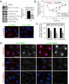
The loss of Cx43 at the BTB by RNAi hinders junction reassembly after calcium repletion. Sertoli cells freshly isolated from 20-day-old rat testes were cultured for 3 d, forming an intact epithelium with an established functional TJ-permeability barrier when assessed by transepithelial electrical resistance (TER) across the cell epithelium; and ultrastructures of TJ, basal ES and desmosome-like junction were also detected when examined by electron microscopy. Thus, the Sertoli cell epithelium used for this experiment mimicked the BTB in vivo. Thereafter, cells were transfected with 50–80 nM of nontargeting control (Ctrl) siRNA duplexes or Cx43 siRNA duplexes for 24 h. Calcium switch was performed ∼1.5 d after the transfection. (A) There was a 50% knockdown of Cx43 in cells transfected with Cx43 siRNA duplexes ∼2 d posttransfection. The levels of occludin, CAR, N-cadherin, and ZO-1 remained unchanged, illustrating the specificity of this RNAi experiment and that there was no off-target effect following the silencing of Cx43 in the Sertoli cell epithelium. The fluorescence micrographs shown in the bottom panel in (A) also support findings of the immunoblot analysis shown in the upper panel since there was a considerable loss of Cx43 at the cell-cell interface after the knockdown of Cx43 by RNAi. (B) After calcium repletion, Sertoli cell epithelium transfected with Cx43 siRNA duplexes displayed a significantly slower rebound of the disrupted TJ barrier when compared with cells transfected with non-targeting Ctrl siRNA duplexes. (C and D) Approximately 5 h after calcium ion repletion, Sertoli cells transfected with siRNA duplexes were fixed for dual-labeled immunofluorescence analysis to investigate the localization of occludin, ZO-1, and N-cadherin. In Sertoli cells with a knockdown of Cx43, considerably fewer junction proteins were found at the cell–cell interface, illustrating a disruption in the kinetics of junction reassembly in Sertoli cells with a knockdown of Cx43. Micrographs in the second and fourth columns in D are the corresponding grayscale images of the true-color images on the left, in order to better depict changes in protein localization. A representative dataset from 4 independent experiments is shown. (Scale bar, 40 μm.) *P < 0.05; **P < 0.01; statistical difference from the Ctrl group was tested using Student's t test in A and C and ANOVA analysis with posthoc two-tailed Dunnett's test in B.
Fig. 3.
Fig. 3.
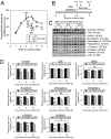
Bisphenol A (BPA) causes a transient disruption of the BTB integrity and changes in the steady-state levels of proteins at the BTB in primary Sertoli cell cultures. (A) In the TER experiment, Sertoli cells were treated with vehicle control (0.1% ethanol, V Ctrl) or BPA (200 μM) on day 3 of culture. BPA-treated Sertoli cells displayed a drastic reduction of the TJ-permeability barrier from 8 h posttreatment onward, when compared with the V Ctrl. In one treatment group, BPA was removed from the Sertoli cell epithelium after 24 h treatment to allow the “resealing” of the disrupted TJ barrier. A representative dataset is shown (n = 4). (BD) SCs were treated with V Ctrl or BPA for 24 h. In the BPA treatment group, BPA was removed after 24 h and cells were rinsed and replenished with normal culture medium. Cell lysates were collected 0, 8, and 24 h after BPA removal to analyze changes in the protein levels of various junction markers of TJ, basal ES and desmosome-like junction. The cytoskeletal protein levels, namely vimentin and actin, serve as loading control. Representative immunoblots are shown in C, where n = 3–5. The densitometry results are summarized in D. The BPA-treated group from each time point was normalized and compared against the V Ctrl group of the corresponding time point. Protein levels of the BPA-treated group were also compared at different times after BPA removal to assess the degree of recovery. At 0 h after BPA removal, the disruptive effect of BPA on junction protein level was observed for integral membrane proteins of TJ (occludin and JAM-A), basal ES (N-cadherin) and desmosome-like junction (desmoglein-2), and basal ES adaptors (α- and β-catenins). Junction proteins, including JAM-A, N-cadherin, desmoglein-2, and β-catenin, show a significant rebound from the lowered protein level after BPA removal. These data thus illustrate the reversible effect of BPA on junction integrity and junction protein levels. *P < 0.05; **P < 0.01; statistical difference from the V Ctrl group was tested using ANOVA analysis with a posthoc Tukey/Kramer test.
Fig. 4.
Fig. 4.
Changes in the distribution of junction proteins following treatment of Sertoli cell epithelium with BPA. The distribution of occludin/ZO-1 (A), N-cadherin/β-catenin (B), α-catenin (C), and Cx43 (D) in SCs was studied after the treatment and removal of BPA. During BPA treatment, fewer junction proteins were observed at the cell–cell interface. After BPA removal, reappearance of these junction proteins was observed at the cell–cell interface. These data again illustrate the reversibility of junction disruption induced by BPA. (Scale bars, 40 μm.)
Fig. 5.
Fig. 5.
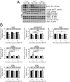
Changes in the steady-state protein levels of connexins and ERK following treatment of Sertoli cells with BPA and its removal. Sertoli cells cultured at 0.5 × 10⁶ cells/cm2 on Matrigel-coated multiwell plates were treated with BPA as depicted in Fig 3B. Cx43 and its phosphorylated form pCx43-Ser368 show a drastic reduction in protein levels after 24 h BPA treatment but only pCx43-Ser368 shows a significant recovery from the reduction at 8 h after BPA removal (B). The protein level of Cx26 remains unchanged by BPA treatment. Its level was lowered at 24 h after BPA removal when other junction proteins recover, displaying the differential effect of BPA on connexins expressed by Sertoli cells. The ERK signaling pathway was induced during BPA treatment but quickly went back to its normal level after BPA removal. Representative immunoblots are shown in A, where n = 3–4. The densitometry of the junction proteins is analyzed and summarized in B. *P < 0.05; **P < 0.01; statistical difference from the Ctrl group was tested using ANOVA analysis with a posthoc Tukey/Kramer test.
Fig. 6.
Fig. 6.
A dye transfer assay to study the gap junction communication after BPA treatment. Freshly isolated Sertoli cells from 20-d-old rat testes were cultured at 0.15 × 10⁶ cells/cm2 on Matrigel-coated glass-bottom dishes for 2 d before their use for this assay. Cells were labeled with calcein AM and then treated with 0.1% ethanol (V Ctrl) or 200 μM BPA for 1–2 h. The dye transfer assay, as described in Materials and Methods, involves photobleaching of a single Sertoli cell and then tracking the transfer of calcein into the photobleached cell (fluorescence recovery) to assess the degree of gap junction communication. At 150 s after photobleaching, Sertoli cells treated with BPA display a lower fluorescence recovery. These data illustrate that BPA can disrupt the gap junction communication in Sertoli cells after 1–2 h treatment. The result reported herein is representative of data from three independent experiments with similar findings. *P < 0.001.
Fig. 7.
Fig. 7.
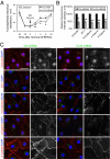
A study using the BPA model to examine the role of Cx43 in TJ-permeability barrier reassembly in Sertoli cell epithelium. After ≈1.5 d posttransfection with Ctrl or Cx43 siRNA duplexes, cells were treated with 200 μM BPA for 24 h to induce junction restructuring. (A) Upon BPA removal, the resealing of the disrupted TJ-permeability barrier occurred in cells transfected with either nontargeting control or Cx43 siRNA duplexes, but at a significantly slower rate in cells with a Cx43 knockdown. (B and C) Changes in the distribution of proteins at the Sertoli–Sertoli cell interface were examined by immunofluorescence microscopy. Junction proteins, including occludin, N-cadherin, ZO-1, α-catenin, and β-catenin, relocalized back to the cell surface after 24 h of BPA removal in the control silencing group. However, cells with a knockdown of Cx43 displayed a disrupted relocalization of these junction proteins back to the cell–cell interface. These data illustrate that the knockdown of Cx43 impaired junction reassembly after BPA-induced Sertoli cell TJ-permeability barrier disruption. Micrographs in the second and fourth columns in C are the corresponding grayscale images of the true-color images on the left, in order to better depict changes in protein localization. Representative data from four independent experiments are shown. (Scale bar, 40 μm.)

Similar articles

Cited by

References

    1. Cheng CY, Mruk DD. Cell junction dynamics in the testis: Sertoli-germ cell interactions and male contraceptive development. Physiol Rev. 2002;82:825–874. - PubMed
    1. Cheng CY, Mruk DD. A local autocrine axis in the testes that regulate spermatogenesis. Nat Rev Endocrinol. 2010;6:380–395. - PMC - PubMed
    1. Vogl AW, Vaid KS, Guttman JA. The Sertoli cell cytoskeleton. Adv Exp Med Biol. 2008;636:186–211. - PubMed
    1. Dbouk HA, Mroue RM, El-Sabban ME, Talhouk RS. Connexins: A myriad of functions extending beyond assembly of gap junction channels. Cell Commun Signal. 2009;7:4. - PMC - PubMed
    1. Pointis G, Gilleron J, Carette D, Segretain D. Physiological and physiopathological aspects of connexins and communicating gap junctions in spermatogenesis. Philos Trans R Soc Lond B Biol Sci. 2010;365:1607–1620. - PMC - PubMed

Publication types

LinkOut - more resources