. 2010 Oct 6;68(1):73-86.
doi: 10.1016/j.neuron.2010.09.022.

Snapin-regulated late endosomal transport is critical for efficient autophagy-lysosomal function in neurons

Affiliations

Snapin-regulated late endosomal transport is critical for efficient autophagy-lysosomal function in neurons

Qian Cai et al. Neuron. .

Abstract

Neuron maintenance and survival require late endocytic transport from distal processes to the soma where lysosomes are predominantly localized. Here, we report a role for Snapin in attaching dynein to late endosomes through its intermediate chain (DIC). snapin(-/-) neurons exhibit aberrant accumulation of immature lysosomes, clustering and impaired retrograde transport of late endosomes along processes, reduced lysosomal proteolysis due to impaired delivery of internalized proteins and hydrolase precursors from late endosomes to lysosomes, and impaired clearance of autolysosomes, combined with reduced neuron viability and neurodegeneration. The phenotypes are rescued by expressing the snapin transgene, but not the DIC-binding-defective Snapin-L99K mutant. Snapin overexpression in wild-type neurons enhances late endocytic transport and lysosomal function, whereas expressing the mutant defective in Snapin-DIC coupling shows a dominant-negative effect. Altogether, our study highlights new mechanistic insights into how Snapin-DIC coordinates retrograde transport and late endosomal-lysosomal trafficking critical for autophagy-lysosomal function, and thus neuronal homeostasis.


Figures

Figure 1
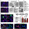
Figure 1. Aberrant Accumulation of Immature Lysosomes in snapin (−/−) Neurons
(A) Cortical neurons from snapin (+/+) and (−/−) embryos were co-immunostained at DIV7 with antibodies against MAP2 and LAMP-1. (B) Re-introducing snapin rescued accumulated LAMP-1-labeled organelles in snapin (−/−) neurons. Neurons were transfected with pIRES-GFP-Snapin or pIRES-GFP as a control at DIV4 and co-immunostained for LAMP-1 and MAP2 at DIV8. Arrows indicate transfected neurons. (C, D) Immature lysosomes in cortical neuron cultures. Late endosomes/immature lysosomes labeled by anti-CI-MPR-immunogold were clustered in the soma (C) and processes (D) of snapin (−/−) neurons at DIV7. Right panels (C) and lower panel (D) are close-up views of the boxed regions. (E) Increased LAMP-1 and LAMP-2 in snapin (−/−) cortical neurons. 40-µg lysates were sequentially detected in the same membrane with antibodies including markers for early endosomes (EEA1), Golgi (p115), and ER (Calnexin). (F) Schematic diagram of cathepsin D maturation during trafficking from TGN through late endosomes to lysosomes. (G) Impaired maturation of cathepsin D in snapin (−/−) mouse brain. 40-µg brain homogenates from E18 embryos were immunoblotted with antibodies against cathepsin D and p115. Percent change in the intermediate chains (interm) and mature forms of the heavy (HC) and light chain (LC) of cathepsin D was normalized against that of wild-type littermates. ** p<0.01. Error bars: s.e.m. Student’s t test. (H) Reduced cathepsin D activity in snapin (−/−) cortical neurons. Neurons were loaded with Bodipy-FL-pepstatin A followed by co-staining with antibodies against MAP2 and LAMP-1. Note that reduced active cathepsin D signals (green) were co-localized with lysosomes (red) in (−/−) cells. Scale bars in panels C and D: 500 nm; in other panels: 10 µm. (also see Figure S1).
Figure 2
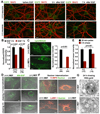
Figure 2. Snapin Deletion Impairs Late Endocytic Trafficking and Lysosomal Degradation
(A, B) Reduced degradation of internalized EGFR in snapin (−/−) cortical neurons. Cortical neurons were co-immunostained with MAP2 and EGFR before or 3 h or 7 h after EGF incubation (A). The EGFR mean intensity was normalized against MAP2 from the same neurons and expressed as ratios relative to the mean intensity at 3 h EGF incubation (B). (C) Reduced acidic lysosomes in snapin (−/−) MEFs labeled by fluorescence pH indicator LysoSensor (green). Mean intensity (right panel) of LysoSensor fluorescence in snapin (−/−) MEFs was normalized to those in (+/+) controls. (D, E) Pulse-chase assays showing impaired degradation of Alexa-488-EGF in snapin (−/−) MEFs. The mean intensity of 488-EGF after a 3-hr chase was normalized against that after the 30-min pulse (E). (F) Deleting snapin impaired delivery of internalized dextran to lysosomes. Note that most dextran signals were retained in CI-MPR-labeled late endosomes. (G) Electron microscopy showing impaired degradation of BSA-gold in snapin (−/−) MEFs. Gold particles were flocculated in the late endocytic compartments of snapin (+/+) cells due to BSA degradation, while BSA-gold conjugates remained discrete within the organelles of the snapin (−/−) MEFs. The total number of cells quantified is indicated in parentheses under bar graphs. Scale bars in G: 100 nm; in other panels: 10 µm. Error bars: s.e.m. Student’s t test, (also see Figure S2).
Figure 3
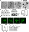
Figure 3. Snapin Deficiency Reduces Autolysosome Clearance
(A) Electron micrographs of hippocampi from snapin (+/+) and (−/−) embryos (E18). Clustered Avd-like structures with higher electron density (arrows) were consistently found in snapin (−/−) hippocampus. Images were selected near perinuclear regions. (B) Immunogold EM of cultured snapin (−/−) cortical neurons (DIV7) revealed LAMP-1-labeled structures similar to Avds in snapin (−/−) mouse hippocampi and suggestive of autolysosomes. (C) Enhanced LC3-II in snapin (−/−) brain homogenates (BH, 40 µg) and MEF lysates (20 µg) cultured in complete medium (CM) or starvation medium (HBSS) for 3 hr. (D, E) Biochemical analysis showing reduced clearance of autolysosome substrates p62 and LC3-II in snapin (−/−) cortical neurons. Neurons were treated with DMSO or the lysosomal inhibitors (LIs) leupeptin and pepstatin A (20 µM each) for 24 hrs. 40-µg lysates were sequentially immunobloted with antibodies against p62, LC3, Snapin and p115. The intensity of LC3-II bands (gels=4) was normalized to p115 and expressed as a ratio relative to (+/+) Ctrl (**: p<0.01) (E). Treating (−/−) neurons with LIs failed to significantly increase LC3-II level (p=0.07). (F, G) Cell biological analysis showing reduced turnover of GFP-LC3-labeled organelles in snapin (−/−) cortical neurons. Neurons were transfected with GFP-LC3 at DIV5 and treated with DMSO or LIs (leupeptin and pepstatin A, 20 µM each) at DIV6 for 24 hrs. GFP-LC3 signals were recruited into vesicular structures in snapin (+/+) neurons following LI treatment. Treating snapin (−/−) neurons with LIs failed to significantly increase LC3-II puncta (p=0.19) (G). (H) Increased p62 and LC3-II in autolysosomes from snapin (−/−) brains. LAMP-1-associated membranous organelles were immuno-isolated from light membrane fractions of snapin (+/+) and (−/−) mouse brains with Dyna magnetic beads coated with an anti-LAMP-1 antibody. Relative purity was assessed by measuring LAMP-1 and p115 levels. Scale bars in F: 10 µm, (also see Figure S3).
Figure 4
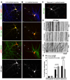
Figure 4. Impaired Retrograde Transport of Late Endosomes in snapin (−/−) Cortical Neurons
(A–C) Representative images showing distribution patterns of Rab7-labeled late endosomes in snapin (+/+) (A), (−/−) (B), and rescued cortical neurons (C). Neurons were co-transfected with mRFP and YFP-Rab7 at DIV6 followed by immunostaining with an anti-MAP2 antibody at DIV7. Late endosomes in snapin (−/−) neurons appear as large clusters along processes, while in (+/+) and rescued neurons they display as a much smaller punctate pattern. Arrows indicate axons. MAP*: blue MAP2 signal was converted to green for better color comparison. (D) Representative kymographs showing the mobility of axonal late endosomes during 12-min recordings in snapin (+/+), (−/−) and rescued neurons. Vertical lines represent stationary organelles; oblique lines or curves to the right represent anterograde movements and lines to the left indicate retrograde transport. Re-introducing HA-Snapin into (−/−) neurons recruits stationary organelles into the retrograde mobile pool. (E) Relative mobility of axonal late endosomes in snapin (+/+), (−/−) and rescued neurons. Data were quantified from the total number of axonal late endosomes (LE) in neurons (n) from >3 experiments, as indicated in parentheses. Scale bars: 10 µm. Error bars: s.e.m. Student’s t test, (also see Figure S4 and Videos 1–4).
Figure 5
Figure 5. Snapin Is Required for Tethering Dynein to the Late Endocytic Membrane
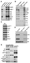
(A, B) Snapin associates with late endocytic compartments. Snapin- or LAMP-1-associated organelles were immuno-isolated from light membrane fractions of snapin (+/+) and (−/−) mouse livers with Dyna magnetic beads coated with either anti-Snapin (A) or anti-LAMP-1 (B) antibody, or normal IgG as a control. Bead-bound organelles were resolved by PAGE and sequentially detected with antibodies on the same membranes after stripping between applications of each antibody. (C, D) Deleting snapin has no observable effect on dynein motor expression in brain homogenates (BH) (50 µg) (C) or on assembly of DIC into the dynein heavy chain (DHC) (D). DIC-DHC complex was immunoprecipitated with a DIC antibody from 500 µg BH of snapin (+/+) and (−/−) mice. (E) Deleting snapin reduces association of dynein with late endocytic organelles. Dynein-associated membranous organelles were immuno-isolated from light membrane fractions with anti-DIC-coated Dyna magnetic beads. Reduced LAMP-1 and LAMP-2 (in boxes) were associated with dynein-bound organelles from snapin (−/−) mice.
Figure 6
Figure 6. Snapin Directly Interacts with Dynein DIC
(A) Coomassie Blue staining of GST-Snapin pull-down from rat brain homogenates. Mass spectrometry of the 74-kDa band (in red box) matches the rat DIC sequence. (*indicates degradation of GST-Snapin). (B) GST-Snapin pulls down dynein and dynactin from rat brain homogenates (BH). (C) Immunoprecipitation of Snapin with DIC from BH with an anti-Snapin antibody, normal IgG or pre-immune serum as controls, followed by sequential blotting with antibodies as indicated. (D, E) Snapin-DIC complex was immunoprecipitated by antibodies against either Snapin (D) or GFP (E) from COS cells expressing HA-Snapin and GFP-DIC. (F) Binding of GST-DIC to His-Snapin full-length (FL) but not to its N-terminal half (NT). (G) Snapin-binding sequence. DIC has an N-terminal coiled-coil domain required for binding to p150Glued, a middle Snapin-binding domain, and a C-terminal half containing 7 WD repeats involved in DHC binding.
Figure 7
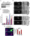
Figure 7. DIC–Binding Defective Snapin Mutant Fails to Rescue Late Endosomal Transport and Lysosomal Biology
(A) GST-DIC pull-down showing defective binding to Snapin-V92K and L99K mutants. (B) Representative images showing distribution of YFP-Rab7-labeled late endosomes (left) and kymographs showing their axonal mobility (right) in snapin (−/−) neurons co-transfected with YFP-Rab7 and HA-Snapin, HA-Snapin-L99K, or HA vector control. (C) Representative images and kymographs showing a dominant-negative effect of Snapin-L99K on axonal retrograde transport of late endosomes in (+/+) neurons. (D) Relative mobility of axonal late endosomes in (+/+) or (−/−) neurons expressing Snapin or Snapin-L99K or those expressing the Snapin-C66A, a defective binding mutant for synaptotagmin I and SNAP-25. (E) Expressing Snapin-L99K in snapin (−/−) cortical neurons failed to rescue accumulated immature lysosomes. Neurons were transfected with pIRES-GFP-Snapin-L99K followed by immunostaining with antibodies against LAMP-1 and MAP2. Arrow indicates transfected neuron. The total number of neurons examined is indicated in parentheses from >3 experiments. Scale bars: 10 µm. Error bars: s.e.m. *:p<0.001. Student’s t test, (also see Figure S5).
Figure 8
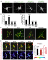
Figure 8. Snapin Enhances Formation of Tubular Lysosomes or Endolysosomes by Interacting with DIC
(A, B) Snapin, but neither its L99K mutant nor the Snapin-binding domain of the DIC108–268 transgene, enhances formation of tubular lysosomes (indicated by red arrows) in cortical neurons (A). Number of tubular lysosomes per neuron (B) was counted in neurons co-expressing GFP-LAMP-1 with HA-Snapin, HA-Snapin-L99K, HA-DIC108–268 or HA vector control. (C, D) Snapin, but neither its L99K mutant nor DIC108–268, facilitates formation of endolysosomes in the soma of cortical neurons (C). Lysosomes were labeled by GFP-LAMP-1 and late endosomes were marked by a short-chase loading of dextran-546. Average fluorescence ratios of GFP-LAMP-1 / dextran-546 were calculated from individual vesicular (arrowheads) or tubular (arrows) endolysosomes (D). (E, F) Snapin expression in COS cells enhances late endocytic membrane trafficking. Representative time-lapse images demonstrate dynamic tubular lysosome formation via fusion between late endosomes (green) and lysosomes (red) within the indicated time period in cells expressing HA-Snapin (E). Rhodamine-Dextran fluorescence levels are also shown in scaled pseudocolor. Arrows indicate tubulation or fusion events. Note that the minus-end is pointed toward the lower left side. The number and average length of tubular lysosomes in cells expressing Snapin were quantified relative to controls (F). Scale Bars in A and C: 10 µm; in E: 5 µm. Student’s t test, (also see Figure S6 and Videos 5 and 6).

Comment in

Similar articles

Cited by

References

    1. Bjørkøy G, Lamark T, Brech A, Outzen H, Perander M, Overvatn A, Stenmark H, Johansen T. p62/SQSTM1 forms protein aggregates degraded by autophagy and has a protective effect on huntingtin-induced cell death. J. Cell Biol. 2005;171:603–614. - PMC - PubMed
    1. Bright NA, Reaves BJ, Mullock BM, Luzio JP. Dense core lysosomes can fuse with late endosomes and are re-formed from the resultant hybrid organelles. J. Cell Sci. 1997;110:2027–2040. - PubMed
    1. Bright NA, Gratian MJ, Luzio JP. Endocytic delivery to lysosomes mediated by concurrent fusion and kissing events in living cells. Curr. Biol. 2005;15:360–365. - PubMed
    1. Buxton P, Zhang XM, Walsh B, Sriratana A, Schenberg I, Manickam E, Rowe T. Identification and characterization of Snapin as a ubiquitously expressed SNARE-binding protein that interacts with SNAP23 in non-neuronal cells. Biochem. J. 2003;375:433–440. - PMC - PubMed
    1. Chen CS, Chen WN, Zhou M, Arttamangkul S, Haugland RP. Probing the cathepsin D using a BODIPY FL-pepstatin A: applications in fluorescence polarization and microscopy. J Biochem. Biophys. Methods. 2000;42:137–151. - PubMed

Publication types

MeSH terms

LinkOut - more resources