

Extreme loss of immunoreactive p-Akt and p-Erk1/2 during routine fixation of primary breast cancer
- PMID: 20920193
- PMCID: PMC3096968
- DOI: 10.1186/bcr2719
Extreme loss of immunoreactive p-Akt and p-Erk1/2 during routine fixation of primary breast cancer
Abstract
Introduction: Very few studies have investigated whether the time elapsed between surgical resection and tissue fixation or the difference between core-cut and excision biopsies impact on immunohistochemically measured biomarkers including phosphorylated proteins in primary breast cancer. The aim of this study was to characterize the differences in immunoreactivity of common biomarkers that may occur (a) due to tissue handling at surgery and (b) between core-cuts and resected tumours.
Methods: Core-cuts taken from surgical breast cancer specimens immediately after resection (sample A) and after routine X-ray of the excised tumour (sample B) were formalin-fixed and paraffin-embedded and compared to the routinely fixed resection specimen (sample C). The variation in immunohistochemical expression of Ki67, oestrogen receptor (ER), progesterone receptor (PgR), human epidermal growth factor 2 (HER2), p-Akt and p-Erk were investigated.
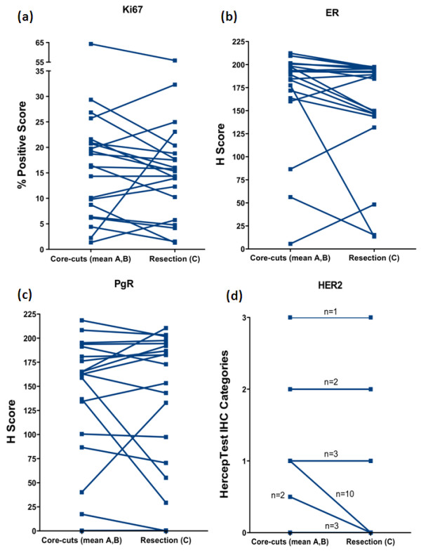
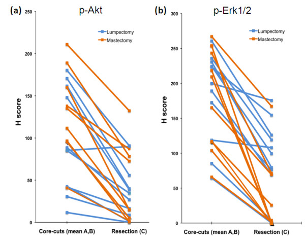
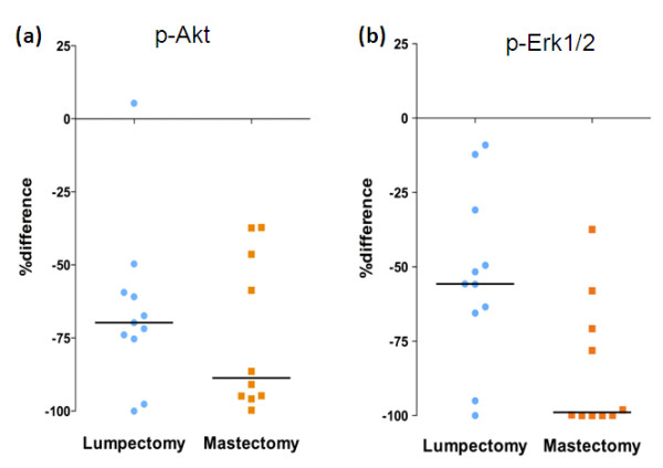
Results: Twenty-one tissue sets with adequate tumour were available. Median time between collection of core-cuts A and B was 30 minutes (range 20 to 80). None of the markers showed significant differences between samples A and B. Similarly, Ki67, ER, PgR and HER2 did not differ significantly between core-cuts and main resection specimen although there was a trend for lower resection values for ER (P=0.06). However, p-Akt and p-Erk1/2 were markedly lower in resections than core-cuts (median 27 vs 101 and 69 vs 193, respectively; both P<0.0001 [two-sided]). This difference was significantly greater in mastectomy than lumpectomy specimens for p-Erk1/2 (P=0.01).
Conclusions: The delay in fixation in core-cuts taken after post-operative X-ray of resection specimens has no significant impact on expression of Ki67, ER, PgR, HER2, p-Akt or p-Erk1/2. However extreme loss of phospho-staining can occur during routine fixation of resection specimens. These differences are likely attributable to suboptimal fixation and may have major repercussions for clinical research involving these markers.
Figures
Similar articles
-
Comparison of protein expression between formalin-fixed core-cut biopsies and surgical excision specimens using a novel multiplex approach.Breast Cancer Res Treat. 2019 Jun;175(2):317-326. doi: 10.1007/s10549-019-05163-6. Epub 2019 Feb 22. Breast Cancer Res Treat. 2019. PMID: 30796652 Free PMC article.
-
Quantitative assessment of effect of preanalytic cold ischemic time on protein expression in breast cancer tissues.J Natl Cancer Inst. 2012 Dec 5;104(23):1815-24. doi: 10.1093/jnci/djs438. Epub 2012 Oct 22. J Natl Cancer Inst. 2012. PMID: 23090068 Free PMC article.
-
Prognostic relevance of activated Akt kinase in node-negative breast cancer: a clinicopathological study of 99 cases.Mod Pathol. 2004 Jan;17(1):15-21. doi: 10.1038/modpathol.3800002. Mod Pathol. 2004. PMID: 14631376
-
Delay to Formalin Fixation (Cold Ischemia Time) Effect on Breast Cancer Molecules.Am J Clin Pathol. 2018 Mar 7;149(4):275-292. doi: 10.1093/ajcp/aqx164. Am J Clin Pathol. 2018. PMID: 29471352 Review.
-
The effects of under 6 hours of formalin fixation on hormone receptor and HER2 expression in invasive breast cancer: a systematic review.Am J Clin Pathol. 2014 Jul;142(1):16-22. doi: 10.1309/AJCP96YDQSTYBXWU. Am J Clin Pathol. 2014. PMID: 24926080 Review.
Cited by
-
FAK inhibition alone or in combination with adjuvant therapies reduces cancer stem cell activity.NPJ Breast Cancer. 2021 May 28;7(1):65. doi: 10.1038/s41523-021-00263-3. NPJ Breast Cancer. 2021. PMID: 34050172 Free PMC article.
-
Post-translational modifications of nuclear receptors and human disease.Nucl Recept Signal. 2012;10:e001. doi: 10.1621/nrs.10001. Epub 2012 Feb 27. Nucl Recept Signal. 2012. PMID: 22438791 Free PMC article. Review.
-
Impaired coordination between signaling pathways is revealed in human colorectal cancer using single-cell mass cytometry of archival tissue blocks.Sci Signal. 2016 Oct 11;9(449):rs11. doi: 10.1126/scisignal.aah4413. Sci Signal. 2016. PMID: 27729552 Free PMC article.
-
SRC drives growth of antiestrogen resistant breast cancer cell lines and is a marker for reduced benefit of tamoxifen treatment.PLoS One. 2015 Feb 23;10(2):e0118346. doi: 10.1371/journal.pone.0118346. eCollection 2015. PLoS One. 2015. PMID: 25706943 Free PMC article.
-
Proteomic modulation in breast tumors after metformin exposure: results from a "window of opportunity" trial.Clin Transl Oncol. 2017 Feb;19(2):180-188. doi: 10.1007/s12094-016-1521-1. Epub 2016 Jun 15. Clin Transl Oncol. 2017. PMID: 27305912 Free PMC article.
References
-
- Dowsett M. Preoperative models to evaluate endocrine strategies for breast cancer. Clin Cancer Res. 2003;9:502s. - PubMed
-
- Wolff AC, Hammond ME, Schwartz JN, Hagerty KL, Allred DC, Cote RJ, Dowsett M, Fitzgibbons PL, Hanna WM, Langer A, McShane LS, Paik S, Pegram MD, Perez EA, Press MF, Rhodes A, Sturgeon C, Taube SE, Tubbs R, Vance GH, Vijver M, Wheeler TM, Hayes DF. American Society of Clinical Oncology/College of American Pathologists Guideline Recommendations for Human Epidermal Growth Factor Receptor 2 Testing in Breast Cancer. J Clin Oncol. 2007;25:118–145. doi: 10.1200/JCO.2006.09.2775. - DOI - PubMed
Publication types
MeSH terms
Substances
Grants and funding
LinkOut - more resources
Full Text Sources
Other Literature Sources
Medical
Research Materials
Miscellaneous
