. 2010 Nov;6(8):1090-106.
doi: 10.4161/auto.6.8.13426.

p62/SQSTM1 is required for Parkin-induced mitochondrial clustering but not mitophagy; VDAC1 is dispensable for both

Affiliations

p62/SQSTM1 is required for Parkin-induced mitochondrial clustering but not mitophagy; VDAC1 is dispensable for both

Derek Narendra et al. Autophagy. 2010 Nov.

Abstract

Mitochondria sustain damage with aging, and the resulting mitochondrial dysfunction has been implicated in a number of diseases including Parkinson disease. We recently demonstrated that the E3 ubiquitin ligase Parkin, which is linked to recessive forms of parkinsonism, causes a dramatic increase in mitophagy and a change in mitochondrial distribution, following its translocation from the cytosol to mitochondria. Investigating how Parkin induces these changes may offer insight into the mechanisms that lead to the sequestration and elimination of damaged mitochondria. We report that following Parkin’s translocation from the cytosol to mitochondria, Parkin (but not a pathogenic mutant) promotes the K63-linked polyubiquitination of mitochondrial substrate(s) and recruits the ubiquitin- and LC3-binding protein, p62/SQSTM1, to mitochondria. After its recruitment, p62/SQSTM1 mediates the aggregation of dysfunctional mitochondria through polymerization via its PB1 domain, in a manner analogous to its aggregation of polyubiquitinated proteins. Surprisingly and in contrast to what has been recently reported for ubiquitin-induced pexophagy and xenophagy, p62 appears to be dispensable for mitophagy. Similarly, mitochondrial-anchored ubiquitin is sufficient to recruit p62 and promote mitochondrial clustering, but does not promote mitophagy. Although VDAC1 (but not VDAC2) is ubiquitinated following mitochondrial depolarization, we find VDAC1 cannot fully account for the mitochondrial K63-linked ubiquitin immunoreactivity observed following depolarization, as it is also observed in VDAC1/3-/- mouse embryonic fibroblasts. Additionally, we find VDAC1 and VDAC3 are dispensable for the recruitment of p62, mitochondrial clustering and mitophagy. These results demonstrate that mitochondria are aggregated by p62, following its recruitment by Parkin in a VDAC1-independent manner. They also suggest that proteins other than p62 are likely required for mitophagy downstream of Parkin substrates other than VDAC1.


Figures
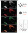
Figure 1
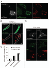
Figure 1
Wild-type Parkin, but not a pathogenic mutant, promotes mitochondrial aggregation. (A) HeLa cells co-transfected with DsRed-Mito and EYFP-Parkin or EYFP-Parkin R275W were treated with 10 µM CCCP for 3 hrs and immunostained for the mitochondrial protein Tom20. (B) Eight circles of equal area have different compaction indexes when dispersed and when aggregated, as measured by ImageJ. The compaction index is ratio of the shortest perimeter possible for an object with the same area as the object of interest divided by the actual perimeter of the object of interest (see Materials and Methods for details). (C) A midplane image for EYFP-Parkin or EYFP-Parkin R275W expressing cells and neighboring untransfected cells treated as in (A) were obtained, the area and perimeter of mitochondria within the cell were measured using ImageJ, and the compaction index was calculated. ≥8 cells per experiment were measured for each condition in at least two independent experiments. Scale bars in images = 10 µm.
Figure 2
Figure 2
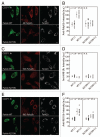
Parkin K63-polyubiquitinates proteins on depolarized mitochondria by confocal imaging. (A) HeLa cells transfected with wild-type EC FP-Parkin (green) or the translocation-competent mutant ECFP-Parkin R275W (green) were treated with CCCP for 1 hr and immunostained for polyubiquitinated protein (red) and Tom20 (white), a mitochondrial marker. Both wild-type Parkin and Parkin R275W co-localize with Tom20. However, polyubiquitinated protein co-localized with depolarized mitochondria only in cells expressing wild-type Parkin. (B) Polyubiquitinated protein fluorescent intensity co-localizing with mitochondria was measured for cells expressing wild-type Parkin or Parkin R275W (WT [+] or R275W [+], respectively) and for untransfected control cells (WT [−] or R275W [−], respectively) from the same fields. ≥8 cells per experiment were measured for each condition in at least two independent experiments. (C) HeLa cells transfected and treated as in (A) were immunostained for K48 polyubiquitinated protein (red) and Tom20 (white). (D) Cells treated as in (C) were analyzed as in (B). (E) HeLa cells transfected and treated as in (A) were immunostained for K63 polyubiquitinated protein (red) and Tom20 (white). (F) Cells treated and immunostained as in (E) were analyzed as described in (B). Scale bars in images = 10 µm.
Figure 3
Figure 3
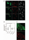
Ubiquitin-binding adaptor p62 is recruited to depolarized mitochondria in cells expressing Parkin. (A) HeLa cells transfected with wild-type EYFP-Parkin (green) or EYFP-Parkin R275W (green) were treated with DMSO or CCCP for 1 hr and immunostained for p62 (red) and Tom20 (white). (B) Pearson correlation coefficients for p62 intensity vs. Tom20 intensity were measured for cells expressing wild-type Parkin [WT (+)] or Parkin R275W [R275W (+)] and for untransfected control cells from the same fields [WT (−) and R275W (−), respectively]. ≥8 cells per experiment were measured for each condition in at least two independent experiments. (C) Cells transfected as in (A) were treated with CCCP for 1 hr and immunostained for p62. Scale bars in images = 10 µm.
Figure 4
Figure 4
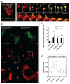
p62 is necessary for mitochondrial aggregation but not mitophagy. (A) HeLa cell co-transfected with GFP-p62 (green) and untagged Parkin in a 1:4 ratio was labeled with Mitrotracker Red (red) and imaged live following addition of 10 µM CCCP at time point 0. Left image depicts the whole cell at time point 0. Changes in morphology of two clusters of mitochondria are shown in the left (L) and right (R) series of images. (A, top right) the left series depicts the fragmentation of thread-like mitochondria from 0 to 70 minutes following CCCP. p62 is recruited to groups of fragmented mitochondria between 20 and 40 minutes. When groups of fragmented mitochondria that co-localize with p62 (yellow) come into contact they begin to move as a single object (a and b at 50 minutes and ab and c at 70 minutes). (A, bottom right) the right series shows p62 labeled structures forming between perinuclear mitochondria as they are compressed together over time. The white line in first image depicts the border of the nucleus. (B) p62+/+ or p62−/− cells transfected with EYFP-Parkin (green) were treated with 20 µM of CCCP for 24 hrs. While mitochondria formed aggregates in p62+/+ cells, mitochondrial failed to form aggregates in p62−/− cells. (C) Cells treated as in (B) were scored for mitochondrial morphology for ≥50 cells/condition in ≥3 independent experiments. Morphology was considered “normal” if it was similar to untransfected cells in the same field and “few dispersed,” if mitochondria appeared reduced in number but no aggregates were apparent. *indicates p-value <0.05. **indicates p-value <0.01. (D) Midplane images of cells treated as in (B) were analyzed using the compaction index as described in (Fig. 1B and C) for ≥8 cells/condition from ≥2 independent experiments. Scale bars in images = 10 µm.
Figure 5
Figure 5
Acute depletion of p62 with RNAi does not inhibit Parkin-induced mitophagy. (A) Lysates from HeLa cells transfected with p62 siRNA or control siRNA for three consecutive days were separated on SDS-PAGE gels and immunoblotted for p62 and VDAC1, which served as a loading control. (B) Cells from (A) plated onto slides and transiently transfected with EYFP-Parkin (green) were treated with CCCP for 24 hrs and immunostained for p62 (red) and the mitochondrial protein Tom20 (white). Scale bar in last image = 10 µm. (C) cells in (B) were scored for the presence of mitochondria.
Figure 6
Figure 6
Mitochondrial aggregation requires the PB1 domain, but not the LIR domain, of p62. (A) Schematic depicting the domain structure of p62. The PB1 domain (which contains residue D69) is responsible for p62 polymerization, while the LIR domain binds LC3 on autophagosomes and the UBA domain binds ubiquitin. (B) p62−/− cells co-transfected with unlabeled Parkin and EGFP, EGFP-p62 wild-type, EGFP-p62 D69A or EGFP-p62 ΔLIR were treated with 20 µM of CCCP for 24 hrs. (C) Cells treated as in (B) were scored for the presence of mitochondrial aggregates for ≥50 cells/condition in ≥3 independent experiments. Scale bars in images = 10 µm.
Figure 7
Figure 7
Anchoring ubiquitin to outer mitochondrial membrane is sufficient for p62 recruitment and mitochondrial clustering but not mitophagy. (A) HeLa cells, which lack Parkin protein expression, were co-transfected with ubiquitin G76V fused to tandem FKBP12 domains and mitochondrial anchor constructs fused to the FRB domain. The cells were subsequently treated with the rapalog AP21967 for 24 hrs. Rapalog induced the translocation of ubiquitin to mitochondria secondary to heterodimerization of the FKBP and FRB domains in the presence of the rapalog. Cells were immunostained with Tom20 and the cells were scored for the absence of mitochondria. ≥50 cells per condition were counted. (B) HeLa cells co-transfected with EGFP-Ubiquitin G76V-FKBP or FKBP-EGFP and FRB-Fis1 (92–152) were treated with rapalog and CCCP for 24 hrs and immunostained for p62 (red) and the mitochondrial protein Tom20 (white). Scale bar in last image = 10 µm. (C) Cells treated as in (B) were scored for the presence of p62 positive and p62 negative mitochondrial clumps. ≥50 cells per condition were counted.
Figure 8
Figure 8
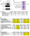
VDAC1 but not VDAC2 is ubiquitinated following mitochondrial uncoupling in the presence of Parkin. (A) Lysates from HeLa cells stably expressing EYFP-Parkin and treated with CCCP for 0, 2 or 5 hrs were separated on 12–22% SDS-PAGE gels and immunoblotted with an antibody against VDAC1 (anti-porin 31HL mAB6) or Tubulin. The predicted positions of unmodified VDAC1 and mono- and di-ubiquitinated forms of VDAC1 are indicated. (B) VDAC1 immunoaffinity purified with mAB6 from HeLa cell lysates following treatment with CCCP for 5 hrs was separated on SDS-PAGE gels and stained with Coomassie blue dye. Approximate positions of five bands cut for protein identification are indicated. (C) Table summarizing peptides identified from bands 1–5. (D) Table depicting relative abundance of three unique VDAC2 peptides and three homologous VDAC1 peptides in bands 1–5. Integrated MS ion intensities for each peptide were normalized to that observed in control band 3. Values greater than 2, indicating a 2-fold increase over control band 3, are highlighted in yellow. (E) Table depicting relative abundance of two unique ubiquitin peptides, TITLE and TLS. Integrated MS ion intensities for each peptide were normalized as in (D). Values greater than 2, indicating a 2-fold increase over control band 3, are highlighted in yellow.
Figure 9
Figure 9
Mitochondria-associated proteins other than VDAC1 and VDAC3 are K63-polyubiquitinated in a Parkin-dependent manner; and VDAC1 and VDAC3 are dispensable for p62 recruitment to mitochondria, mitochondrial sequestration and Parkin-induced mitophagy. (A) VDAC1/3−/− mouse embryonic fibroblasts (MEFs) transiently expressing EYFP-Parkin (white) and treated with 20 µM CCCP for 3 hrs were immunostained for K63 polyubiquitinated proteins (green) and the mitochondrial protein, cytochrome c (red). (B) VDAC1/3+/+ and VDAC1/3−/− MEFs transiently expressing EYFP-Parkin (green) were treated with CCCP for 24 hrs and immunostained for the mitochondrial protein Tom20 (red). (C) Cells in (B) scored for mitochondrial phenotype. (D) Cells treated as in (B) were immunostained for p62 (green) and Tom20 (red). Scale bar in last image = 10 µm.
Figure 10
Figure 10
Microtubule network is dispensible for Parkin-induced mitophagy. (A) HeLa cells transfected with EYFP-Parkin were treated with DMSO or 10 µM nocodazole for 3 hrs and then immunostained for α-tubulin (green). Nocodazole treatment led to complete disruption of the microtubule network. (B) HeLa cells transfected with EYFP-Parkin (blue) were pretreated with DMSO or 10 µM nocodazole for 3 hrs and then 10 µM CCCP was added for an additional 24 hrs. Cells were immunostained for Tom20 (red) and α-tubulin (green). (C) Cells treated as in (B) were scored for presence of mitochondria for ≥50 cells/condition in ≥3 independent experiments. Scale bars in images = 10 µm.

Similar articles

Cited by

References

    1. Wallace DC. A mitochondrial paradigm of metabolic and degenerative diseases, aging and cancer: a dawn for evolutionary medicine. Annu Rev Genet. 2005;39:359–407. - PMC - PubMed
    1. Orr WC, Sohal RS. Extension of life-span by over-expression of superoxide dismutase and catalase in Drosophila melanogaster. Science. 1994;263:1128–1130. - PubMed
    1. Trifunovic A, Hansson A, Wredenberg A, Rovio AT, Dufour E, Khvorostov I, et al. Somatic mtDNA mutations cause aging phenotypes without affecting reactive oxygen species production. Proc Natl Acad Sci USA. 2005;102:17993–17998. - PMC - PubMed
    1. Tatsuta T, Langer T. Quality control of mitochondria: protection against neurodegeneration and ageing. EMBO J. 2008;27:306–314. - PMC - PubMed
    1. Ekstrand MI, Terzioglu M, Galter D, Zhu S, Hofstetter C, Lindqvist E, et al. Progressive parkinsonism in mice with respiratory-chain-deficient dopamine neurons. Proc Natl Acad Sci USA. 2007;104:1325–1330. - PMC - PubMed

MeSH terms

LinkOut - more resources