. 2010 Nov 15;16(22):5458-68.
doi: 10.1158/1078-0432.CCR-10-0712. Epub 2010 Oct 1.

Transduction of tumor-specific T cells with CXCR2 chemokine receptor improves migration to tumor and antitumor immune responses

Affiliations

Transduction of tumor-specific T cells with CXCR2 chemokine receptor improves migration to tumor and antitumor immune responses

Weiyi Peng et al. Clin Cancer Res. .

Abstract

Purpose: One of the most important rate-limiting steps in adoptive cell transfer (ACT) is the inefficient migration of T cells to tumors. Because melanomas specifically express the chemokines CXCL1 and CXCL8 that are known to facilitate the CXCR2-dependent migration by monocytes, our aim is to evaluate whether introduction of the CXCR2 gene into tumor-specific T cells could further improve the effectiveness of ACT by enhancing T-cell migration to tumor.

Experimental design: In this study, we used transgenic pmel-1 T cells, which recognize gp100 in the context of H-2Db, that were transduced with luciferase gene to monitor the migration of transferred T cells in vivo. To visualize luciferase-expressing T cells within a tumor, a nonpigmented tumor is required. Therefore, we used the MC38 tumor model, which naturally expresses CXCL1.

Results: Mice bearing MC38/gp100 tumor cells treated with CXCR2/luciferase-transduced pmel-1 T cells showed enhanced tumor regression and survival compared with mice receiving control luciferase-transduced pmel-1 T cells. We also observed preferential accumulation of CXCR2-expressing pmel-1 T cells in the tumor sites of these mice using bioluminescence imaging. A similar enhancement in tumor regression and survival was observed when CXCR2-transduced pmel-1 T cells were transferred into mice bearing CXCL1-transduced B16 tumors compared with mice treated with control pmel-1 T cells.

Conclusions: These results implicate that the introduction of the CXCR2 gene into tumor-specific T cells can enhance their localization to tumors and improve antitumor immune responses. This strategy may ultimately enable personalization of cancer therapies based on chemokine expression by tumors.


Figures

Figure 1
Figure 1
Human melanoma tumors express ligands for CXCR2, but tumor-infiltrating T cells lack CXCR2 expression. (A) Paraffin-embedded, melanoma lymph node metastases were analyzed by immunohistochemical staining for chemokine CXCL8 and CXCL1. Representative staining is shown at 40X magnification. (B) CXCR2 expression on PBMCs, as determined by flow cytometry. PBMCs from five healthy donors were stained with anti-CD3 and anti-CXCR2. (C) FACS analysis of CXCR2 expression on tumor-infiltrating lymphocytes (TILs). TILs isolated from four melanoma patients were stained with anti-CD4, CD8 and CXCR2. Representative results of all stained samples were shown.
Figure 2
Figure 2
Generation of gp100-expressing MC38 tumor cell lines. (A) Schematic representation of viral vector containing human full-length gp100, an internal ribosomal entry site, and the mIL-4R gene. The construct was transduced into MC38 cells, which were then sorted based on IL-4R expression, as shown in right subpanel. (B) IFN-γ secretion by pmel-1 T cells in response to MC38/gp100 tumors. Pmel-1 T cells were cocultured with the indicated murine tumor cell lines. Twenty-four hours later, IFN-γ concentrations in the supernatants were detected by ELISA. B16 melanoma cells pretreated with IFN-γ were used as a positive control. (C) Growth of MC38/gp100 tumors in C57BL/6 albino mice. C57BL/6 albino mice were s.c. injected with 5 × 10⁵ MC38 or MC38/gp100. Tumor sizes were measured every 3 d. (D) Treatment of MC38/gp100 tumors with pmel-1 T cells. C57BL/6 albino mice were s.c. injected with 5 × 10⁵ MC38 or MC38/gp100. Six days later, all mice were received 350cGy irradiation. Seven days later, 5×10⁶ (5M) pmel-1 T cells were transferred into tumor-bearing mice, along with 5 × 10⁵ gp100 peptide-pulsed DC, both by intravenous injection, and systemic IL-2 treatment. Tumor-bearing mice receiving irradiation only served as the control group. Tumor sizes were measured every 3 d. Pmel-1 T cells suppressed the growth of MC38/gp100 tumors, but not MC38 tumors. Data are plotted as the mean of five mice per group + SEM. Results are representative of two experiments. (E) Pmel-1 T cells migrate specifically to MC38/gp100 tumors. C57BL/6 albino mice were subcutaneously injected with 5×10⁵ MC38/gp100 in the right flank. The same number of MC38 tumor cells was implanted on the left flank. Seven days after tumor implantation, 1×10⁶ sorted luciferase expressing pmel-1 T cells were transferred to tumor-bearing mice. Six days after adoptive transfer, the intensity of luciferase signaling was measured. Data shown were representative of five mice.
Figure 3
Figure 3
Generation and functional characteristics of CXCR2-expressing pmel-1 T cells. (A) Schematic representation of viral vector containing mCXCR2. Long terminal repeat from the murine stem cell PCMV virus drives high-level, constitutive expression of mCXCR2 in pmel-1 T cells. (B) FACS analysis of transduced pmel-1 T cells. Pmel-1 T cells were transduced with modified OFL-GFP alone or OFL-GFP and mCXCR2. Cells were sorted based on the expression of GFP or CXCR2. Sorted cells were cultured for 3 d and used for ACT. Before transfer, the purity of transferred cells was evaluated by flow cytometry. (C) Cytotoxic reactivity of CXCR2-expressing pmel-1 T cells against MC38/gp100 tumor. (D) IFN-γ secretion by CXCR2-expressing pmel-1 T cells in response to MC38/gp100 tumors.
Figure 4
Figure 4
Enhanced migration of CXCR2-expressing T cells along CXCL1 gradients. (A) Migration of CXCR2-expressing T cells along recombinant CXCL1 gradients. C57BL/6 T cells were transduced with either OFL-GFP or OFL-GFP and mCXCR2 and placed into the upper wells of a Transwell system, with CXCL1 in the lower wells at the indicated concentrations. One hour later, D-luciferin was added to the lower wells. T cell migration to the lower well was quantitated by measuring luminescent activity. (B) CXCL1 production by murine tumor lines. Cells (2 × 10⁶) from the indicated murine cell lines were plated in 6-well plates; 24 h later, CXCL1 in supernatants was detected by ELISA. (C) Migration of CXCR2-expressing T cells along tumor cell-derived CXCL1 gradients. C57BL/6 T cells were transduced with either OFL-GFP or OFL-GFP and mCXCR2 and placed into the upper wells of a Transwell system. Conditioned cell culture medium from tumor cells was loaded into the lower wells. One hour after incubation, the cell number in the lower wells was counted. (D) Proliferation of CXCR2-expressing pmel-1 T cells upon TCR stimulation in the presence or absence of CXCL1. CXCR2-expressing T cells or OFL-expressing T cells were stimulated with anti-CD3 and irradiated splenocytes in the presence or absence of recombinant CXCL1. T-cell proliferation was evaluated by 3[H] thymidine incorporation assay. (E) IFN-γ secretion by CXCR2-expressing pmel-1 T cells upon TCR stimulation in the presence or absence of CXCL1. CXCR2-expressing T cells or OFL-expressing T cells were stimulated with anti-CD3 and irradiated splenocytes in the presence or absence of recombinant CXCL1.
Figure 5
Figure 5
Enhanced migration of CXCR2-expressing pmel-1 T cells to tumor sites. (A) In vivo trafficking of OFL-expressing pmel-1 T cells. OFL-expressing pmel-1 T cells (1 × 10⁶) were transferred into mice bearing established 7-day MC38/gp100 tumors. DC vaccine and IL-2 treatment were performed as previous described. Imaging was performed at indicated time points after T cell transfer. (B) Increased accumulation of CXCR2-expressing pmel-1 T cells at the tumor site. Pmel-1 T cells (1 × 10⁶) expressing either OFL alone or CXCR2 with OFL were transferred into mice bearing established MC38/gp100 tumors. Imaging was performed at day 6 after T cell transfer. Data shown were from representative mice. (C) Quantitative imaging analysis of transferred T cells in tumor-bearing mice. Intensities of the luciferase signal at tumor sites in all tumor-bearing mice are depicted. (D) Percentage of Thy1.1+ pmel-1 T-cells of the CD8+ T cells in the peripheral blood on day 6 after T cell transfer. Thy1.1 is a congenic cell surface marker for transferred pmel-1 T cells. Data for (C) and (D) are plotted as the mean of eight mice per group + SEM. Results are representative of three experiments.
Figure 6
Figure 6
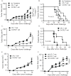
Enhanced suppression of established tumors upon ACT with CXCR2-expressing pmel-1 T cells. Groups of mice were implanted with 5 × 10⁵ tumor cells on day 0, subjected to 350 cGy TBI at day 6, followed on day 7 by ACT with pmel-1 T cells expressing either OFL alone or OFL and CXCR2, along with DC vaccination and systemic IL-2. (A) Tumor growth curve of MC38/gp100 tumor-bearing mice receiving ACT. (B) Kaplan-Meier survival curves of MC38/gp100 tumor-bearing mice treated with ACT. (C) Tumor growth curve and (D) Kaplan-Meier survival curves of B16-CXCL1 tumor-bearing mice treated with ACT with CXCR2-transduced pmel-1 T cells. Tumor growth was monitored every 3 d and plotted as means + SEM. P values in panels (B) and (D) were determined by two-way ANOVA. In both tumor models, CXCR2 transduction enhanced the ability of the T cells to mediate tumor regression and increased survival. Similar results were obtained in repeated experiments. In (A) and (B), N=8-9 for each treatment group; in (C) and (D), n=5 for each treatment group. Results are representative of two experiments. (E) Tumor growth curve of B16 tumor-bearing mice receiving ACT. N=3 for each treatment group. (F) Tumor growth curve of B16-CXCL1 tumor-bearing mice receiving ACT using different numbers of OFL or CXCR2 expressing pmel-1 T cells. Similar protocols for tumor challenge and ACT treatment were followed. Tumor-bearing mice were infused with either 1 × 10⁶ (1M), 5× 10⁶ (5M) OFL expressing T cells, or 1× 10⁶ CXCR2 expressing T cells on day 7 after tumor challenge. N= 3 to 5 mice for each treatment group.

Similar articles

Cited by

References

    1. Dudley ME, Wunderlich JR, Yang JC, et al. Adoptive cell transfer therapy following non-myeloablative but lymphodepleting chemotherapy for the treatment of patients with refractory metastatic melanoma. J Clin Oncol. 2005;23:2346–57. - PMC - PubMed
    1. Wrzesinski C, Paulos CM, Kaiser A, et al. Increased Intensity Lymphodepletion Enhances Tumor Treatment Efficacy of Adoptively Transferred Tumor-specific T Cells. J Immunother. 2009 - PMC - PubMed
    1. Rosenberg SA, Dudley ME. Adoptive cell therapy for the treatment of patients with metastatic melanoma. Current opinion in immunology. 2009;21:233–40. - PMC - PubMed
    1. Pockaj BA, Sherry RM, Wei JP, et al. Localization of 111indium-labeled tumor infiltrating lymphocytes to tumor in patients receiving adoptive immunotherapy. Augmentation with cyclophosphamide and correlation with response. Cancer. 1994;73:1731–7. - PubMed
    1. Fisher B, Packard BS, Read EJ, et al. Tumor localization of adoptively transferred indium-111 labeled tumor infiltrating lymphocytes in patients with metastatic melanoma. J Clin Oncol. 1989;7:250–61. - PubMed

Publication types

MeSH terms

Associated data