. 2010 Sep 23;6(9):e1001121.
doi: 10.1371/journal.ppat.1001121.

Ebolavirus is internalized into host cells via macropinocytosis in a viral glycoprotein-dependent manner

Affiliations

Ebolavirus is internalized into host cells via macropinocytosis in a viral glycoprotein-dependent manner

Asuka Nanbo et al. PLoS Pathog. .

Abstract

Ebolavirus (EBOV) is an enveloped, single-stranded, negative-sense RNA virus that causes severe hemorrhagic fever with mortality rates of up to 90% in humans and nonhuman primates. Previous studies suggest roles for clathrin- or caveolae-mediated endocytosis in EBOV entry; however, ebolavirus virions are long, filamentous particles that are larger than the plasma membrane invaginations that characterize clathrin- or caveolae-mediated endocytosis. The mechanism of EBOV entry remains, therefore, poorly understood. To better understand Ebolavirus entry, we carried out internalization studies with fluorescently labeled, biologically contained Ebolavirus and Ebolavirus-like particles (Ebola VLPs), both of which resemble authentic Ebolavirus in their morphology. We examined the mechanism of Ebolavirus internalization by real-time analysis of these fluorescently labeled Ebolavirus particles and found that their internalization was independent of clathrin- or caveolae-mediated endocytosis, but that they co-localized with sorting nexin (SNX) 5, a marker of macropinocytosis-specific endosomes (macropinosomes). Moreover, the internalization of Ebolavirus virions accelerated the uptake of a macropinocytosis-specific cargo, was associated with plasma membrane ruffling, and was dependent on cellular GTPases and kinases involved in macropinocytosis. A pseudotyped vesicular stomatitis virus possessing the Ebolavirus glycoprotein (GP) also co-localized with SNX5 and its internalization and infectivity were affected by macropinocytosis inhibitors. Taken together, our data suggest that Ebolavirus is internalized into cells by stimulating macropinocytosis in a GP-dependent manner. These findings provide new insights into the lifecycle of Ebolavirus and may aid in the development of therapeutics for Ebolavirus infection.


Conflict of interest statement

The authors have declared that no competing interests exist.

Figures
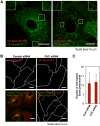
Figure 1
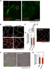
Figure 1. Internalization of DiI-labeled Ebola virions is independent of the clathrin -mediated endocytic pathway.
(A) DiI-labeled Ebola virions (red) do not co-localize with eGFP-labeled CCPs. DiI-EbolaΔVP30 virions (left panel) or DiI-Ebola VLPs (right panel) were adsorbed to Vero cells expressing CLCa-eGFP for 30 min on ice. Cells were then incubated for 15 min at 37°C and the co-localization of DiI-labeled viral particles with eGFP-labeled CCPs was analyzed by using confocal microscope. Insets show enlargements of the boxed areas. Scale bars, 10 µm. (B) Effect of clathrin-heavy chain down-regulation on the internalization of DiI-labeled Ebola virions. Vero cells were transfected with control siRNA (left panels) or CHC siRNA (right panels) to down-regulate CHC expression. The efficiency of CHC down-regulation was analyzed by immunofluorescent staining 48 h post-transfection (red; lower panels); the effect of siRNA on Alexa Fluor 633-Tf is apparent (green; lower panels). Labeled Ebola VLPs were adsorbed to the siRNA-transfected cells for 30 min on ice 48 h post-transfection. After incubation for 2 h at 37°C, surface-bound virions were removed by the addition of trypsin for 5 min at 37°C and the internalization of Ebola VLPs was analyzed by using confocal laser scanning microscope (upper panels). Outlines of individual cells were drawn. Scale bars, 10 µm. (C) Quantitative analysis of the internalization of DiI-labeled Ebola virions in siRNA-transfected Vero cells. The number of DiI-virions in 10 individual siRNA-transfected cells was measured. Each experiment was performed in triplicate and the results are presented as the mean ± SD.
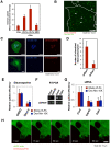
Figure 2
Figure 2. Internalization of DiI-labeled EBOV particles is independent of the caveolae-mediated endocytic pathway.
(A) DiI-labeled EBOV particles do not co-localize with eGFP-labeled caveolae. DiI-EbolaΔVP30 virions (left panel) or DiI-Ebola VLPs (right panel) were adsorbed to Cav1-eGFP-expressing Vero cells for 30 min on ice. The cells were then incubated for 15 min at 37°C and the co-localization of DiI-labeled viral particles with eGFP-labeled caveolae was analyzed by using confocal laser scanning microscope. Insets show enlargements of the boxed areas. Scale bars, 10 µm. (B) Effect of Cav1 down-regulation on the internalization of DiI-labeled Ebola virions. Vero cells were transfected with control siRNA (left panels) or siRNA to down-regulate Cav1 expression (right panels). The efficiency of Cav1 down-regulation was analyzed by use of immunofluorescent staining 48 h post-transfection (lower panels) and western blot analysis (C). Labeled Ebola VLPs were adsorbed to the siRNA-transfected cells for 30 min on ice 48 h post-transfection. After incubation for 2 h at 37°C, surface-bound virions were removed by the addition of trypsin for 5 min at 37°C and the internalization of Ebola VLPs was analyzed by using confocal laser scanning microscope (upper panels). Outlines of individual cells were drawn. Scale bars, 10 µm. (D) Quantitative analysis of the internalization of DiI-labeled Ebola virions in siRNA-transfected Vero cells. The internalized DiI-virions were analyzed in 10 individual siRNA-transfected cells. Each experiment was performed in triplicate and the results are presented as the mean ± SD. (E) Internalization of DiI-labeled Ebola virions in cells lacking Cav1. DiI-labeled EbolaΔVP30 virions were adsorbed to Cav1-deficient Huh7 cells for 30 min on ice. The internalization of DiI-EbolaΔVP30 virions was analyzed 2 h after the temperature shift to 37°C. Outlines of individual cells were drawn. Scale bar, 10 µm. (F) Effect of dynasore on the internalization of DiI-labeled Ebola virions. Vero cells were treated with DMSO (left panel) or dynasore (right panel) for 30 min at 37°C. Labeled Ebola VLPs were adsorbed to the cells for 30 min on ice and incubated for 2 h at 37°C in the presence of DMSO or dynasore. Surface-bound virions were removed by trypsin and the internalization of DiI-virions was analyzed by using confocal laser scanning microscope. Dynasore treatment interfered with the internalization of Alexa Fluor 633-Tf (green in right panel), attesting to its functionality. Scale bars, 10 µm. (G) Quantitative analysis of the internalization of DiI-labeled Ebola virions in dynasore-treated Vero cells. The internalized DiI-virions were analyzed in 10 individual DMSO- or dynasore-treated cells. Each experiment was performed in triplicate and the results are presented as the mean ± SD.
Figure 3
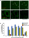
Figure 3. Internalized DiI-EBOV particles co-localize with the macropinosome marker sorting nexin (SNX) 5.
(A) Time-lapse analysis of the co-localization of DiI-labeled viral particles with eGFP-SNX5. DiI-EbolaΔVP30 virions (upper panels) or DiI-influenza virus (lower panels) were adsorbed to eGFP-SXN5-expressing Vero cells for 30 min on ice. The cells were then incubated at 37°C and time-lapse images were acquired at 20-second intervals over a period of 20 min by using confocal laser scanning microscope. Still frames at the indicated times (min) after the temperature shift to 37°C are shown. Virions co-localizing with SNX5 are indicated by arrows. Scale bars, 5 µm. (B) Co-localization efficiency of EBOV particles with SNX5. Shown are the co-localization efficiencies of DiI-EbolaΔVP30 (blue bars), DiI-Ebola VLPs (yellow bars), and DiI-influenza virus (red bars) with eGFP-SXN5 at the indicated time points after the temperature shift to 37°C. The number of DiI-labeled virions co-localized with eGFP-SNX5-positive vesicles was measured in 10 individual cells and the percentage of co-localization in the total DiI-virions is shown for each time point. Each experiment was performed in triplicate and the results are presented as the mean ± SD.
Figure 4
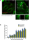
Figure 4. Internalized DiI-labeled EBOV particles are transported to endosomes.
(A) Internalized DiI-labeled Ebola virions are transported to Rab7-positive vesicles. DiI-Ebola virions were adsorbed to eGFP-Rab7-expressing Vero cells for 30 min on ice. The cells were then incubated at 37°C and images were acquired at the indicated time points. Shown are representative images at 0 (left panel) and 120 min (right panel) after the temperature shift. DiI-labeled virions that co-localize with Rab7-positive vesicles are indicated by arrows. Insets show enlargements of the boxed areas. Scale bars, 10 µm. (B) Co-localization efficiency of EBOV particles with Rab7-positive vesicles. The co-localization efficiencies of DiI-EbolaΔVP30 virions (blue bars) and -Ebola VLPs (yellow bars) with Rab7-positive vesicles were analyzed at the indicated time points. The number of DiI-labeled virions co-localized with eGFP-Rab7-positive vesicles was measured in 10 individual cells and the percentage of co-localization in the total DiI-virions is shown for each time point. Each experiment was performed in triplicate and the results are presented as the mean ± SD.
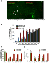
Figure 5
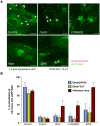
Figure 5. Effect of macropinocytosis inhibitors on the co-localization of DiI-labeled viral particles with Rab7-positive vesicles.
Vero cells expressing eGFP-Rab7 were pretreated with cytochalasin D (CytoD), wortmannin (Wort), LY294002, or EIPA for 30 min at 37°C as described in the Materials and Methods. DiI-EbolaΔVP30 virions, DiI-Ebola VLPs and DiI-influenza virus were adsorbed to the cells for 30 min on ice. The cells were then incubated at 37°C in the presence of inhibitors for 2 h. As a control, DMSO-treated cells were incubated with labeled EBOV particles. Representative images of the co-localization of DiI-EbolaΔVP30 virions with eGFP-Rab7 acquired 2 h after the temperature shift are shown (A). DiI-labeled EbolaΔVP30 virions that co-localize with eGFP-Rab7-positive vesicles are indicated by arrows. Scale bars, 10 µm. (B) shows a graphic representation of the data. The number of DiI-labeled EbolaΔVP30 virions (blue bars), Ebola VLPs (yellow bars) and influenza virions (red bars) co-localized with eGFP-Rab7-positive vesicles was measured in 10 individual cells and the percentage of co-localization in the total DiI-virions is shown for each time point. Each experiment was carried out in triplicate and the results are presented as the mean ± SD.
Figure 6
Figure 6. Macropinocytosis-associated events occur during Ebola virion internalization.
(A) The effect of the internalization of DiI-labeled Ebola VLPs on dextran uptake. Vero cells were incubated with 0.5 mg/ml Alexa Fluor 647-Dex Mw 10K in the absence or presence of Ebola VLPs for 60 min at 37°C. The uptake of Alexa Fluor 647-Dex Mw 10K was analyzed by using flow cytometry. The effect of EIPA pretreatment was assessed in parallel. Each experiment was performed in triplicate and the results are presented as the mean ± SD. (B) Co-localization of internalized DiI-labeled Ebola VLPs and Dex Mw 10K. DiI-Ebola VLPs were adsorbed to Vero cells for 30 min on ice. The cells were cultured in the presence of 0.5 mg/ml Alexa Fluor 647-Dex Mw 10K for 10 min at 37°C. Co-localization of DiI-virions (red) and Alexa Fluor-Dex Mw 10K (green) was analyzed by using confocal laser scanning microscope. Co-localized virions are shown by arrows. Outlines of individual cells were drawn. Scale bar, 10 µm. (C) Effect of a dominant-negative form of Rac1 on the internalization of DiI-labeled Ebola virions. The eGFP-fused, wild-type Rac1 (wtRac1, upper panels) or the dominant-negative form of Rac1 (dnRac1, lower panels) was expressed in Vero cells. DiI-labeled Ebola VLPs were adsorbed to the cells for 30 min on ice. After incubation for 2 h at 37°C, surface-bound virions were removed by trypsin and the internalization of DiI-virions was analyzed by using confocal laser scanning microscope. Expression of dnRac1 interfered with the internalization of Alexa Fluor 647-Dex Mw 10K (blue; lower middle panel), attesting to its functionality. Scale bars, 10 µm. (D) Quantitative analysis of the internalization of DiI-labeled Ebola virions in wtRac1 or dnRac1-expressed Vero cells. The internalized DiI-virions were measured in 10 individual wtRac1 or dnRac1-expressed cells. Each experiment was performed in triplicate and the results are presented as the mean ± SD. (E) Effect of PKC inhibitors on the internalization of DiI-labeled Ebola virions. Vero cells were treated with DMSO or staurosporine (Stauro) for 30 min at 37°C. Labeled Ebola VLPs were adsorbed to the cells for 30 min on ice and incubated for 2 h at 37°C in the absence or presence of inhibitor. Surface-bound virions were removed by trypsin and the internalization of DiI-virions was analyzed by using confocal laser scanning microscope. The internalized DiI-virions were analyzed in 10 individual DMSO- or staurosporine-treated cells (red bars). The efficiency of Alexa Fluor-Dex Mw 10K uptake in inhibitor-treated cells was measured by using flow cytometry (blue bars). Each experiment was performed in triplicate and relative uptake efficiencies are presented as the mean ± SD (red bars). Staurosporine treatment interfered with the internalization of Alexa Fluor 633-Tf (blue bars), attesting to its functionality. (F) The down-regulation of Cdc42 and Pak1 by siRNA. The efficiencies of Cdc42 and Pak1 knock-down were assessed by use of RT-PCR. Total cellular RNA was isolated from siRNA-transfected Vero cells 48 h post-transfection by using the TRI reagent (Sigma-Aldrich) according to the manufacturer's instructions. cDNA synthesis was performed with Molony murine leukemia virus RTase using a random hexamer (Invitrogen) according to the manufacturer's protocol. PCR was carried out for 25–30 cycles consisting of a DNA denaturing step for 30 s at 94°C, annealing for 30 s at 55°C, and extension for 1 min at 72°C by use of Taq DNA polymerase (Promega). Glyceraldehyde-3-phosphate dehydrogenase (G3PDH) was used as an endogenous control. The oligonucleotides used for amplification of individual genes are shown in Table S1. (G) Effect of down-regulation of Cdc42 and Pak1 on the internalization of DiI-labeled Ebola virions. Vero cells were transfected with control (Cont) non-targeting siRNA or siRNA to down-regulate Cdc42 and Pak1 expression. Labeled Ebola VLPs were adsorbed to the siRNA-transfected cells for 30 min on ice, 48 h post-transfection. After incubation for 2 h at 37°C, surface-bound virions were removed by trypsin for 5 min at 37°C and the internalization of Ebola VLPs was analyzed by using confocal laser scanning microscope, and the number of DiI-virions in 10 individual siRNA-transfected cells was measured. The efficiency of Alexa Fluor-Dex Mw 10K uptake in siRNA-transfected cells was measured by use of flow cytometry (blue bars). Each experiment was performed in triplicate and the relative uptake efficiencies are presented as the mean ± SD. (H) The internalization of Ebola virions is associated with plasma membrane ruffling. DiI-EbolaΔVP30 virions were adsorbed to eGFP-actin-expressing Vero cells for 30 min on ice. The cells were then incubated at 37°C and time-lapse images were acquired at 15-second intervals over a period of 10 min by using confocal laser scanning microscope. Still frames at the indicated times (sec) after the temperature shift to 37°C are shown. Scale bar, 10 µm.
Figure 7
Figure 7. Macropinocytotic internalization of Ebola virions is GP-dependent.
(A) Co-localization of SNX5 with VSV pseudotyped with EBOV GP. Labeled VSV particles pseudotyped with EBOV GP (DiI-VSVΔ*G-GP) or VSV G (DiI-VSVΔ*G-G) were adsorbed to eGFP-SNX5-expressing Vero cells for 30 min on ice. The cells were then incubated at 37°C and time-lapse images were acquired at 20-second intervals over a period of 30 min by using confocal laser scanning microscope. Still frames of DiI-VSVΔ*G-GP (left panel) and DiI-VSVΔ*G-G (right panel) at 10 min after the temperature shift are shown. DiI-pseudovirions that co-localize with eGFP-SNX5 are indicated by arrows. Scale bars, 10 µm. (B) Graphic representation of the co-localization of EBOV GP-pseudotyped VSV virions with Rab7-positive vesicles. Co-localization of DiI-VSVΔ*G-GP (green bars) with Rab7-positive vesicles was analyzed at the indicated time points as indicated in the Materials and Methods. Experiments were performed in triplicate and the results are presented as the mean ± standard deviation. Results obtained for DiI-EbolaΔVP30 (blue bars) and DiI-VSVΔ*G-G (red bars) are shown for comparison. (C) Effect of macropinocytosis inhibitors on the co-localization of DiI-labeled VSV pseudovirions with eGFP-Rab7-positive vesicles. Vero cells expressing eGFP-Rab7 were pretreated with CytoD, Wort, LY294002 or EIPA for 30 min at 37°C; control cells were treated with DMSO. DiI-labeled VSVΔ*G-GP (green bars) or VSVΔ*G-G (red bars) were adsorbed to cells for 30 min on ice. The cells were then incubated at 37°C in the presence of inhibitors for 2 h. Co-localization of DiI-pseudovirions with eGFP-Rab7-positive vesicles was analyzed as described in the Materials and Methods. Experiments were carried out in triplicate and the results are presented as the mean ± standard deviation. (D) Effect of macropinocytosis inhibitors on the infectivity of VSV pseudovirions. Vero cells were treated with individual inhibitors for 30 min at 37°C and infected with VSVΔG*-GP (green bars) or VSVΔG*-G (red bars) in the presence of the inhibitor. 1 h post-infection, surface-bound virions were removed by trypsin and the cells were cultured for 24 h in the absence of inhibitors. The infection efficiency of each pseudovirus was determined by measuring the number of GFP-positive cells using with conventional fluorescent microscope. Each experiment was performed in triplicate and the relative infection efficiencies are presented as the mean ± SD.
Figure 8
Figure 8. Model of GP-dependent EBOV cell entry.
For EBOV cell entry, the binding of GP to cellular receptor(s) may activate cellular actin modulators (PI3K, small GTPases, PKC and Pak1), which trigger the actin-dependent membrane ruffling that leads to macropinocytosis. The virions are then internalized via macropinocytosis. Macropinosomes containing the virions are eventually fused to Rab7-positive late endosomes/lysosome (late maturation), resulting in the fusion of the viral envelope with the endosomal membrane in a low pH- and cathepsin-dependent manner.

Similar articles

Cited by

References

    1. Conner SD, Schmid SL. Regulated portals of entry into the cell. Nature. 2003;422:37–44. - PubMed
    1. Sieczkarski SB, Whittaker GR. Dissecting virus entry via endocytosis. J Gen Virol. 2002;83:1535–1545. - PubMed
    1. Marsh M, Helenius A. Virus entry: open sesame. Cell. 2006;124:729–740. - PMC - PubMed
    1. Marsh M, Helenius A. Adsorptive endocytosis of Semliki Forest virus. J Mol Biol. 1980;142:439–454. - PubMed
    1. Matlin KS, Reggio H, Helenius A, Simons K. Infectious entry pathway of influenza virus in a canine kidney cell line. J Cell Biol. 1981;91:601–613. - PMC - PubMed

Publication types

MeSH terms