

Portable filter-based microdevice for detection and characterization of circulating tumor cells
- PMID: 20876796
- PMCID: PMC2955786
- DOI: 10.1158/1078-0432.CCR-10-1105
Portable filter-based microdevice for detection and characterization of circulating tumor cells
Abstract
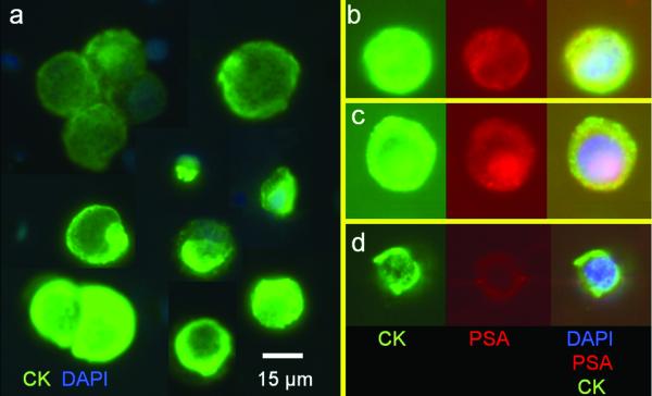
Purpose: Sensitive detection and characterization of circulating tumor cells (CTC) could revolutionize the approach to patients with early-stage and metastatic cancer. The current methodologies have significant limitations, including limited capture efficiency and ability to characterize captured cells. Here, we report the development of a novel parylene membrane filter-based portable microdevice for size-based isolation with high recovery rate and direct on-chip characterization of captured CTC from human peripheral blood.
Experimental design: We evaluated the sensitivity and efficiency of CTC capture in a model system using blood samples from healthy donors spiked with tumor cell lines. Fifty-nine model system samples were tested to determine the recovery rate of the microdevice. Moreover, 10 model system samples and 57 blood samples from cancer patients were subjected to both membrane microfilter device and CellSearch platform enumeration for direct comparison.
Results: Using the model system, the microdevice achieved >90% recovery with probability of 95% recovering at least one cell when five are seeded in 7.5 mL of blood. CTCs were identified in 51 of 57 patients using the microdevice, compared with only 26 patients with the CellSearch method. When CTCs were detected by both methods, greater numbers were recovered by the microfilter device in all but five patients.
Conclusions: This filter-based microdevice is both a capture and analysis platform, capable of multiplexed imaging and genetic analysis. The microdevice presented here has the potential to enable routine CTC analysis in the clinical setting for the effective management of cancer patients.
©2010 AACR.
Figures


PBS only (square);
PBS with 50k LNCaP cells (circle);
PBS with 50k LNCaP cells fixed in 1% acetone (triangle);
50% human blood (triangle);
50% human blood with 25k LNCaP cells (square);
50% blood fixed in 1% NBF (triangle);
50% blood and 25k LNCaP cells fixed in 1% NBF (triangle);
50% blood and 25k LNCaP cells fixed in 2% NBF (hexagon);
50% blood and 25k LNCaP cells fixed in 5% NBF. Dash line part of curve 9 was caused by severe blocking of filter so that the filtration could not be completed (star);
100% human blood (pentagon).

Comment in
-
Reporting the capture efficiency of a filter-based microdevice: a CTC is not a CTC unless it is CD45 negative--letter.Clin Cancer Res. 2011 May 1;17(9):3048-9; author reply 3050. doi: 10.1158/1078-0432.CCR-10-3234. Clin Cancer Res. 2011. PMID: 21536548 No abstract available.
Similar articles
-
A cancer detection platform which measures telomerase activity from live circulating tumor cells captured on a microfilter.Cancer Res. 2010 Aug 15;70(16):6420-6. doi: 10.1158/0008-5472.CAN-10-0686. Epub 2010 Jul 27. Cancer Res. 2010. PMID: 20663903 Free PMC article.
-
Membrane microfilter device for selective capture, electrolysis and genomic analysis of human circulating tumor cells.J Chromatogr A. 2007 Aug 31;1162(2):154-61. doi: 10.1016/j.chroma.2007.05.064. Epub 2007 May 29. J Chromatogr A. 2007. PMID: 17561026
-
A microfluidic chip integrated with a high-density PDMS-based microfiltration membrane for rapid isolation and detection of circulating tumor cells.Biosens Bioelectron. 2015 Sep 15;71:380-386. doi: 10.1016/j.bios.2015.04.080. Epub 2015 Apr 24. Biosens Bioelectron. 2015. PMID: 25950932
-
Microdevice in Cellular Pathology: Microfluidic Platforms for Fluorescence in situ Hybridization and Analysis of Circulating Tumor Cells.Anal Sci. 2015;31(9):867-73. doi: 10.2116/analsci.31.867. Anal Sci. 2015. PMID: 26353951 Review.
-
Circulating tumor cells in breast cancer: detection systems, molecular characterization, and future challenges.Clin Chem. 2011 Sep;57(9):1242-55. doi: 10.1373/clinchem.2011.165068. Epub 2011 Jul 22. Clin Chem. 2011. PMID: 21784769 Review.
Cited by
-
Incorporation of lateral microfiltration with immunoaffinity for enhancing the capture efficiency of rare cells.Sci Rep. 2020 Aug 26;10(1):14210. doi: 10.1038/s41598-020-71041-7. Sci Rep. 2020. PMID: 32848184 Free PMC article.
-
Combining electrochemical sensors with miniaturized sample preparation for rapid detection in clinical samples.Sensors (Basel). 2014 Dec 30;15(1):547-64. doi: 10.3390/s150100547. Sensors (Basel). 2014. PMID: 25558994 Free PMC article. Review.
-
Benchtop technologies for circulating tumor cells separation based on biophysical properties.Biomed Res Int. 2015;2015:239362. doi: 10.1155/2015/239362. Epub 2015 Apr 21. Biomed Res Int. 2015. PMID: 25977918 Free PMC article. Review.
-
Cytometric comparisons between circulating tumor cells from prostate cancer patients and the prostate-tumor-derived LNCaP cell line.Phys Biol. 2012 Feb;9(1):016002. doi: 10.1088/1478-3975/9/1/016002. Epub 2012 Feb 3. Phys Biol. 2012. PMID: 22306736 Free PMC article.
-
Successful chemoimmunotherapy against hepatocellular cancer in a novel murine model.J Hepatol. 2017 Jan;66(1):75-85. doi: 10.1016/j.jhep.2016.07.044. Epub 2016 Aug 9. J Hepatol. 2017. PMID: 27520877 Free PMC article.
References
-
- Lugo TG, Braun S, Cote RJ, Pantel K, Rusch V. Detection and measurement of occult disease for the prognosis of solid tumors. J Clin Oncol. 2003;21:2609–15. - PubMed
-
- Schabel FM., Jr Rationale for adjuvant chemotherapy. Cancer. 1977;39:2875–82. - PubMed
-
- Pantel K, Alix-Panabieres C, Riethdorf S. Cancer micrometastases. Nat Rev Clin Oncol. 2009 - PubMed
-
- Cristofanilli M, Budd GT, Ellis MJ, et al. Circulating tumor cells, disease progression, and survival in metastatic breast cancer. N Engl J Med. 2004;351:781–91. - PubMed
Publication types
MeSH terms
Substances
Grants and funding
LinkOut - more resources
Full Text Sources
Other Literature Sources
