. 2010 Oct 25;207(11):2331-41.
doi: 10.1084/jem.20101074. Epub 2010 Sep 27.

Helminth secretions induce de novo T cell Foxp3 expression and regulatory function through the TGF-β pathway

Affiliations

Helminth secretions induce de novo T cell Foxp3 expression and regulatory function through the TGF-β pathway

John R Grainger et al. J Exp Med. .

Abstract

Foxp3-expressing regulatory T (T reg) cells have been implicated in parasite-driven inhibition of host immunity during chronic infection. We addressed whether parasites can directly induce T reg cells. Foxp3 expression was stimulated in naive Foxp3⁻ T cells in mice infected with the intestinal helminth Heligmosomoides polygyrus. In vitro, parasite-secreted proteins (termed H. polygyrus excretory-secretory antigen [HES]) induced de novo Foxp3 expression in fluorescence-sorted Foxp3⁻ splenocytes from Foxp3-green fluorescent protein reporter mice. HES-induced T reg cells suppressed both in vitro effector cell proliferation and in vivo allergic airway inflammation. HES ligated the transforming growth factor (TGF) β receptor and promoted Smad2/3 phosphorylation. Foxp3 induction by HES was lost in dominant-negative TGF-βRII cells and was abolished by the TGF-β signaling inhibitor SB431542. This inhibitor also reduced worm burdens in H. polygyrus-infected mice. HES induced IL-17 in the presence of IL-6 but did not promote Th1 or Th2 development under any conditions. Importantly, antibody to mammalian TGF-β did not recognize HES, whereas antisera that inhibited HES did not affect TGF-β. Foxp3 was also induced by secreted products of Teladorsagia circumcincta, a related nematode which is widespread in ruminant animals. We have therefore identified a novel pathway through which helminth parasites may stimulate T reg cells, which is likely to be a key part of the parasite's immunological relationship with the host.


Figures
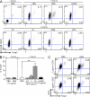
Figure 1.
Figure 1.
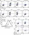
HES increases the percentage of CD4+Foxp3+ T cells in mitogen-stimulated splenocyte cultures. (A) Representative plots of CD25 versus Foxp3 expression, gated on CD4+ T cells, from C57BL/6 splenocytes cultured in the presence of PBS alone, 2 µg/ml Con A, or combinations of Con A with pathogen products. Con A was added to cultures 30 min after pathogen products, and flow cytometry was performed 48 h later. Top row, PBS alone, Con A, Con A plus 10 µg/ml HES, and Con A plus 10 µg/ml of heat-inactivated (hi) HES. Heat inactivation was performed for 30 min at 100°C. Bottom row, Con A plus 10 µg/ml Propionibacterium acnes extract (Pa), 10 µg/ml Salmonella typhimurium extract (St), 1 µg/ml Pam-3-CSK4, or 1 µg/ml LPS. (B) Percentage of Foxp3+ cells within the CD4+ T cell population of splenocytes exposed to the indicated stimuli. Data represent mean ± SD from three replicate cultures with cells from individual C57BL/6 mice. Results of Student’s t test: **, P < 0.005. (C) Foxp3 induction in splenocytes, from naive mice, exposed to PBS or HES in combination with low (1 µg/ml) or high (10 µg/ml) doses of α-CD3 antibody. Data are representative of at least three experiments performed using different batches of HES.
Figure 2.
Figure 2.
HES shows TGF-β–like activity and directly induces Foxp3 expression in CD4+ T cells. (A) Foxp3 and GATA3 expression in FACS-sorted naive Foxp3-eGFP–negative CD4+ T cells cultured for 3 d with splenic DC in the presence of α-CD3 alone and in combination with HES, rhTGF-β1, or Th2-polarizing cytokines. (B) Foxp3 and T-Bet expression in FACS-sorted naive Foxp3-eGFP–negative CD4+ T cells, cultured as in A with HES, rhTGF-β1, or Th1-polarizing cytokines. (C) Proliferation measured by CFSE labeling of FACS-sorted naive Foxp3-eGFP–negative CD4+ T cells after 3 d of culture with splenic DC in the presence of α-CD3 alone and in combination with HES or rhTGF-β1. Error bars represent SEM. (D) Intracellular IL-17A and Foxp3 expression in FACS-sorted naive Foxp3-eGFP–negative CD4+ T cells cultured for 4 d with splenic DC in the presence of α-CD3, with or without IL-6 and in combination with HES or PBS control. Data are representative of at least three similar experiments performed using different batches of HES.
Figure 3.
Figure 3.
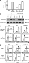
HES activates TGF-β–responsive cells, induces SMAD-2/3 phosphorylation, and induces Foxp3 expression through the TGF-β signaling pathway. (A) HES activates the TGF-β–responsive reporter cell line MFB-F11, inducing expression of alkaline phosphatase activity. Error bars represent SEM of triplicate wells of MFB-F11 cells stimulated with each treatment. (B) Phosphorylation of SMAD-2/3 signaling molecules, activated by TGF-β ligation, was measured in C57BL/6 CD4+ cells stimulated in the absence (−) or presence of plate-bound anti-CD3/anti-CD28, together with PBS, 5 ng/ml rhTGF-β1, or 10 μg/ml HES. Cell lysates were probed by Western blotting with phospho-SMAD-2/3–specific antibody (top) and then stripped and reprobed with antibody to SMAD-2/3 peptide backbone. The figure represents a single determination, with internal replication (with and without anti-CD3/CD28). (C) CD4+ cells were purified from WT C57BL/6 or dominant-negative TGF-βRII mice and stimulated with plate-bound anti-CD3/anti-CD28 in the presence of IL-2, with either 10 µg/ml HES or 5 ng/ml rhTGF-β1. After 72 h, Foxp3 expression was analyzed by flow cytometry. (D) WT CD4+ cells were cultured as in C but were treated with 5 µM of the ALK-5/TGF-βRI inhibitor SB431542 (or DMSO as a control) alongside HES or rhTGF-β1. After 72 h, Foxp3 expression was analyzed by flow cytometry. Results shown in A, C, and D, are representative of at least three similar experiments performed with different batches of HES.
Figure 4.
Figure 4.
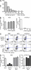
HES and mammalian TGF-β are not recognized or cross-inhibited by specific antibodies to each other. (A) ELISA assay sensitive to 15 pg/ml rhTGF-β1 fails to react with native HES or acid-activated (AA) HES. (B) HES activation of MFB-F11 cells, calibrated by a standard curve of rhTGF-β, is not diminished even when preincubated with 100 µg/ml of the pan–TGF-β antibody 1D11 (left). In contrast, rhTGF-β1 activity is dramatically inhibited by as little as 1 µg/ml 1D11 (right). Data are representative of three similar experiments performed using different batches of HES. (C) Induction of Foxp3 expression in CD4+ T cells by rhTGF-β1, but not by HES, is ablated by preincubation with 30 µg/ml 1D11. Data are representative of three similar experiments performed using different batches of HES. (D) Day-28 sera from H. polygyrus (Hp)–infected mice, but not naive mice, blocks the ability of HES to activate MFB-F11 cells (left). In contrast, rhTGF-β1 activity was not affected by infection sera (right). HES or rhTGF-β1 were preincubated for 60 min with the indicated antibody before testing on MFB-F11 cells. Data are representative of three similar experiments. Error bars in A, C, and D represent SEM of triplicate wells of MFB-F11 cells stimulated with each treatment.
Figure 5.
Figure 5.
Blocking of TGF-β signaling reduces worm burden, and ES from a related nematode parasite has TGF-β–like and Foxp3-inducing activity. (A) WT C57BL/6 mice were infected with 200 H. polygyrus L3 cells by gavage. On days 35, 37, and 39, mice were intraperitoneally injected with 200 µl of a 1:1 DMSO/PBS solution containing 2 mg/ml SB431542 or 200 µl of DMSO/PBS alone. Mice were sacrificed on day 40 of infection and adult worms in the lumen of the gut counted. Data are combined from two identical experiments, each with four to five mice per group. Data points show individual mice and bars represent mean values. Results of Student’s t test: *, P < 0.05. (B) ES from 10 µg/ml T. circumcincta fourth-stage larvae (TcL4ES) is able to activate the MFB-F11 TGF-β reporter assay, and this activity is unaffected by high levels (100 µg/ml) of α–TGF-β (1D11) antibody. The equivalent ES from 10 µg/ml H. contortus (HcL4ES) had no detectable TGF-β activity. Error bars represent SEM. (C) 30 µg/ml TcL4ES induces significant Foxp3 expression after 72 h in purified CD4+ cultures stimulated with plate-bound anti-CD3/anti-CD28 and exogenous IL-2. In contrast, 30 µg/ml HcL4ES has no significant effect. Results presented in B and C are representative of three similar experiments.
Figure 6.
Figure 6.
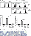
HES-induced Foxp3+ T reg cell are functionally suppressive in vitro and in vivo in a Th2 environment. (A) The response of CD4+Foxp3 CFSE-labeled index responder T cells stimulated with α-CD3 for 4 d was measured by flow cytometry with and without additional unlabeled suppressor cells induced in vitro. Left, CFSE-labeled CD4+Foxp3 index responder T cells alone. Right, CFSE-labeled responder cells mixed with graded numbers of induced Foxp3+ T cells generated in vitro by co-culture of FACS-sorted CD4+Foxp3 T cells with HES or rhTGF-β1. Data are representative of three similar experiments performed using different batches of HES. (B) C57BL/6 mice were sensitized to OVA by two intraperitoneal (i.p.) injections with 10 µg OVA adsorbed to Alum 10 d apart. On days 16 and 19, 106 HES or rhTGF-β1 iT reg cells (generated as in A) were transfered intravenously in 200 µl PBS. Control mice were injected with 200 µl PBS alone. 1 d after each iT reg cell transfer, mice were given soluble OVA by direct tracheal inoculation. Lavage cells were recovered 24 h after the final airway challenge, and total cellularity (left) and eosinophilia (right) were enumerated. Data points represent results from individual mice and are representative of two identical experiments., and bars represent mean values. (C) Histological sectioning of lung tissues from allergen-sensitized mice receiving HES or rhTGF-β1 iT reg cells, as described in B. Bars, 100 µm.
Figure 7.
Figure 7.
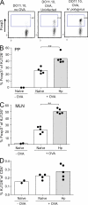
H. polygyrus infection amplifies de novo Foxp3 expression among OVA-specific DO11.10 T cells. Male BALB/c mice were seeded on day 1 of infection with 106 eGFP-negative CD4+ T cells from (Foxp3-eGFP.BALB/c × DO11.10) F1 male mice and given 1.5% OVA in drinking water from day 2. MLN and PP were harvested at day 7. (A) Representative flow cytometry plots of MLN DO11.10 cells identified with clonotypic antibody KJ126 in control mice, those receiving cells alone, and those given both cells and oral OVA in the absence or presence of infection. (B) Percentage of KJ126+CD4+ T cells expressing Foxp3 in the MLN. **, P < 0.01. (C) Percentage of KJ126+CD4+ T cells expressing Foxp3 in the PP. **, P < 0.01. (D) Percentage of CD4+ T cells expressing KJ126 in the MLNs of cell recipients given cells alone, or oral OVA with or without infection. Results shown are representative of two similar experiments, with control groups of two mice and OVA-treated groups of five mice in each case. Data points represent individual mice and bars represent mean values.

Similar articles

Cited by

References

    1. Akdis M., Verhagen J., Taylor A., Karamloo F., Karagiannidis C., Crameri R., Thunberg S., Deniz G., Valenta R., Fiebig H., et al. 2004. Immune responses in healthy and allergic individuals are characterized by a fine balance between allergen-specific T regulatory 1 and T helper 2 cells. J. Exp. Med. 199:1567–1575 10.1084/jem.20032058 - DOI - PMC - PubMed
    1. Anthony R.M., Urban J.F., Jr., Alem F., Hamed H.A., Rozo C.T., Boucher J.L., Van Rooijen N., Gause W.C. 2006. Memory T(H)2 cells induce alternatively activated macrophages to mediate protection against nematode parasites. Nat. Med. 12:955–960 10.1038/nm1451 - DOI - PMC - PubMed
    1. Attisano L., Wrana J.L. 2002. Signal transduction by the TGF-β superfamily. Science. 296:1646–1647 10.1126/science.1071809 - DOI - PubMed
    1. Babu S., Blauvelt C.P., Kumaraswami V., Nutman T.B. 2006. Regulatory networks induced by live parasites impair both Th1 and Th2 pathways in patent lymphatic filariasis: implications for parasite persistence. J. Immunol. 176:3248–3256 - PubMed
    1. Baumgart M., Tompkins F., Leng J., Hesse M. 2006. Naturally occurring CD4+Foxp3+ regulatory T cells are an essential, IL-10-independent part of the immunoregulatory network in Schistosoma mansoni egg-induced inflammation. J. Immunol. 176:5374–5387 - PubMed

Publication types

MeSH terms

Substances