

The nonstructural proteins of Nipah virus play a key role in pathogenicity in experimentally infected animals
- PMID: 20856799
- PMCID: PMC2939873
- DOI: 10.1371/journal.pone.0012709
The nonstructural proteins of Nipah virus play a key role in pathogenicity in experimentally infected animals
Abstract
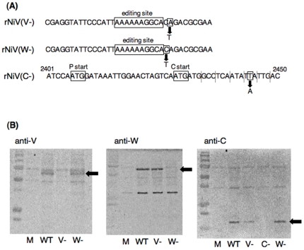
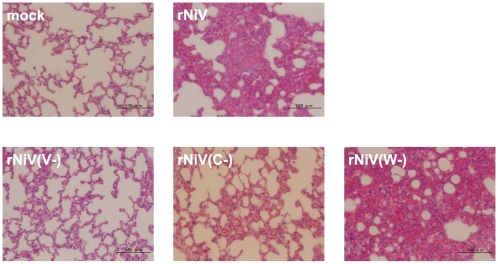
Nipah virus (NiV) P gene encodes P protein and three accessory proteins (V, C and W). It has been reported that all four P gene products have IFN antagonist activity when the proteins were transiently expressed. However, the role of those accessory proteins in natural infection with NiV remains unknown. We generated recombinant NiVs lacking V, C or W protein, rNiV(V-), rNiV(C-), and rNiV(W-), respectively, to analyze the functions of these proteins in infected cells and the implications in in vivo pathogenicity. All the recombinants grew well in cell culture, although the maximum titers of rNiV(V-) and rNiV(C-) were lower than the other recombinants. The rNiV(V-), rNiV(C-) and rNiV(W-) suppressed the IFN response as well as the parental rNiV, thereby indicating that the lack of each accessory protein does not significantly affect the inhibition of IFN signaling in infected cells. In experimentally infected golden hamsters, rNiV(V-) and rNiV(C-) but not the rNiV(W-) virus showed a significant reduction in virulence. These results suggest that V and C proteins play key roles in NiV pathogenicity, and the roles are independent of their IFN-antagonist activity. This is the first report that identifies the molecular determinants of NiV in pathogenicity in vivo.
Conflict of interest statement
Figures

Similar articles
-
Nipah Virus C and W Proteins Contribute to Respiratory Disease in Ferrets.J Virol. 2016 Jun 24;90(14):6326-6343. doi: 10.1128/JVI.00215-16. Print 2016 Jul 15. J Virol. 2016. PMID: 27147733 Free PMC article.
-
Establishment of a Nipah virus rescue system.Proc Natl Acad Sci U S A. 2006 Oct 31;103(44):16508-13. doi: 10.1073/pnas.0606972103. Epub 2006 Oct 19. Proc Natl Acad Sci U S A. 2006. PMID: 17053073 Free PMC article.
-
Distinct and overlapping roles of Nipah virus P gene products in modulating the human endothelial cell antiviral response.PLoS One. 2012;7(10):e47790. doi: 10.1371/journal.pone.0047790. Epub 2012 Oct 19. PLoS One. 2012. PMID: 23094089 Free PMC article.
-
[Study of pathogenicity of Nipah virus and its vaccine development].Uirusu. 2014;64(1):105-12. doi: 10.2222/jsv.64.105. Uirusu. 2014. PMID: 25765986 Review. Japanese.
-
Hendra and Nipah viruses: why are they so deadly?Curr Opin Virol. 2012 Jun;2(3):242-7. doi: 10.1016/j.coviro.2012.03.006. Epub 2012 Apr 5. Curr Opin Virol. 2012. PMID: 22483665 Review.
Cited by
-
Structural basis for importin alpha 3 specificity of W proteins in Hendra and Nipah viruses.Nat Commun. 2018 Sep 12;9(1):3703. doi: 10.1038/s41467-018-05928-5. Nat Commun. 2018. PMID: 30209309 Free PMC article.
-
Nipah Virus Disease: Epidemiological, Clinical, Diagnostic and Legislative Aspects of This Unpredictable Emerging Zoonosis.Animals (Basel). 2022 Dec 31;13(1):159. doi: 10.3390/ani13010159. Animals (Basel). 2022. PMID: 36611767 Free PMC article. Review.
-
Birth and pathogenesis of rogue respiratory viruses.Annu Rev Pathol. 2015;10:449-71. doi: 10.1146/annurev-pathol-012414-040501. Epub 2014 Nov 24. Annu Rev Pathol. 2015. PMID: 25423349 Free PMC article. Review.
-
Animal models of disease shed light on Nipah virus pathogenesis and transmission.J Pathol. 2015 Jan;235(2):196-205. doi: 10.1002/path.4444. J Pathol. 2015. PMID: 25229234 Free PMC article. Review.
-
In silico designing of an epitope-based peptide vaccine cocktail against Nipah virus: an Indian population-based epidemiological study.Arch Microbiol. 2023 Nov 13;205(12):380. doi: 10.1007/s00203-023-03717-3. Arch Microbiol. 2023. PMID: 37955744
References
Publication types
MeSH terms
Substances
LinkOut - more resources
Full Text Sources
