. 2010 Oct;17(10):1210-7.
doi: 10.1038/nsmb.1901. Epub 2010 Sep 19.

Escherichia coli RecBC helicase has two translocase activities controlled by a single ATPase motor

Affiliations

Escherichia coli RecBC helicase has two translocase activities controlled by a single ATPase motor

Colin G Wu et al. Nat Struct Mol Biol. 2010 Oct.

Abstract

E. coli RecBCD is a DNA helicase with two ATPase motors (RecB, a 3'→5' translocase, and RecD, a 5'→3' translocase) that function in repair of double-stranded DNA breaks. The RecBC heterodimer, with only the RecB motor, remains a processive helicase. Here we examined RecBC translocation along single-stranded DNA (ssDNA). Notably, we found RecBC to have two translocase activities: the primary translocase moves 3'→5', whereas the secondary translocase moves RecBC along the opposite strand of a forked DNA at a similar rate. The secondary translocase is insensitive to the ssDNA backbone polarity, and we propose that it may fuel RecBCD translocation along double-stranded DNA ahead of the unwinding fork and ensure that the unwound single strands move through RecBCD at the same rate after interaction with a crossover hot-spot indicator (Chi) sequence.


Figures

Figure 1
Figure 1. RecBCD and RecBC structures
RecB (orange), RecC (blue), and RecD (green) subunits are indicated. (a). Ribbon diagram of a RecBCD–DNA complex ,. (b). Cartoon depiction of a RecBC–DNA complex. RecB motor, nuclease, and arm domains are indicated along with the catalytically dead RecC motor and nuclease domains. The paths of the 3′- and 5′-terminated unwound ssDNA are shown.
Figure 2
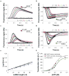
Figure 2. RecB translocates with 3′ to 5′ directionality along ssDNA
(a) RecB monomer translocation kinetics for a series of 5′–Cy3–(dT)L DNA (Supplementary Table 1, DNA I–VIII). Cy3 fluorescence from DNA alone (DNA I) is shown in filled black circles. (b). RecB monomer translocation kinetics for a series of 5′–OG–(dT)L substrates. OG (Oregon Green) fluorescence from DNA alone (DNA I) is shown in filled black circles. Smooth black curves in panels A and B are simulated time courses using Equation S1 and the best fit kinetic parameters (Table 1). (c). Time course obtained with RecB and (dT)54 –Cy3–3′ (Supplementary Table 1, DNA IX). (d). Time course obtained with RecBC and 5′–Cy3–(dT)54 (filled circles) or (dT)54–Cy3–3′ (opened circles).
Figure 3
Figure 3. RecBC displays both a primary (3′ to 5′) and secondary (5′ to 3′) translocase activity
RecBC translocation time courses obtained using the DNA substrates depicted which possesses a 24 bp duplex with a high affinity (twin-dT6) RecBC loading site on one end and either 5′– or 3′–(dT)L ssDNA extensions on the other end. (a). DNA substrates labeled with Cy3 on the 5′ end of the (dT)L extension. (b). DNA substrates labeled with fluorescein (F) on the 5′ end of the (dT)L extension. (c). DNA substrates labeled with Cy3 on the 3′ end of the (dT)L extension. (d). DNA substrates labeled with fluorescein (F) on the 3′ end of the (dT)L extension. Smooth black curves in panels a d are simulated time courses using Equation S2 and the kinetic parameters in Table 1. (e). Dependence of the lag time on ssDNA extension length, L. Cy3 data from panel a (opened circles) (Lag time = 0.00110 L + 0.0095) (909 ± 51 nt s−1). Fluorescein data from panel b (filled circles) (Lag time = 0.000971 L + 0.0110) (1,030 ± 53 nt s−1). Cy3 data from panel c (opened squares) (Lag time = 0.00101 L + 0.0093) (990 ± 49 nt s−1). Fluorescein data from panel d (filled squares) (Lag time = 0.000843 L + 0.0134) (1,187 ± 61 nt s−1). (f). [ATP] dependence of RecBC translocation rates (from lag time analyses) for the (circles)-primary (3′ to 5′) (Vmax = 946 ± 64 nt s−1, KM = 203 ± 32 μM); and (squares)-secondary (5′ to 3′) (Vmax = 1,055 ± 75 nt s−1, KM = 123 ± 28 μM) translocases. (triangles)-Effects of [ATP] on RecB monomer translocation (Vmax = 860 ± 53 nt s−1, KM = 125 ± 38 μM). Smooth curves represent fits to the Michaelis-Menton equation (Eq. S4) and the best fit parameters summarized in Table 1.
Figure 4
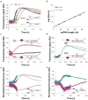
Figure 4. The primary RecBC translocase site remains bound to ssDNA upon reaching a 5′-end, while its secondary translocase continues
(a) RecBC translocation along a partial duplex substrate with a 5′ to 3′ dT60 ssDNA extension, doubly labeled with Cy3 and Cy5 as shown. Time course monitoring FRET as Cy5 fluorescence (filled circles) (due to exciting Cy3 donor) shows that the 3′-terminated ssDNA remains bound to RecBC while the secondary translocase continues; × Translocation time course for the same DNA, but without the Cy5 label (opened squares). (b). RecBC translocation along a partial duplex substrate with a 3′ to 5′ dT60 ssDNA extension doubly labeled with Cy3 and Cy5 as shown. Time course monitoring FRET as Cy5 fluorescence (filled circles) (due to exciting Cy3 donor) shows dissociation of the 5′-terminated ssDNA; Translocation time course for the same DNA, but without the Cy5 label (opened squares).
Figure 5
Figure 5. primary and secondary RecBC translocases operate simultaneously along two ssDNA extensions
Translocation of RecBC along DNA substrates containing a 24 bp duplex region with high affinity RecBC loading site on one end and two (dT)L extensions of equal length (L = 15, 30, 45, 50, 75 nucleotides) and labeled with Cy3 on one of the two ends as depicted. (a). Normalized time courses for DNA substrates 5′–Cy3 labeled DNA (filled circles) and 3′–Cy3 labeled DNA (opened circles) for L = 30, 45, and 75 nucleotides. (b). Lag time analyses of time courses; 5′–Cy3 labeled DNA (filled circles) (Lag time = 0.00149 L + 0.0098) (671 ± 47 nt s−1). 3′–Cy3 labeled DNA (opened circles) (Lag time = 0.00161 L + 0.0016) (621 ± 43 nt s−1). (c). Backbone polarity of the ssDNA extension along which the primary (3′ to 5′) translocase operates is reversed using a 5′–5′ linkage at the position indicated (red X). DNA I - top strand end-labeled with Cy3; DNA II - bottom strand end-labeled with Cy3. (d). Backbone polarity of the ssDNA extension along which the secondary translocase operates is reversed using a 5′–5′ linkage at the position indicated (red X); DNA III - top strand end-labeled with Cy3; DNA IV - bottom strand end-labeled with Cy3. (e). FRET experiment monitoring Cy3 (purple circles) and Cy5 fluorescence (blue circles) performed using the DNA substrate double labeled with Cy3 and Cy5 as depicted. Red and black squares show the time courses for a DNA possessing both ssDNA extensions, but containing only a Cy3 fluorophore (as in panel (b), with L = 60)). (f). FRET experiment monitoring Cy3 (purple squares) and Cy5 fluorescence (blue squares) performed as in panel (e), but with a DNA substrate in which the backbone polarity of the top ssDNA extension was reversed using a 3′–3′ linkage at the position indicated (red X).
Figure 6
Figure 6. RecBC re-initiation of DNA unwinding after a ssDNA gap
DNA substrates contain a proximal 24 bp duplex with a RecBC loading site (twin dT6 fork), a ssDNA gap of length, L = 2, 21 or 41 nucleotides, followed by a distal 40 bp duplex DNA. The ssDNA connecting the proximal and distal duplexes runs either 3′ to 5 (panel a) or 5′ to 3′ (panel b) relative to the RecBC loading site. (a). Single round time course for RecBC unwinding the proximal 24 bp duplex (diamonds) and the distal 40 bp duplex connected by a 3′ to 5′ ssDNA with L = 2 nt (circles), 21 nt (squares), or 41 nt (triangles). Smooth curves indicate fits to Scheme 3 (Eq. S3) using the parameters (mtkt = 928 ± 38 nt s−1; mUkU = 396 ± 15 bp s−1 (constrained)). (b). Single round time course for RecBC unwinding the proximal 24 bp duplex (diamonds) and the distal 40 bp duplex connected by a 5′ to 3′ ssDNA with L = 2 nt (circles), 21 nt (squares), or 41 nt (triangles). Smooth curves indicate fits to Scheme 3 (Eq. S3) using the parameters (mtkt = 919 ± 42 nt s−1; mUkU = 396 ± 15 bp s−1 (constrained)). Models for re-initiation of DNA unwinding by RecBC after traversing a ssDNA gap in either strand are shown below each panel.

Comment in

Similar articles

Cited by

References

    1. Dillingham MS, Kowalczykowski SC. RecBCD enzyme and the repair of double-stranded DNA breaks. Microbiol Mol Biol Rev. 2008;72:642–671. - PMC - PubMed
    1. Singleton MR, Dillingham MS, Wigley DB. Structure and Mechanism of Helicases and Nucleic Acid Translocases. Annu Rev Biochem. 2007;76:23–50. - PubMed
    1. Dillingham MS, Spies M, Kowalczykowski SC. RecBCD enzyme is a bipolar DNA helicase. Nature. 2003;423:893–897. - PubMed
    1. Taylor AF, Smith GR. RecBCD enzyme is a DNA helicase with fast and slow motors of opposite polarity. Nature. 2003;423:889–893. - PubMed
    1. Rigden DJ. An inactivated nuclease-like domain in RecC with novel function: implications for evolution. BMC Struct Biol. 2005;5:9. - PMC - PubMed

Publication types

MeSH terms

LinkOut - more resources