. 2010 Dec 2;29(48):6343-56.
doi: 10.1038/onc.2010.366. Epub 2010 Sep 13.

Suppression of Her2/neu expression through ILK inhibition is regulated by a pathway involving TWIST and YB-1

Affiliations
Free PMC article

Suppression of Her2/neu expression through ILK inhibition is regulated by a pathway involving TWIST and YB-1

J Kalra et al. Oncogene. .
Free PMC article

Abstract

In a previous study it was found that the therapeutic effects of QLT0267, a small molecule inhibitor of integrin-linked kinase (ILK), were influenced by Her2/neu expression. To understand how inhibition or silencing of ILK influences Her2/neu expression, Her2/neu signaling was evaluated in six Her2/neu-positive breast cancer cell lines (LCC6(Her2), MCF7(Her2), SKBR3, BT474, JIMT-1 and KPL-4). Treatment with QLT0267 engendered suppression (32-87%) of total Her2/neu protein in these cells. Suppression of Her2/neu was also observed following small interfering RNA-mediated silencing of ILK expression. Time course studies suggest that ILK inhibition or silencing caused transient decreases in P-AKT(ser473), which were not temporally related to Her2/neu downregulation. Attenuation of ILK activity or expression was, however, associated with decreases in YB-1 (Y-box binding protein-1) protein and transcript levels. YB-1 is a known transcriptional regulator of Her2/neu expression, and in this study it is demonstrated that inhibition of ILK activity using QLT0267 decreased YB-1 promoter activity by 50.6%. ILK inhibition was associated with changes in YB-1 localization, as reflected by localization of cytoplasmic YB-1 into stress granules. ILK inhibition also suppressed TWIST (a regulator of YB-1 expression) protein expression. To confirm the role of ILK on YB-1 and TWIST, cells were engineered to overexpress ILK. This was associated with a fourfold increase in the level of YB-1 in the nucleus, and a 2- and 1.5-fold increase in TWIST and Her2/neu protein levels, respectively. Taken together, these data indicate that ILK regulates the expression of Her2/neu through TWIST and YB-1, lending support to the use of ILK inhibitors in the treatment of aggressive Her2/neu-positive tumors.


Figures
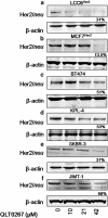
Figure 1
Figure 1
Her2/neu expression following treatment of various breast cancer cell lines with QLT0267. Expression of total Her2/neu in (a) LCC6Her2, (b) MCF7Her2, (c) BT474, (d) KPL4, (e) SKBR3 and (f) JIMT-1 cells treated with QLT0267 was determined using western blot analysis. Cells were treated for 24 h with 10, 21 or 42 μ QLT0267. Subsequently, cells were lysed, proteins were isolated and 50 μg whole-cell lysates were separated on 10% SDS–PAGE gels as described in the Materials and methods. Membranes were probed for Her2/neu and β-actin. In all six cell lines, increasing concentrations of QLT0267 inhibited the expression of total Her2/neu. At 42 μ, total Her2/neu is decreased by 69, 86.5, 49, 47, 63, and 32% (n=3) in LCC6Her2, MCF7Her2, BT474, KPL4, SKBR3 and JIMT-1 cells, respectively.
Figure 2
Figure 2
(a) Pathway analysis of SKBR3 cells transiently nucleofected with 2 μg of ILK siRNA using the Amaxa Nucleofector. Whole-cell lysates (50 μg) harvested from cells at 24, 48, 72 and 96 h post transfection were separated on 10% SDS–PAGE gels. Resulting western blots were probed for ILK, Her2/neu, AKT, PAKTser473 and β-actin to verify loading. ILK expression was decreased by 49, 66, 66 and 79% at 24, 48, 72 and 96 h, respectively. PAKTser473 was suppressed by 79% at 24 h where ILK silencing was at 49%. At 48 h of treatment with ILK siRNA, SKBR3 cells exhibit a 66% suppression of ILK. At this and later time points, PAKTser473 expression is similar to control cells. Total Her2/neu expression was reduced by 71% at 96 h of treatment with ILK siRNA when compared with the Neg siRNA (n=3). (b) SKBR3 cells were treated with 42 μ QLT0267 for 6, 18 or 24 h. Subsequently, cells were lysed, 50 μg of protein was isolated and then separated on 10% SDS–PAGE gels. Resulting western blots were probed for Her2/neu, PAKTser473 and β-actin to verify loading. Treatment with QLT0267 suppressed PAKTser473 in all cell lines at a time point earlier than that observed to suppress Her2/neu. PAKTser473 was decreased at 6 h, whereas Her2/neu levels decreased substantially at 24 h, where PAKTser473 begins to increase. (c) SKBR3 cells were treated with 42 μ QLT0267 for 24 h or transfected with ILK siRNA. Subsequently, RNA was isolated from cells and reverse transcribed. Her2/neu was amplified from complementary DNA (cDNA) using quantitative reverse transcriptase–PCR (RT–qPCR) and PCR. A 9.8- and 2.5-fold decrease of Her2/neu transcript was observed when cells were treated using QLT0267 or ILK siRNA.
Figure 3
Figure 3
Inhibition of ILK activity or expression influences YB-1 transcription and subcellular localization. (a) SKBR3 cells were transiently nucleofected with 4 μg ILK siRNA. Subsequently, cells were lysed and 50 μg of protein was isolated from samples at 24 and 48 h, separated on a 10% SDS–PAGE gel and probed for ILK, Her2/neu, YB-1 and β-actin to verify loading. ILK expression was substantially silenced when SKBR3 cells were treated with 4 μg of ILK siRNA for both 24 and 48 h. Cells exhibit a 96% decrease in total Her2/neu expression after 48 h, at which time YB-1 expression is reduced by 74%. (b) YB-1 transcript levels were analyzed in SKBR3 cells treated with QLT0267 or nucleofected with 4 μg ILK siRNA for 48 h using PCR. A 9.9-fold and 6.5-fold decrease in YB-1 transcript was observed in QLT0267-treated and ILK-silenced cells, respectively, when compared with control. (c) SKBR3 cells were transfected with a YB-1 promoter/luciferase construct and treated with QLT0267 or vehicle control (PTE) for 24 h. A significant reduction in YB-1 promoter activity of 50% is achieved when cells are treated with QLT0267 when compared with untreated controls (P<0.05) (d) SKBR3 cells grown on coverslips were treated with 42 μ QLT0267 for 24 h, fixed with 4% paraformaldehyde (PFA) and then stained for YB-1. Immunofluorescent images show that treatment of SKBR3 cells trigger a decrease in YB-1 protein (red) as well as a change in localization to granular structures in the cytoplasm (white arrows). Hoechst staining was used to counter stain nuclei (blue). Bar, 5 μm.
Figure 4
Figure 4
Overexpression of ILK in SKBR3 cells increases YB-1 expression and nuclear localization. (a) SKBR3 cells were stably nucleofected with an empty pIRES-hrGFP II vector or one containing a FLAG-tagged ILK gene. Cells overexpressing ILKWT or the empty vector were analyzed for ILK, FLAG and β-actin protein expression in whole-cell lysates using western blot analysis. Cells transfected with ILK exhibited double bands when blots were probed for ILK indicating the endogenous protein (lower band) and the FLAG-tagged exogenous protein (upper band). FLAG protein was readily detected in ILK-transfected cells. (b) SKBR3vector and SKBR3ILKWT cells were grown on coverslips, fixed, and then stained for YB-1 (red) and the nucleus (blue). SKBR3vector cells exhibit a diffuse and mainly punctuate cytoplasmic pattern of YB-1 staining whereas SKBR3ILKWT cells exhibit a mainly nuclear or peri-nuclear localization of YB-1. (c) A total of 50 μg of cytoplasmic and nuclear protein lysate fractions harvested from SKBR3vector and SKBR3ILKWT cells were separated on SDS–PAGE gels, transferred to nitrocellulose membranes and probed with anti-YB-1, and anti-vinculin and CREB to verify purity of cytoplasmic and nuclear fractions, respectively. The resulting western analysis showed that YB-1 increased by fourfold in the nuclear fraction and by 254% when considering cytoplasmic and nuclear fractions together in cells transfected with ILK when compared with vector-transfected cells.
Figure 5
Figure 5
Inhibition of ILK activity or expression regulates TWIST expression. (a) SKBR3 cells were treated with 42 μ QLT0267, Neg siRNA or ILK siRNA. Subsequently, cells were lysed, protein was isolated and then separated on a 10% SDS–PAGE gel. Resulting western blots were probed for ILK, TWIST and β-actin. TWIST protein is reduced by 98% in SKBR3 cells treated with QLT0267 or nucleofected with ILK siRNA when compared with controls (untreated or Neg siRNA, respectively). (b) SKBR3 cells were transiently nucleofected with Control or 4 μg TWIST siRNA for 96 h. Subsequently, cells were lysed, protein was isolated from samples, separated on a 10% SDS–PAGE gel and probed for Her2/neu, YB-1, TWIST and β-actin. Silencing of TWIST is seen at 96 h. With a 40% silencing of TWIST, YB-1 is decreased by 47% and Her2/neu total protein is reduced by 70%.
Figure 6
Figure 6
Overexpression of ILK increases TWIST expression through activation of STAT-3. (a) SKBR3 cells were stably nucleofected with an empty pIRES-hrGFP II vector or one containing a FLAG-tagged ILK gene. Cells overexpressing ILKWT or the empty vector were analyzed for ILK, TWIST and β-actin protein expression in whole-cell lysates using western blot analysis. Cells transfected with ILK exhibited double bands when blots were probed for ILK, indicating the endogenous protein (lower band) and the FLAG-tagged exogenous protein (upper band). Overexpression of ILK was shown to increase TWIST by 1.9-fold at the protein level. (b) SKBR3vector and SKBR3ILKWT cells were grown on coverslips, fixed with 4% paraformaldehyde (PFA) SKBR3ILKWT and then stained for TWIST (red). Nuclei were counterstained with Hoechst (blue). Immunofluorescent analysis showed that SKBR3ILKWT cells have a substantial increase in TWIST staining. (c) Protein lysates were collected from SKBR3vector and SKBR3ILKWT cells, cytoplasmic fractions were separated from nuclear fractions and run out on SDS–PAGE gels. The resulting western analysis showed that P-STAT-3se705 increased by twofold in the nuclear fraction of cells transfected with ILK. Interestingly ILK overexpressing cells exhibit a 225% increase in total STAT when considering cytoplasmic and nuclear fractions together in cells transfected with ILK when compared with vector-transfected cells.
Figure 7
Figure 7
Proposed working model of the ILK-centered regulation of Her2/neu through multiple mechanisms involving YB-1. ILK is able to phosphorylate many downstream effectors that have the potential to regulate YB-1 transcription, translation and subcellular localization. Known kinases that phosphorylate YB-1 inducing its nuclear translocation include GSK-3 (Coles et al., 2005), ERK (Coles et al., 2005), RSK (Stratford et al., 2008) and AKT (Sutherland et al., 2005; Oda et al., 2007; To et al., 2007; Bader and Vogt, 2008). ILK activates AKT through phosphorylation of serine 473 (Delcommenne et al., 1998; Persad et al., 2000; McDonald et al., 2008a, 2008b). ILK inhibits GSK-3β through phosphorylation on serine 9 (Delcommenne et al., 1998; Troussard et al., 1999). When active, GSK-3 ubiquinates hypoxia-inducible factor-1α (Hif1α), leading to its degradation (Mottet et al., 2003; Flugel et al., 2007). However, when Hif1α is activated, which can occur through AKT/mTOR (Pore et al., 2006), it translocates to the nucleus and can bind to the HRE segment on the TWIST promoter, leading to an increase in TWIST protein (Gort et al., 2008; Peinado and Cano, 2008; Yang and Wu, 2008; Yang et al., 2008). Thus, ILK can regulate the expression of TWIST by phosphorylating AKT, leading to activation of Hif1α. Alternatively, ILK can also phosphorylate STAT-3 on serine 705 (Yau et al., 2005; Fuchs et al., 2008). Activated STAT-3 translocates to the nucleus and induces the expression of TWIST (Ling and Arlinghaus, 2005; Lo et al., 2007; Cheng et al., 2008b). TWIST binds to E-box regions in the promoter sequence of YB-1 initiating its transcription (Shiota et al., 2008a, 2008b, 2009). YB-1 nuclear localization is associated with increased expression of Her2/neu (Wu et al., 2006; Fujii et al., 2008; Kashihara et al., 2009). When ILK is inhibited, several of these pathways could potentially lead to decreased transcription of YB-1 and cytosolic sequestration in stress granules. When YB-1 is depleted in the nucleus, transcription of Her2/neu is decreased (Wu et al., 2006; Fujii et al., 2008; Kashihara et al., 2009). In the cytoplasm, YB-1 may bind to YB-1 and Her2/neu mRNA, inhibiting their translation (Skabkina et al., 2003, 2004, 2005).

Similar articles

Cited by

References

    1. Ahmed N, Riley C, Oliva K, Stutt E, Rice GE, Quinn MA. Integrin-linked kinase expression increases with ovarian tumour grade and is sustained by peritoneal tumour fluid. J Pathol. 2003;201:229–237. - PubMed
    1. Anderson P, Kedersha N. RNA granules: post-transcriptional and epigenetic modulators of gene expression. Nat Rev Mol Cell Biol. 2009a;10:430–436. - PubMed
    1. Anderson P, Kedersha N. Stress granules. Curr Biol. 2009b;19:R397–R398. - PubMed
    1. Ansieau S, Bastid J, Doreau A, Morel AP, Bouchet BP, Thomas C, et al. Induction of EMT by twist proteins as a collateral effect of tumor-promoting inactivation of premature senescence. Cancer Cell. 2008;14:79–89. - PubMed
    1. Bader AG, Vogt PK. Phosphorylation by Akt disables the anti-oncogenic activity of YB-1. Oncogene. 2008;27:1179–1182. - PubMed

Publication types

MeSH terms