. 2010 Nov;84(22):11634-45.
doi: 10.1128/JVI.01321-10. Epub 2010 Sep 8.

SUMOylation promotes PML degradation during encephalomyocarditis virus infection

Affiliations

SUMOylation promotes PML degradation during encephalomyocarditis virus infection

Bouchra El McHichi et al. J Virol. 2010 Nov.

Abstract

The promyelocytic leukemia (PML) protein is expressed in the diffuse nuclear fraction of the nucleoplasm and in matrix-associated structures, known as nuclear bodies (NBs). PML NB formation requires the covalent modification of PML to SUMO. The noncovalent interactions of SUMO with PML based on the identification of a SUMO-interacting motif within PML seem to be required for further recruitment within PML NBs of SUMOylated proteins. RNA viruses whose replication takes place in the cytoplasm and is inhibited by PML have developed various strategies to counteract the antiviral defense mediated by PML NBs. We show here that primary fibroblasts derived from PML knockout mice are more sensitive to infection with encephalomyocarditis virus (EMCV), suggesting that the absence of PML results in an increase in EMCV replication. Also, we found that EMCV induces a decrease in PML protein levels both in interferon-treated cells and in PMLIII-expressing cells. Reduction of PML was carried out by the EMCV 3C protease. Indeed, at early times postinfection, EMCV induced PML transfer from the nucleoplasm to the nuclear matrix and PML conjugation to SUMO-1, SUMO-2, and SUMO-3, leading to an increase in PML body size where the viral protease 3C and the proteasome component were found colocalizing with PML within the NBs. This process was followed by PML degradation occurring in a proteasome- and SUMO-dependent manner and did not involve the SUMO-interacting motif of PML. Together, these findings reveal a new mechanism evolved by EMCV to antagonize the PML pathway in the interferon-induced antiviral defense.


Figures

FIG. 1.
FIG. 1.
EMCV infection led to a decrease in the PML level in PMLIII-expressing cells or in IFN-treated cells. (A) Alteration of PML NB in EMCV-infected cells. CHO-PMLIII cells were left uninfected or were infected for 2 and 12 h with EMCV at an MOI of 1. Double-immunofluorescence staining was performed using monoclonal anti-PML antibody visualized by Alexa Fluor 594 and rabbit antiviral protein antibodies followed by Alexa Fluor 488 labeling. (B) CHO-PMLIII cells were infected for 12 h at an MOI of 0.1, 0.2, 1, 5, or 10. (C) CHO-PMLIII or U373MG-PMLIII cells were infected at an MOI of 5 for 2, 4, or 6 h. The different total cell extracts from uninfected and infected cells were analyzed by Western blotting and revealed by rabbit anti-PML antibody. The same blots were reprobed with anti-actin antibody. Molecular size markers are indicated on the left. (D) EMCV infection led to PML decrease in IFN-treated U373MG cells. U373MG cells, treated with 1,000 units/ml of IFN-α for 24 h, were not infected or were infected with EMCV at an MOI of 5 for 12 or 16 h. The different total cell extracts were analyzed by Western blotting and revealed by rabbit anti-PML antibody. The same blot was reprobed with anti-Sp100, anti-PKR, or anti-actin antibodies. Molecular size markers are indicated on the right. The unmodified form of PML is indicated by an arrowhead, and the cleavage product is indicated by an arrow.
FIG. 2.
FIG. 2.
(A) EMCV infection did not alter PML mRNA levels. CHO cells transfected with the empty vector (CHO-pSG5) or overexpressing PMLIII were not infected or were infected with EMCV at an MOI of 5. Total RNA extracted as described in Materials and Methods at 4 and 8 h postinfection for cells overexpressing PMLIII and at 8 h postinfection for control samples (20 μg of RNA by lane) was analyzed for PML and GAPDH. (B) The proteasome inhibitor did not alter viral protein expression and abrogated EMCV-induced PMLIII degradation. Total cell extracts were prepared from CHO-PMLIII cells noninfected or infected for 4 h with EMCV at an MOI of 5 in the absence or presence of epoxomicin. Twenty micrograms of protein extract of each sample was analyzed by Western blotting using antiviral protein antibodies (left panel; virus antigens are indicated), anti-PML, or anti-Actin antibodies (right panel); the unmodified form of PML is indicated by an arrowhead. (C) Confocal microscopy analysis of PML and 20S in EMCV-infected cells. U373MG-PMLIII cells were not infected or were infected with EMCV at an MOI of 5 for 2 h. Double-immunofluorescence staining was performed using monoclonal anti-PML antibody visualized by Alexa Fluor 594 and rabbit anti-20S antibody followed by Alexa Fluor 488 labeling.
FIG. 3.
FIG. 3.
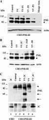
3Cpro expression induced PML cleavage and degradation. (A) CHO-PMLIII cells were infected with VV, VV-P1, VV-P2, or VV-P3 at an MOI of 1 for 8 h. (B) CHO-PMLIII cells were infected with VV, VV-3AB, VV-3C, and VV-3D at an MOI of 1 for 8 h. (C) CHO-PMLIII cells were infected with VV or VV-3C at an MOI of 1 for 12 h (lanes 2, 3, and 4) or 16 h (lanes 5 and 6); extracts from CHO cells transfected with pcDNA 3C vector were used as a control for 3Cpro expression (lane 1). Total cell extracts were analyzed by Western blotting using anti-PML, anti-actin, or anti-3C antibodies. The unmodified form of PML is indicated by an arrowhead, and the cleavage products are indicated by arrows.
FIG. 4.
FIG. 4.
(A) EMCV infection shifted PML toward the nuclear matrix. RIPA-soluble (R) and -insoluble (P) fractions from CHO-PMLIII cells, not infected or infected with EMCV at MOIs of 1 and 5 for 2 h, were analyzed by Western blotting with anti-PML and anti-3C antibodies. The unmodified form of PML is indicated by an arrowhead, and the cleavage product is indicated by an arrow. (B) PML localization in IFN-treated cells. Total extracts from U373MG cells untreated or treated with IFN-α or IFN-γ for 24 h were analyzed by Western blotting using anti-PML antibody (left panel). EMCV infection induced the transfer of PML from the nucleoplasm to the nuclear matrix in IFN-treated cells (right panel). U373MG cells were untreated or treated with IFN-α or IFN-γ for 24 h and then not infected or infected with EMCV at an MOI of 5 for 2 h. RIPA-soluble (R) and -insoluble (P) fractions from the different samples were analyzed by Western blotting with anti-PML antibody.
FIG. 5.
FIG. 5.
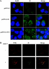
3Cpro localization in transfected cells and in infected cells stably expressing PMLIII. (A) HeLa cells were transfected with pcDNA3, pcDNA3-3Cpro, or pcDNA3-3CD. Cells were fixed 18 h later, and immunofluorescence was performed with anti-3C antibody. DAPI staining is shown. (B) 3Cpro colocalized with PML within the NBs. Double-immunofluorescence staining was performed on nuclear matrix isolated from CHO-PMLIII cells infected with EMCV at an MOI of 5 for 2 h using monoclonal anti-PML antibody visualized by Alexa Fluor 594 and rabbit anti-3C antibody followed by Alexa Fluor 488.
FIG. 6.
FIG. 6.
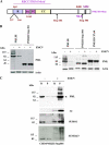
EMCV-induced PML degradation required its RING domain and its C-terminal region. (A) Schematic representation of the domain structure of the PMLIII isoform (residues 1 to 641). PML domains include the RING finger (R), the B1 and B2 boxes, the coiled-coil (CC) motif, the nuclear localization domain (NLS), three SUMOylation sites (K65, K160, and K490), and the SUMO-interacting motif (SIM). The localization of PMLIII mutants is also shown. (B) PMLIII (residues 1 to 641), PMLIII Stop504, and PMLIII C57/60 stably expressed in CHO cells infected with EMCV at an MOI of 5 for 8 h. The different cell extracts were analyzed by Western blotting and revealed by rabbit anti-PML and anti-actin antibodies. (C) Resistance of PMLIII Stop504 to EMCV-induced degradation did not reflect its inability to be targeted to the nuclear matrix. RIPA-soluble and -insoluble fractions from CHO cells stably expressing PMLIII Stop504, left uninfected or infected with EMCV at an MOI of 5 for 2 h, were analyzed by Western blotting with anti-PML, anti-3C, anti-SUMO-1, and anti-SUMO-2/3 antibodies.
FIG. 7.
FIG. 7.
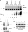
(A) EMCV infection increased PMLIII and PMLIII SIM SUMOylation. U373MG cells stably expressing PMLIII, PMLIII SIM, or PMLIII 3KR were infected with EMCV at an MOI of 5 for 2 h. (B) EMCV-induced PML degradation necessitated its covalent but not its noncovalent binding to SUMO. U373MG cells stably expressing PMLIII, PMLIII SIM, or PMLIII 3KR were infected with EMCV at an MOI of 5 for 8 h. The different cell extracts were analyzed by Western blotting and revealed by rabbit anti-PML or anti-actin antibodies. The unmodified form of PML is indicated by an arrowhead.
FIG. 8.
FIG. 8.
(A) EMCV infection increased the conjugation of PML to SUMO. HEK293 cells were cotransfected with PMLIII-expressing vector and pcDNA3, His6-tagged SUMO-1, -SUMO-2, or SUMO-3, and cell extracts from uninfected or infected cells for 2 h at an MOI of 5 were purified on Ni2+-NTA-agarose beads. The inputs and the purified extracts were analyzed by Western blotting using anti-PML antibody. (B and C) Role of SUMO paralogs in EMCV-induced PML degradation. U373MG-PMLIII cells transfected with control siRNA scramble or siRNA specific for SUMO-1 or SUMO-2/3 were infected with EMCV at an MOI of 5 for 8 h. Cells extracts were analyzed by Western blotting with anti-SUMO-1 and anti-SUMO-2/3 (B) or anti-PML and anti-actin antibodies (C). The unmodified form of PML is indicated by an arrowhead.
FIG. 9.
FIG. 9.
Absence of PML expression in PML−/− MEFs resulted in increased expression of viral proteins. WT MEFs and PML−/− MEFs were infected with EMCV for 8 h at different MOIs. Protein extracts were analyzed by Western blotting and revealed by antiviral protein antibodies.

Similar articles

Cited by

References

    1. Aminev, A. G., S. P. Amineva, and A. C. Palmenberg. 2003. Encephalomyocarditis viral protein 2A localizes to nucleoli and inhibits cap-dependent mRNA translation. Virus Res. 95:45-57. - PubMed
    1. Aminev, A. G., S. P. Amineva, and A. C. Palmenberg. 2003. Encephalomyocarditis virus (EMCV) proteins 2A and 3BCD localize to nuclei and inhibit cellular mRNA transcription but not rRNA transcription. Virus Res. 95:59-73. - PubMed
    1. Amineva, S. P., A. G. Aminev, A. C. Palmenberg, and J. E. Gern. 2004. Rhinovirus 3C protease precursors 3CD and 3CD′ localize to the nuclei of infected cells. J. Gen. Virol. 85:2969-2979. - PubMed
    1. Arnold, E., M. Luo, G. Vriend, M. G. Rossmann, A. C. Palmenberg, G. D. Parks, M. J. Nicklin, and E. Wimmer. 1987. Implications of the picornavirus capsid structure for polyprotein processing. Proc. Natl. Acad. Sci. U. S. A. 84:21-25. - PMC - PubMed
    1. Barral, P. M., D. Sarkar, P. B. Fisher, and V. R. Racaniello. 2009. RIG-I is cleaved during picornavirus infection. Virology 391:171-176. - PMC - PubMed

Publication types

MeSH terms