Review
. 2010 Oct;20(10):609-17.
doi: 10.1016/j.tcb.2010.08.003. Epub 2010 Aug 31.

DNA methylation and cellular reprogramming

Affiliations
Review

DNA methylation and cellular reprogramming

Daniel D De Carvalho et al. Trends Cell Biol. 2010 Oct.

Abstract

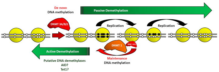
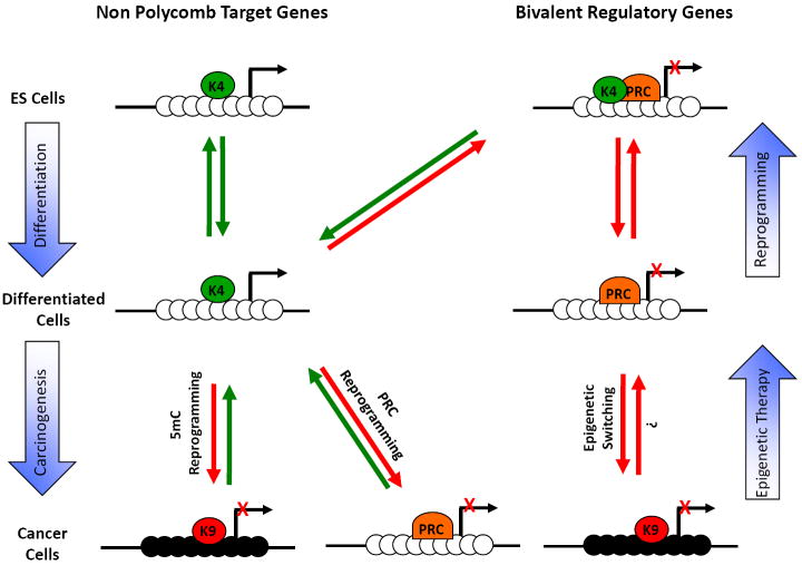
The recent discovery that a small number of defined factors are sufficient to reprogram somatic cells into pluripotent stem cells has significantly expanded our knowledge of the plasticity of the epigenome. In this review we discuss some aspects of cell fate plasticity and epigenetic alterations, with emphasis on DNA methylation during cellular reprogramming. Recent data suggest that DNA methylation is a major barrier to induced pluripotent stem (iPS) cell reprogramming. The demethylating agent 5-azacytidine can enhance the efficiency of iPS cells generation and the putative DNA demethylase protein activation-induced cytidine deaminase (AID/AICDA) can erase DNA methylation at pluripotency gene promoters, thereby allowing cellular reprogramming. Elucidation of the epigenetic changes taking place during cellular reprogramming will enhance our understanding of stem cell biology and facilitate therapeutic applications.


Figures

Figure 1
Figure 1. Cell fusion experiments showing the importance of DNA methylation as an epigenetic barrier for reprogramming
(a) Human fibroblasts can be fused with mouse muscle cells to generate heterokaryons and the plethora of muscle transcription factors can induce the expression of human muscle genes that were repressed in fibroblasts. On the other hand, the fusion of HeLa human cancer cells with murine muscle cells cannot induce human muscle genes unless treated with the demethylating drug 5-Azacytidine prior to cell fusion [4, 5]. (TF): transcription factor. (b) Human fibroblasts can also be fused with murine ES cells to generate heterokaryons and the factors present in the ES cells can reprogram the human Oct4 and Nanog promoters, causing DNA demethylation and gene expression. On the other hand, this reprogramming cannot occur when the mouse putative DNA demethylase, AID is silenced by RNAi [60].
Figure 2
Figure 2. DNA methylation and demethylation
De novo DNA methylation patterns are established by catalytically active methyltransferases DNMT3A and DNMT3B. This process is enhanced in the presence of the catalytically inactive DNMT3L. During replication, the original DNA methylation pattern is maintained largely by the activity of DNMT1, with some participation by DNMT3A and DNMT3B. DNA methylation patterns can be erased by DNA demethylation, through active or passive processes. Active demethylation can occur by enzymatic replacement of a methylated cytosine with an unmethylated residue without the requirement of cell division. Several enzymes, such as AID and Tet1, are proposed to play roles as DNA demethylases. On the other hand, passive demethylation can occur during successive replications, when the activity of maintenance DNA methylation is abrogated. Black circles represent methylated CpG sites and open circles represent unmethylated CpG sites.
Figure 3
Figure 3. Epigenome landscaping during differentiation and carcinogenesis
(a) CpG rich regions: In embryonic stem cells, most CpG rich regions contained within promoters are marked by H3K4me3 and can be subdivided in two groups. The first group comprises genes that are not Polycomb targets and are usually expressed in ES cells. The second group comprises Polycomb target genes. This group adopts a bivalent chromatin structure and is usually repressed in ES cells. During cell differentiation, the bivalent chromatin structure becomes monovalent and some genes retain only the H3K4me3 mark (K4) while others retain only the H3K27me3 mark (PRC). Interestingly, during reprogramming towards pluripotency, most of these genes reacquire the bivalent structure [73, 74]. During carcinogenesis, a group of expressed genes become de-novo repressed, either by DNA methylation (5mC reprogramming) or by histone methylation (PRC reprogramming). In addition, another group of genes already repressed by Polycomb gains a more stable repression by DNA methylation (epigenetic switching) [85]. The gain of DNA methylation is usually accompanied by H3K9 methylation (K9). Black circles are methylated CpG sites and white circles are unmethylated. (b) CpG poor regions: In ES cells, pluripotency genes located in non-CGI are marked by H3K4me3, the CpG sites are unmethylated and the genes are expressed. During cell differentiation, these genes gain DNA methylation, the repressive H3K9me3 mark and are not expressed. Tissue specific genes are silenced by DNA methylation in ES cells and during differentiation these genes are expressed in some tissues, while remaining silenced in others. During reprogramming, this group can revert their chromatin structure, becoming very similar to that of ES cells [70]. Black circles are methylated CpG sites and white circles are unmethylated.

Similar articles

Cited by

References

    1. Amabile G, Meissner A. Induced pluripotent stem cells: current progress and potential for regenerative medicine. Trends Mol Med. 2009;15:59–68. - PubMed
    1. Hochedlinger K, Plath K. Epigenetic reprogramming and induced pluripotency. Development. 2009;136:509–523. - PMC - PubMed
    1. Moving AHEAD with an international human epigenome project. Nature. 2008;454:711–715. - PMC - PubMed
    1. Chiu CP, Blau HM. Reprogramming cell differentiation in the absence of DNA synthesis. Cell. 1984;37:879–887. - PubMed
    1. Chiu CP, Blau HM. 5-Azacytidine permits gene activation in a previously noninducible cell type. Cell. 1985;40:417–424. - PubMed

Publication types

Substances