. 2010 Aug;22(8):2825-37.
doi: 10.1105/tpc.109.072215. Epub 2010 Aug 31.

The cytosolic tail dipeptide Ile-Met of the pea receptor BP80 is required for recycling from the prevacuole and for endocytosis

Affiliations

The cytosolic tail dipeptide Ile-Met of the pea receptor BP80 is required for recycling from the prevacuole and for endocytosis

Bruno Saint-Jean et al. Plant Cell. 2010 Aug.

Abstract

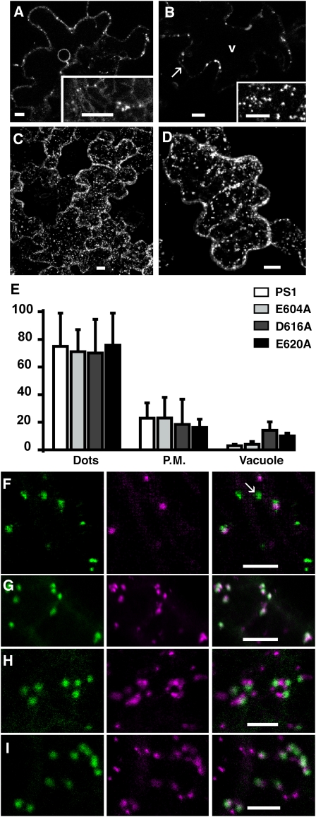
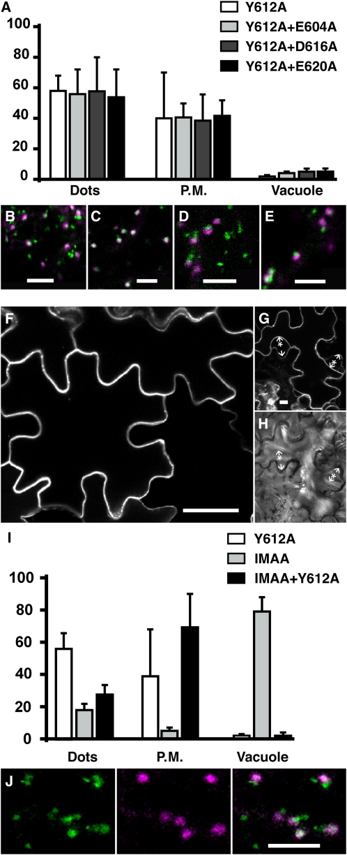
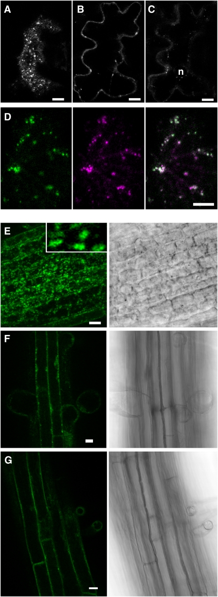
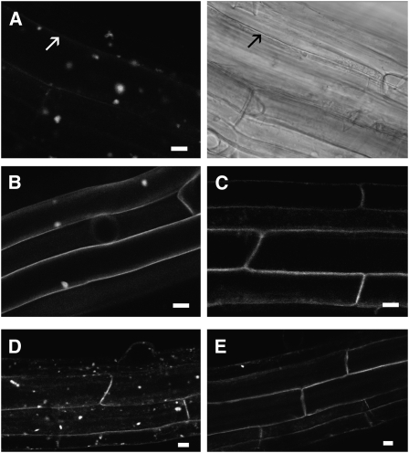
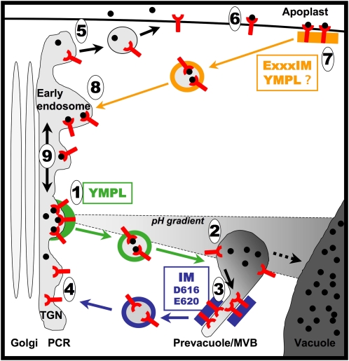
Pea (Pisum sativum) BP80 is a vacuolar sorting receptor for soluble proteins and has a cytosolic domain essential for its intracellular trafficking between the trans-Golgi network and the prevacuole. Based on mammalian knowledge, we introduced point mutations in the cytosolic region of the receptor and produced chimeras of green fluorescent protein fused to the transmembrane domain of pea BP80 along with the modified cytosolic tails. By analyzing the subcellular location of these chimera, we found that mutating Glu-604, Asp-616, or Glu-620 had mild effects, whereas mutating the Tyr motif partially redistributed the chimera to the plasma membrane. Replacing both Ile-608 and Met-609 by Ala (IMAA) led to a massive redistribution of fluorescence to the vacuole, indicating that recycling is impaired. When the chimera uses the alternative route, the IMAA mutation led to a massive accumulation at the plasma membrane. Using Arabidopsis thaliana plants expressing a fluorescent reporter with the full-length sequence of At VSR4, we demonstrated that the receptor undergoes brefeldin A-sensitive endocytosis. We conclude that the receptors use two pathways, one leading directly to the lytic vacuole and the other going via the plasma membrane, and that the Ileu-608 Met-609 motif has a role in the retrieval step in both pathways.


Figures

Figure 1.
Figure 1.
Cytosolic Sequence of BP80 and Mutations Used in This Study. The cytosolic sequence of pea BP80 and its closest Arabidopsis homologs VSR3 and VSR4 were aligned. Amino acids mutated into Ala based on their homology with mammalian signals are shown above the chart. The respective cytosolic domains were fused together with the transmembrane sequence of BP80 to the reporter GFP to produce the unmutated reporter PS1 and mutated reporters E604A, IMAA (I608A plus M609A), Y612A, D616A, and E620A. Numbering of the mutated amino acids was based on the Arabidopsis VSR3 sequence.
Figure 2.
Figure 2.
Mutations of Single Acidic Amino Acids Have Minor Effects on the Localization of the Reporter. Tobacco epidermal cells transiently expressing reporter proteins were observed either 48 (A) or 72 h ([B] to [I]) after transformation. Bars = 10 μm in (A) to (D) and 5 μm in (F) to (I). (A) The unmutated control reporter PS1 was found in spots and the ER at an early stage of expression as seen in a section through the nucleus and in a section at the surface of the cell (inset). (B) After 72 h of expression, the reporter has reached its final destination as seen in a section crossing through the vacuole (v) at the level of the nucleus with little labeling at the plasma membrane (arrow). Typical punctate structures can be better visualized in an optical section through the cytosol (inset). (C) Accumulation of individual z-sections of cells expressing PS1 reporter. (D) Accumulation of individual z-sections from a cell expressing E604A mutated reporter. (E) Relative fluorescence intensity in three chosen locations, dots, plasma membrane (P.M.), and vacuole for the following constructs: control PS1 (white), E604A (light gray), D616A (dark gray), and E620A (black). Error bar stands for sd from 90 to 150 measures for each subcellular location and each construct. (F) to (I) Tobacco epidermal cells transiently coexpressing PS1-based reporter proteins (green) and the Golgi reference ERD2-CFP (purple). The reporter protein was either PS1 (F), E604A (G), D616A (H), or E620A (I). (F) Typical prevacuole labeling (arrow) is separated from Golgi labeling.
Figure 3.
Figure 3.
The Y612A Mutation Increases the Presence of the Reporter at the Plasma Membrane, While the IMAA Mutation Leads to GFP Accumulation in the Vacuole. Tobacco epidermal cells transiently expressing reporter proteins were observed either 72 ([A] to [C]) or 48 h ([D] to [G]) after transformation using a confocal microscope. Bars = 10 μm in (A), (B), (D), and (E) and 5 μm in (F) and (G). (A) Accumulation of individual z-sections from cells expressing Y612A. (B) A single section of cells expressing the IMAA reporter shows a massive fluorescence accumulation in the vacuole (v) leading to a negative staining of the nucleus (arrow). (C) Relative fluorescence intensity in three chosen locations, dots, plasma membrane (P.M.), and vacuole, for the following constructs: control PS1 (white), Y612A (gray), and IMAA (black). Error bar stands for sd from 90 to 150 measures for each subcellular location and each construct. (D) and (E) Single sections at the level of the nucleus (D) or at the surface (E) for cells expressing IMAA show intermediate labeling of spots prior to vacuole accumulation. (F) and (G) Coexpression of IMAA (green) either with a Golgi reference ERD2-CFP ([F]; purple) or with the BP80 ligand Aleu-CFP ([G]; purple).
Figure 4.
Figure 4.
In the Absence of a Functional Tyr Motif, the IMAA Mutation Blocks the Reporter in the Plasma Membrane, While Mutating Acidic Amino Acids Have Minor Effects. Tobacco epidermal cells expressing transiently reporter proteins were observed 72 h after transformation using a confocal microscope. In (A) and (I), error bar stands for sd from 90 to 150 measures for each subcellular location and each construct. Arrow, cell wall; arrow with a star, plasma membrane. Bars = 5 μm in (B) to (E) and (J) and 10 μm in (F) and (G). (A) Relative fluorescence intensity in three chosen locations, dots, plasma membrane (P.M.), and vacuole, for the following constructs: Y612A (white), Y612A+E604A (light gray), Y612A+D616A (dark gray), and Y612A+E620A (black). (B) to (E) Coexpression of the reporter proteins Y612A ([B]; green), Y612A+E604A ([C]; green), Y612A+D616A ([D]; green), or Y612A+E620A ([E]; green) with the Golgi reference ERD2-CFP (purple). (F) to (H) Single confocal section of epidermal cells taken at the level of the nucleus, under normal conditions (F) or after plasmolysis ([G] and [H]). (I) Relative fluorescence intensity in three chosen locations, dots, plasma membrane (P.M.), and vacuole, for the following constructs: Y612A (white), IMAA (gray), and IMAA+Y612A (black). (J) Coexpression of IMAA+Y612A (green) with a Golgi reference ERD2-CFP (purple).
Figure 5.
Figure 5.
A Full-Length VSR Fused to a Fluorescent Protein Partially Localizes in the Plasma Membrane and Colocalizes with the PS1 Reporter (A) to (D) Tobacco epidermal cells transiently expressing reporter proteins were observed 72 h after transformation using a confocal microscope. (A) to (C) Confocal sections of an epidermal cell expressing citrine-AtVSR4 taken either at the surface (A), deeper inside the cell (B), or at the level of the nucleus (C). (D) Coexpression of PS1 fused to CFP (green) with citrine-AtVSR4 (purple). (E) to (G) Arabidopsis plantlets stably expressing citrine-AtVSR4. Roots from 7-d-old plants were observed with a confocal microscope with the bright-field setting (right side) or under fluorescence confocal mode (left side) at different levels: the apex ([E] and inset: 10 times enlarged detail), the middle portion (F), or the top part (G). n, nucleus. Bars = 10 μm.
Figure 6.
Figure 6.
Citrine-AtVSR4 Undergoes BFA-Sensitive Endocytic Recycling One-week-old plants expressing citrine-AtVSR4 were submitted to BFA treatment (50 μM) for 60 min in presence of 50 μM cycloheximide. Cells from the top portion of the root, where the fusion protein is exclusively found in the plasma membrane, were observed using a confocal microscope. Arrow, plasma membrane. Bars = 10 μm. (A) Citrine-AtVSR4 labeling after BFA treatment. The right side shows a bright-field image of the same cells shown on the left. (B) Control plants expressing LTi6a-GFP after BFA treatment. (C) Citrine-AtVSR4 after 60 min of cycloheximide treatment. Plasma membrane labeling of citrine-AtVSR4 had fully recovered after BFA treatment for 10 min (D) and a subsequent wash in cycloheximide for 80 min (E).
Figure 7.
Figure 7.
Proposed Trafficking Model for the VSR BP80. The receptor BP80 uses a major (green and blue arrows) or an alternative pathway (black and orange arrows). In the major pathway, BP80 recognizes the ligand (black circle) at the level of the TGN that is part of a PCR. The exit from the TGN (1) requires the YMPL motif most likely through its interaction with μA-adaptin containing complex (green box and vesicle coat). After fusion of the vesicle to the prevacuole (2), the receptor releases its ligand, possibly due to a pH decrease. The free receptor is then segregated out of the ligand release area (3) most likely by interaction of the IM motif with a retrieval complex (blue box). The free receptor is packed in coated vesicles (vesicle with blue coat) back to the TGN where fusion (4) may require VPS45. The final steps for ligand transport to the vacuole (dashed arrow) do not require BP80 but instead are believed to be maturation processes of the prevacuole/MVB that eventually fuses with the vacuole. The alternative pathway serves to retrieve missorted ligands from the apoplast. The free receptor but also free ligand can exit the PCR (5; black arrows) to reach the plasma membrane by a BFA-sensitive process. At the plasma membrane, the receptor binds a ligand (6) and is endocytosed using the IM dipeptide as part of a dileucine-like motif ExxxIM possibly with the help of the Tyr motif. The endocytosis signal should interact (7) with the retrieval complex (orange box and vesicle coat). Fusion of the vesicle with the early endosome (8) leads to release the receptor-ligand complex in the PCR where the complex can diffuse along the membrane (9) and may enter the main pathway thanks to the Tyr motif (1).

Similar articles

Cited by

References

    1. Ahmed S.U., Bar-Peled M., Raikhel N.V. (1997). Cloning and subcellular location of an Arabidopsis receptor-like protein that shares common features with protein-sorting receptors of eukaryotic cells. Plant Physiol. 114: 325–336 - PMC - PubMed
    1. Ahmed S.U., Rojo E., Kovaleva V., Venkataraman S., Dombrowski J.E., Matsuoka K., Raikhel N.V. (2000). The plant vacuolar sorting receptor AtELP is involved in transport of NH2-terminal propeptide-containing vacuolar proteins in Arabidopsis thaliana. J. Cell Biol. 149: 1335–1344 - PMC - PubMed
    1. Anderson R., Orci L. (1988). A view of acidic intracellular compartments. J. Cell Biol. 106: 539–543 - PMC - PubMed
    1. Batoko H., Zheng H.-Q., Hawes C., Moore I. (2000). A Rab1 GTPase is required for transport between endoplasmic reticulum and Golgi apparatus and for normal Golgi movement in plants. Plant Cell 12: 2201–2217 - PMC - PubMed
    1. Blom N., Gammeltoft S., Brunak S. (1999). Sequence- and structure-based prediction of eukaryotic protein phosphorylation sites. J. Mol. Biol. 294: 1351–1362 - PubMed

Publication types

MeSH terms

Substances

LinkOut - more resources