. 2010 Aug;22(8):2749-67.
doi: 10.1105/tpc.110.076257. Epub 2010 Aug 31.

Arabidopsis VILLIN5, an actin filament bundling and severing protein, is necessary for normal pollen tube growth

Affiliations

Arabidopsis VILLIN5, an actin filament bundling and severing protein, is necessary for normal pollen tube growth

Hua Zhang et al. Plant Cell. 2010 Aug.

Abstract

A dynamic actin cytoskeleton is essential for pollen germination and tube growth. However, the molecular mechanisms underlying the organization and turnover of the actin cytoskeleton in pollen remain poorly understood. Villin plays a key role in the formation of higher-order structures from actin filaments and in the regulation of actin dynamics in eukaryotic cells. It belongs to the villin/gelsolin/fragmin superfamily of actin binding proteins and is composed of six gelsolin-homology domains at its core and a villin headpiece domain at its C terminus. Recently, several villin family members from plants have been shown to sever, cap, and bundle actin filaments in vitro. Here, we characterized a villin isovariant, Arabidopsis thaliana VILLIN5 (VLN5), that is highly and preferentially expressed in pollen. VLN5 loss-of-function retarded pollen tube growth and sensitized actin filaments in pollen grains and tubes to latrunculin B. In vitro biochemical analyses revealed that VLN5 is a typical member of the villin family and retains a full suite of activities, including barbed-end capping, filament bundling, and calcium-dependent severing. The severing activity was confirmed with time-lapse evanescent wave microscopy of individual actin filaments in vitro. We propose that VLN5 is a major regulator of actin filament stability and turnover that functions in concert with oscillatory calcium gradients in pollen and therefore plays an integral role in pollen germination and tube growth.


Figures

Figure 1.
Figure 1.
VLN5 Is Expressed Preferentially in Pollen. The expression pattern of VLN5 throughout the Arabidopsis plant was examined by RT-PCR of separated tissues and with a promoter-reporter fusion. (A) VLN5-specific primers were used to determine tissue expression patterns by RT-PCR. Tubulin2 was used as an internal loading control. The samples are as follows: lane 1, seedling; lane 2, root; lane 3, internode; lane 4, bottom part of stem; lane 5, juvenile leaf; lane 6, adult leaf; lane 7, flower; lane 8, anther; lane 9, pollen; lane 10, silique at early stage; lane 11, silique at late stage. (B) VLN5 promoter activity was determined using GUS as a reporter. Samples include (a) 7-d-old seedling, (b) leaves, (c) stems, (d) flower, and (e) siliques at different stages. Inset: pollen grain and germinated tube. Bar = 10 μm for the pollen tube.
Figure 2.
Figure 2.
Identification of Transcript Levels in T-DNA Insertion Mutants and Transgenic RNAi Lines. (A) Physical structure of the Arabidopsis VLN5 gene. VLN5 contains 22 exons and 21 introns, which are represented by filled boxes and lines, respectively. The position of two independent T-DNA insertion mutants, designated vln5-1 (SAIL_512_F03) and vln5-2 (GABI_225F09), are noted by triangles above the diagram. (B) Three separate pairs of primers, marked by arrows in (A), were designed to identify the level of VLN5 transcripts. The first pair of primers (V5F1 and V5R1) was designed to amplify VLN5 full-length cDNA, which was not present in vln5-1 and vln5-2 mutant plants. The second and third primer pairs, V5F2/V5R2 and V5F3/V5R3, were used to amplify upstream and downstream transcripts, respectively. Flowers from wild-type Col-0 plants and the two homozygous insertion lines were subjected to RT treatment. eIF4A was used as an internal loading control. (C) VLN5 transcripts were reduced significantly in VLN5 RNAi flowers. Flowers from wild-type Col-0 and VLN5 RNAi plants were subjected to quantitative real-time PCR analysis. The expression level of the eIF4A gene was used as an internal control. All data represent the mean value of three biological replications. Error bars represent ± sd (n = 3); **P < 0.01.
Figure 3.
Figure 3.
VLN5 Loss-of-Function Mutant Plants Have Retarded Pollen Tube Growth. (A) to (C) Micrographs of pollen tubes after germination for 2 h in vitro. Pollen was isolated from plants with the following genotypes: wild-type Col-0 (A), homozygous vln5-1 (B), and homozygous vln5-2 (C). Bar in (C) = 100 μm. (D) to (F) Length distribution of pollen tubes: wild-type Col-0 (D), vln5-1 (E), and vln5-2 (F). (G) Measurement of growth rates was performed by tracking individual pollen tubes. (a) to (f) Pairs of images from single pollen tubes selected for measurement: (a) wild-type Col-0 pollen tube and (b) the same pollen tube after 30 min of growth; (c) vln5-1 pollen tube and (d) the same pollen tube after 30 min growth; (e) vln5-2 pollen tube and (f) the same pollen tube after 30 min growth. Bar = 50 μm in (f) for (a) to (f). (g) A plot of pollen tube growth rates shows that vln5 mutant pollen tubes had reduced rates compared with the wild type. Wild-type Col-0, black bar; vln5-1, gray bar; vln5-2, white bar. Error bars represent mean values ± se; n = 200. Pollen tube growth rate of vln5 pollen tubes was significantly different from that of wild-type Col-0 pollen tubes as determined by analysis of variance followed by Dunnett post-hoc multiple comparisons; **P < 0.01.
Figure 4.
Figure 4.
VLN5 Loss of Function Does Not Affect the Amount or Distribution of Actin Filaments in Ungerminated Pollen Grains and Pollen Tubes. Pollen grains and pollen tubes from wild-type Col-0 and VLN5 loss-of-function mutants were subjected to actin filament staining with Alexa-488 phalloidin. More than 50 stained pollen tubes were obtained for each genotype, and typical images for each genotype, which represented >85% of the population, are presented. See Supplemental Figure 7 for more examples. The images of actin staining of pollen grains and pollen tubes are projections of confocal optical sections. Bar = 7.5 μm in (I) for pollen grains and 10 μm in (J) for pollen tubes. (A) Wild-type Col-0 pollen grain. (B) Wild-type Col-0 pollen tube. On the right is a transmitted light image of the corresponding pollen tube. (C) vln5-1 pollen grain. (D) vln5-1 pollen tube and corresponding transmitted light image. (E) vln5-2 pollen grain. (F) vln5-2 pollen tube and corresponding transmitted light image. (G) VLN5 RNAi line 1 pollen grain. (H) VLN5 RNAi line 1 pollen tube and corresponding transmitted light image. (I) VLN5 RNAi line 2 pollen grain. (J) VLN5 RNAi line 2 pollen tube and corresponding transmitted light image.
Figure 5.
Figure 5.
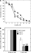
VLN5 Loss of Function Renders Pollen Germination and Pollen Tube Growth Hypersensitive to LatB. (A) Germination of vln5 pollen is more sensitive than wild-type pollen to perturbation of the actin cytoskeleton by LatB treatment. Pollen grains from wild-type Col-0 and vln5 plants were germinated on medium containing various concentrations of LatB. Germination was scored after pollen had germinated for 6 h. The average germination rate versus LatB concentration was plotted. Closed boxes, wild-type Col-0; closed triangles, vln5-1; open triangles, vln5-2. Error bars represent se; n ≥ 500. Asterisks denote values that were significantly different from wild-type Col-0 by χ2 test (P < 0.01). (B) The growth of vln5-1 and vln5-2 pollen tubes was more sensitive than wild-type pollen to LatB treatment. To measure the growth rate of pollen tubes, pollen from wild-type Col-0 and vln5 mutant plants was initially germinated for 2 h in standard germination medium. Individual pollen tubes were subsequently tracked to measure the growth rate of pollen tubes. To determine the effect of LatB on growth rates, 3 nM LatB was added to the germination medium. The growth rate of pollen tubes from wild-type Col-0, vln5-1, and vln5-2 in standard germination medium was normalized to 100%, and the relative growth rate of pollen tubes in 3 nM LatB was plotted. Wild-type Col-0 grew significantly better than did vln5 mutant pollen in the presence of LatB. Error bars represent mean ± se (n > 72); **P < 0.01 (Student’s t test).
Figure 6.
Figure 6.
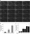
Pollen Grains and Pollen Tubes with Reduced VLN5 Levels Have Unstable Actin Filaments. Ungerminated pollen grains and pollen tubes treated with 100 nM LatB for 30 min were subjected to actin staining with Alexa-488 phalloidin. Transmitted light images of each pollen tube are shown to the right of the fluorescence microscope image. (A) Wild-type Col-0 pollen grain. (B) Wild-type Col-0 pollen tube. (C) vln5-1 pollen grain. (D) vln5-1 pollen tube. (E) vln5-2 pollen grain. (F) vln5-2 pollen tube. Bar = 7.5 μm in (E) for pollen grains and 10 μm in (F) for pollen tubes. (G) Quantification of relative F-actin level in pollen grains treated with 100 nM LatB. The amount of F-actin in treated wild-type Col-0 pollen grains was normalized to 100%. Error bars represent ± sd (n = 10); **P < 0.01 (Student’s t test). (H) Quantification of relative F-actin level in pollen tubes treated with 100 nM LatB. The amount of F-actin in treated wild-type Col-0 pollen tubes was normalized to 100%. Error bars represent ± sd (n > 27); *P < 0.05 (Student’s t test).
Figure 7.
Figure 7.
VLN5 Binds to Actin Filaments with High Affinity. (A) Recombinant VLN5 expressed in bacteria was purified to homogeneity with several chromatography steps. Three micrograms of purified protein separated on an SDS-PAGE gel and stained with Coomassie blue is shown. (B) A high-speed cosedimentation assay was employed to determine the affinity of VLN5 for actin filaments. Three micromolar polymerized actin was incubated with various concentrations of VLN5 and the mixtures were centrifuged at 200,000g for 1 h to separate bound versus unbound VLN5. After centrifugation, equal amounts of pellets (P) and supernatants (S) were separated by SDS-PAGE and stained with Coomassie blue. Samples loaded in lanes 1 to 12 contained actin plus 0 (lanes 1 and 2), 0.1 (lanes 3 and 4), 0.2 (lanes 5 and 6), 0.5 (lanes 7 and 8), 1 (lanes 9 and 10), and 2 μM (lanes 11 and 12) VLN5, whereas lanes 13 and 14 contained 2 μM VLN5 only. (C) Actin filaments were sedimented in the presence of various concentrations of VLN5, as shown in (B). The amount of VLN5 in pellets and supernatants from the various samples was determined with Image J. The concentration of VLN5 in the pellet ([VLN5]bound) as a function of the concentration of VLN5 in the supernatant ([VLN5]free) was plotted, and the data were fitted with a hyperbolic function to calculate the equilibrium dissociation constant (Kd) value. In this representative experiment, the calculated Kd value was 0.55 μM. (D) The Kd value for VLN1, used as a positive control for binding experiments, was determined to be 0.63 μM in this representative experiment. (E) To determine the effects of Ca2+ on VLN5 binding to actin filaments, 1 μM VLN5 was incubated with 3 μM F-actin in the presence of various [Ca2+]free. The percentage of VLN5 that sedimented with actin filaments was plotted as a function of [Ca2+] free. Error bars represent ±sd (n = 3).
Figure 8.
Figure 8.
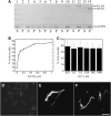
VLN5 Bundles Actin Filaments in a Calcium-Insensitive Manner. A low-speed cosedimentation assay and fluorescence microscopy were employed to determine the bundling activity of VLN5. (A) VLN5 bundles actin filaments in a dose-dependent manner. Reactions containing actin alone, actin plus various concentrations of VLN5, or actin plus 1 μM VLN1 were incubated for 30 min and then sedimented at 13,600g for 30 min. Samples for the supernatant (S) and pellet (P) were separated by SDS-PAGE; lanes 1, 3, 5, 7, 9, 11, and 13 represent supernatant of actin alone, actin plus 50 nM VLN5, actin plus 100 nM VLN5, actin plus 200 nM VLN5, actin plus 500 nM VLN5, actin plus 1 μM VLN5, and actin plus 1 μM VLN1, respectively. Samples in lanes 2, 4, 6, 8, 10, 12, and 14 represent pellet of actin alone, actin plus 50 nM VLN5, actin plus 100 nM VLN5, actin plus 200 nM VLN5, actin plus 500 nM VLN5, actin plus 1 μM VLN5, and actin plus 1 μM VLN1, respectively. (B) Percentage of actin sedimented in the low-speed assay as a function of the concentration of VLN5. (C) VLN5 bundles actin filaments in a calcium-insensitive manner. To determine whether the bundling activity of VLN5 is regulated by calcium, actin filaments were incubated with 1 μM VLN5 in the presence of various [Ca2+]free. The amounts of actin in the pellet were determined in the presence of different [Ca2+]free. The percentage of actin sedimented at 10 nM Ca2+ was 78%, at 100 nM Ca2+ was 71%, at 1 μM Ca2+ was 75%, at 10 μM Ca2+ was 72%, at 100 μM Ca2+ was 71%, and at 1 mM Ca2+ was 72%. Three independent experiments were conducted; error bars represent ±sd. (D) to (F) Micrographs of actin filaments in the presence or absence of villin stained with rhodamine phalloidin. (D) Individual actin filaments in the absence of villin. The image was captured at a 500-ms exposure time. (E) Actin filament bundles formed in the presence of 1 μM VLN5. The image was captured at a 150-ms exposure time. (F) Actin bundles formed in the presence of 1 μM VLN1. The image was captured at a 150-ms exposure time. Bar = 10 μm.
Figure 9.
Figure 9.
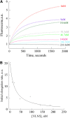
VLN5 Caps the Barbed Ends of Actin Filaments. (A) Preformed F-actin seeds (0.8 μM) were incubated with different concentrations of VLN5, and 1 μM G-actin, saturated with 4 μM human profilin I, was added to initiate actin elongation at the barbed end. Polymerization was monitored by tracking the increase in pyrene-actin fluorescence upon assembly. Concentrations of VLN5, from top to bottom, were 0, 9, 18.6, 93.3, 46.7, 140, 186.7, and 233 nM. A single representative experiment (n = 3) is shown. (B) Initial rates of elongation as a function of VLN5 concentration were plotted for the representative experiment shown in (A). The data were fit with Equation 1 (see Methods) to determine an apparent Kd value of 20.4 nM for VLN5 binding to filament barbed ends.
Figure 10.
Figure 10.
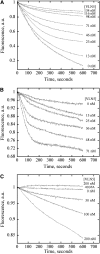
VLN5 Stabilizes Actin Filaments in the Presence of 10 nM Free Ca2+ but Enhances Actin Depolymerization in the Presence of 10 μM Ca2+. (A) VLN5 protects actin filaments from dilution-mediated depolymerization in the presence of 10 nM free Ca2+. In this assay, the amount of F-actin is proportional to pyrene fluorescence. Five micromolar F-actin (100% pyrene labeled) was incubated with various concentrations of VLN5 for 5 min at room temperature and diluted 25-fold into low-ionic strength Buffer G in the presence of 10 nM Ca2+. Under these conditions, actin depolymerization could occur at both pointed and barbed ends of actin filaments. However, the loss of subunits from barbed ends of actin filaments contributes mainly to depolymerization, which was blocked by capping of VLN5. Therefore, VLN5 prevents actin depolymerization in a dose-dependent manner. (B) VLN5 enhances actin depolymerization following dilution in the presence of 10 μM Ca2+. Five micromolar F-actin (50% pyrene labeled) was incubated with various concentrations of VLN5 for 5 min at room temperature and diluted 50-fold in polymerization solution (1× KMEI) in the presence of 10 μM Ca2+. Given that actin depolymerization was induced in polymerization solution and that the final actin concentration drops to the value close to the critical concentration at the barbed end of actin filaments, actin depolymerization mainly occurs at the pointed end and is slow in the absence of VLN5. However, the addition of VLN5 may induce the severing of actin filaments, which increases the number of actin filaments. Although the newly generated barbed ends of actin filaments were capped by VLN5, the increased number of pointed ends would enhance actin depolymerization. (C) VLN5 stabilizes actin filaments from profilin-mediated actin depolymerization in the presence of 2 mM EGTA but enhances profilin-mediated actin depolymerization in the presence of 10 μM free Ca2+.
Figure 11.
Figure 11.
Direct Visualization of VLN5 Severing Activity by Time-Lapse TIRFM. (A) Prepolymerized, 25 nM rhodamine-labeled actin filaments were introduced into a perfusion chamber, where they were attached to the coverglass by NEM-myosin. Five nanomolar VLN5 was injected into the perfusion chamber in the presence of 1 μM free Ca2+. Individual actin filaments showed an increasing number of breaks (arrows) as time elapsed. Bar = 20 μm. See Supplemental Movie 1 online for the entire series. (B) The severing activity of VLN5 is less potent than that of human villin (hVLN). Either 5 nM VLN5 or 0.5 nM hVLN was perfused into a chamber containing 25 nM actin filaments in the presence of 1 μM free Ca2+. The average severing frequency was plotted in the presence or absence of villins. Error bars represent ± se (n = 15). Asterisks represent values that were significantly different from that of the no villin control (*P < 0.05 by a Student’s t test). (C) The severing activity of VLN5 is Ca2+ dependent. Five nanomolar VLN5 in the presence of various concentrations of Ca2+ was perfused into the perfusion chamber and time-lapse images were collected. At least 15 filaments for each experimental treatment were selected to calculate the average severing frequency, and the average severing frequency was plotted against [Ca2+]. Error bars represent ± se; n = 3 for each data set. Asterisks represent values that were significantly different from that of the no villin control (*P < 0.05 by a Student’s t test).

Similar articles

Cited by

References

    1. Amann K.J., Pollard T.D. (2001). Direct real-time observation of actin filament branching mediated by Arp2/3 complex using total internal reflection fluorescence microscopy. Proc. Natl. Acad. Sci. USA 98: 15009–15013 - PMC - PubMed
    1. Andrianantoandro E., Pollard T.D. (2006). Mechanism of actin filament turnover by severing and nucleation at different concentrations of ADF/cofilin. Mol. Cell 24: 13–23 - PubMed
    1. Blanchoin L., Amann K.J., Higgs H.N., Marchand J.B., Kaiser D.A., Pollard T.D. (2000). Direct observation of dendritic actin filament networks nucleated by Arp2/3 complex and WASP/Scar proteins. Nature 404: 1007–1011 - PubMed
    1. Bretscher A., Weber K. (1979). Villin: The major microfilament-associated protein of the intestinal microvillus. Proc. Natl. Acad. Sci. USA 76: 2321–2325 - PMC - PubMed
    1. Cardenas L., Lovy-Wheeler A., Kunkel J.G., Hepler P.K. (2008). Pollen tube growth oscillations and intracellular calcium levels are reversibly modulated by actin polymerization. Plant Physiol. 146: 1611–1621 - PMC - PubMed

Publication types

MeSH terms