. 2010 Oct;78(2):490-505.
doi: 10.1111/j.1365-2958.2010.07346.x. Epub 2010 Sep 2.

Asp3 mediates multiple protein-protein interactions within the accessory Sec system of Streptococcus gordonii

Affiliations

Asp3 mediates multiple protein-protein interactions within the accessory Sec system of Streptococcus gordonii

Ravin Seepersaud et al. Mol Microbiol. 2010 Oct.

Abstract

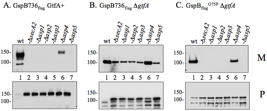
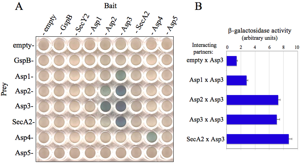
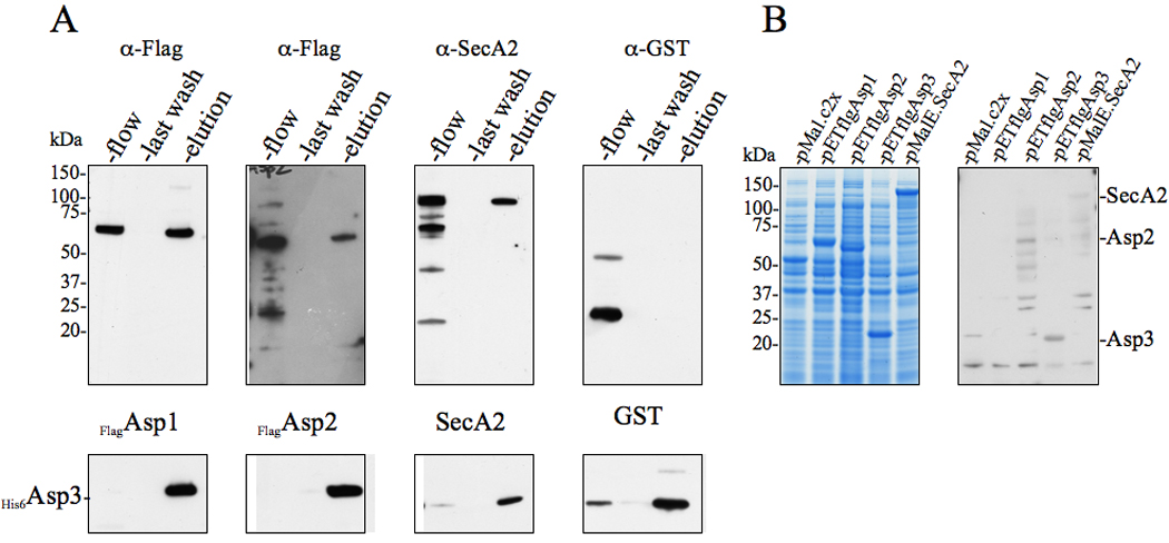
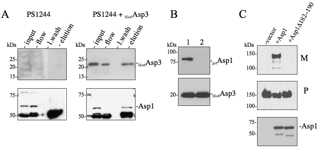
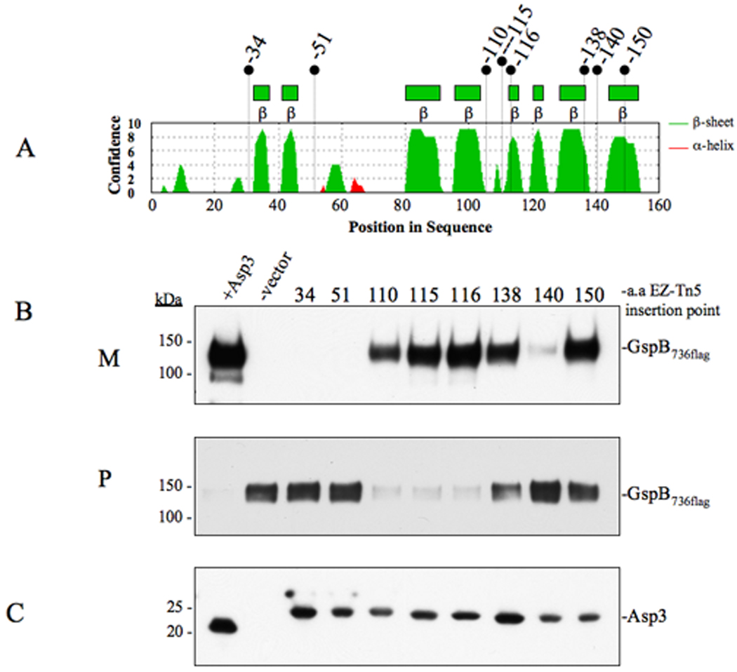
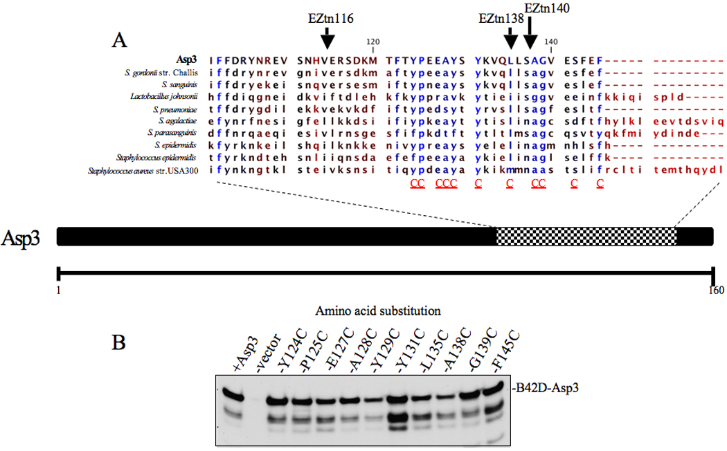
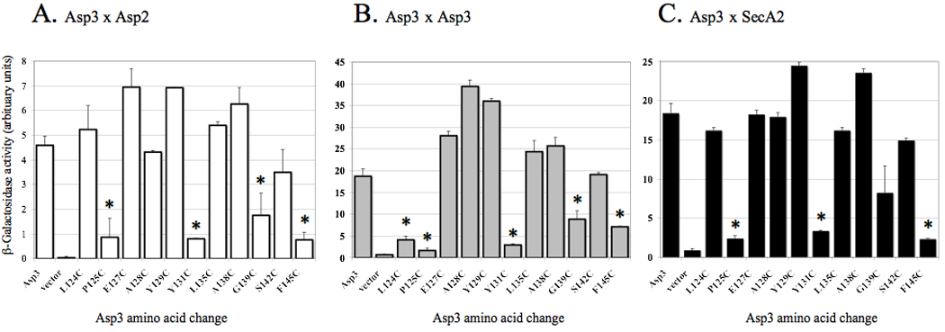
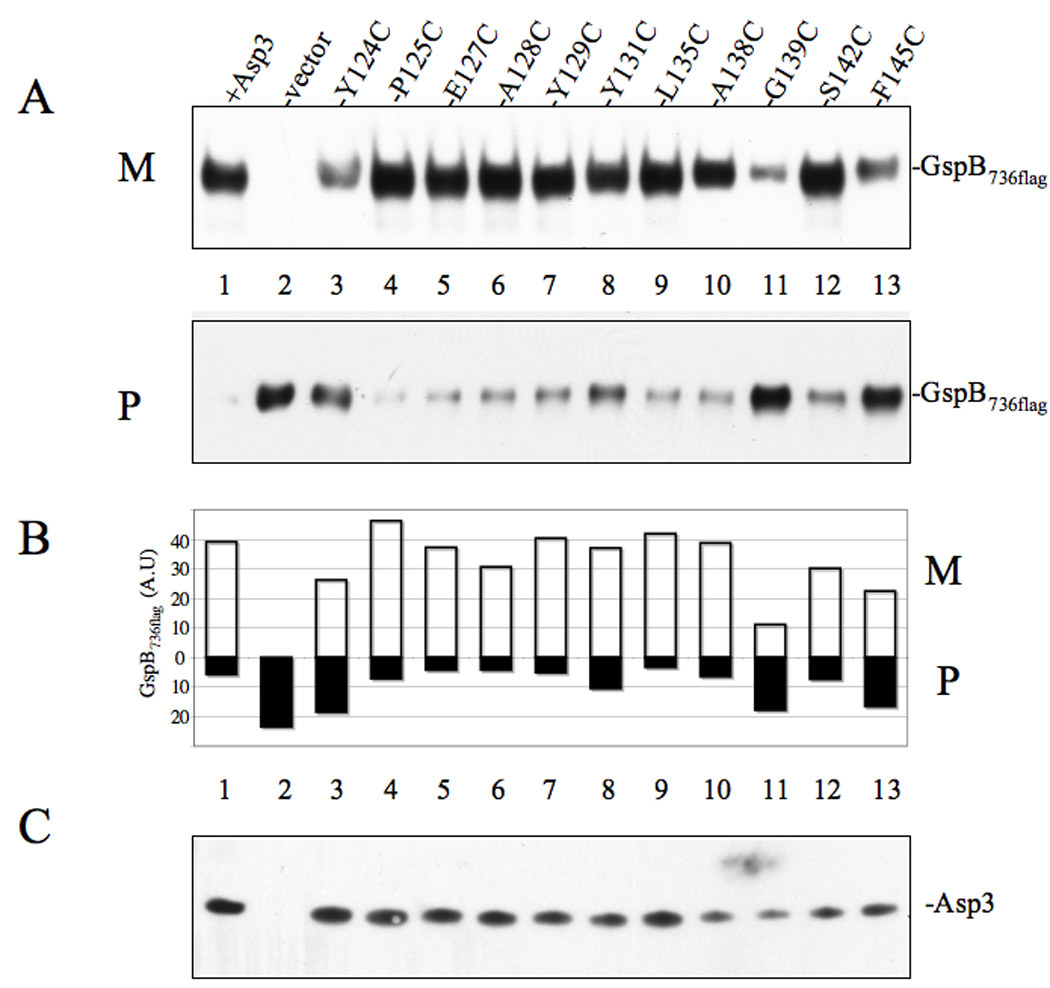
Bacterial binding to human platelets is an important step in the pathogenesis of infective endocarditis. Streptococcus gordonii can mediate its platelet attachment through a cell wall glycoprotein termed GspB ('gordonii surface protein B'). GspB export is mediated by a seven-component accessory Sec system, containing two homologues of the general secretory pathway (SecA2 and SecY2) and five accessory Sec proteins (Asps1-5). Here we show that the Asps are required for optimal export of GspB independent of the glycosylation process. Furthermore, yeast two-hybrid screening of the accessory Sec system revealed interactions occurring between Asp3 and the other components of the system. Asp3 was shown to bind SecA2, Asp1, Asp2 and itself. Mutagenesis of Asp3 identified N- and C-terminal regions that are essential for GspB transport, and conserved residues within the C-terminal domain mediated Asp3 binding to other accessory Sec components. The loss of binding by Asp3 also resulted in an impaired ability of S. gordonii to secrete GspB. These studies indicate that Asp3 is a central element mediating multiple interactions among accessory Sec components that are essential for GspB transport to the cell surface.


Figures

Figure 1
Figure 1. Export of glycosylated and non-glycosylated variants of GspB736flag by accessory Sec deletion strains
Proteins obtained from culture media (M) or lysed protoplasts (P) were separated by SDS-PAGE (3–8%) and probed with anti-FLAG antibody for GspB736flag detection. (A) Export of GspB736flag by the parent strain PS919 (Lane 1) or its accessory Sec deletion variants, PS920 (Lane 2), PS942 (Lane 3), PS944 (Lane 4), PS945 (Lane 5), PS925 (Lane 6), PS924 (Lane 7). (B) Export of nonglycosylated GspB736flag by PS1057 (Lane 1), or by accessory Sec/gtfA double deletion strains PS1063 (Lane 2), PS1060 (Lane 3), PS1061 (Lane 4), PS1062 (Lane 5), PS1059 (Lane 6), PS1058 (Lane 7). (C) Export of nonglycosylated GspB736flagG75P by PS1765 (Lane 1), or by accessory Sec/gtfA double deletion strains PS1208 (Lane 2), PS1768 (Lane 3), PS1769 (Lane 4), PS1770 (Lane 5), PS1771 (Lane 6), PS1772 (Lane 7). Glycosylated GspB is visible as a protein of approximately 140 or 150 kDa in the media or protoplasts respectively, while the non-glycosylated form is detected as a protein of approximately 100 or 110 kDa in size in the media or protoplasts respectively. Breakdown products of both forms of GspB are visible as smaller proteins accumulating within the protoplasts.
Figure 2
Figure 2. Protein-protein binding among accessory Sec components as identified by yeast two-hybrid analysis
(A) Diploid yeast cells containing the indicated plasmid pairs (bait or prey) were grown on selective media containing galactose and X-gal. Cells containing a positive bait and prey interaction were visualized as blue growing colonies. (B) Quantitative β-galactosidase assays of Asp3 binding pairs.
Figure 3
Figure 3. Asp3 forms complexes with Asp1, Asp2, Asp3 and SecA2
(A) Lysates of E. coli BL21 (DE3) co-expressing His6Asp3 and Flag-tagged Asp1, Asp2, or SecA2 were applied to Ni2+ agarose, followed by washing of the columns and elution of the retained material. Lysates of cells co-expressing His6Asp3 and GST served as negative controls for nonspecific binding. The various fractions (flow, last wash, elution) were probed by Western blotting for proteins co-purifying with His6Asp3. Eluted His6Asp3 was detected using anti-His6 antibody (lower panel) and co-purifying proteins were detected with anti-Flag, anti-SecA2 or anti-GST antibodies (upper panel). (B) E. coli BL21 (DE3) lysates expressing MalE, flagAsp1, flagAsp2 flagAsp3 or MalE.SecA2 were electrophoresed in SDS-PAGE (4–12%) and either stained with Coomassie blue (left panel), or transferred to membranes and probed with His6Asp3 (right panel). Binding of Asp3 to the immobilized proteins was detected with anti-His6 antibody.
Figure 4
Figure 4. Effects of Asp3-Asp1 interaction upon the export of GspB736flag
(A) in vivo co-immunoprecipitation of Asp3 with accessory Sec components. S. gordonii PS1244 was complemented in trans with His6Asp3 or vector alone. Clarified lysates were applied to anti-Asp3 resin, followed by washing of the column and elution of the retained material. The various fractions (flow, last wash, elution) were probed by Western blotting for proteins co-purifying with His6Asp3. Eluted His6Asp3 was detected using anti-His6 antibody (upper panel) and the co-purifying protein was detected with anti-Asp1 antisera. (B) Co-expression co-purification of Asp3-Asp1 complexes. Lysates of E. coli BL21 (DE3) co-expressing His6Asp3 with either (1) gstAsp1 or (2) gstAsp1 (Δ182−190) were applied to Ni2+ agarose, followed by washing of the columns and elution of the retained material. Eluted material was probed by Western blotting for proteins co-purifying with His6Asp3. Eluted His6Asp3 was detected using anti-His6 antibody (lower panel) and co-purifying Asp1 was detected with anti-GST antisera (upper panel). (C) Western blot analysis of GspB736flag export from S. gordonii PS1242 derivative strains carrying internal deletions within Asp1. Culture media (M) and protoplasts (P) were collected from exponentially growing strains and prepared as described in the methods and materials. Proteins were separated by SDS-PAGE (3–8%) and following Western blot transfer, GspB736flag was probed with anti-flag antibodies.
Figure 5
Figure 5. Effects of Asp3 domain disruption upon the export of GspB736flag
(A) Secondary structure analysis of Asp3. Locations of EZ-Tn5 insertions are shown above. (B) Western blot analysis of GspB736flag export from PS1244 derivative strains carrying in-frame insertions within Asp3. Culture media (M) and protoplasts (P) were collected from exponentially growing strains and prepared as described in the methods and materials. Proteins were separated by SDS-PAGE (3–8%) and following Western blot transfer, GspB736flag was probed with anti-flag antibody. (C) Western blot detection of Asp3 and the in-frame insertion mutants. Proteins from the protoplast fraction were separated by SDS-PAGE (4–12%) and probed with anti-Asp3 antibody.
Figure 6
Figure 6. C-terminus alignment of Asp3 and its homologues
(A) Conserved amino acid residues present among Asp3 homologues are shown in blue. Residues targeted for cysteine replacement are indicated by a red C below the sequence alignment of the C-terminus. Previously generated Asp3 EZ-Tn5 insertions are shown above the amino acid insertion point. (B) Lysates of S. cerversiae cells over-expressing Asp3 cysteine replacement mutants were separated by SDS-PAGE (4–12%) transferred to nitrocellulose and probed with Asp3 (bottom panel).
Figure 7
Figure 7. Disruption of protein binding between Asp3 mutants and accessory Sec components
Binding of Asp3 cysteine replacement mutants with (A) Asp2, (B) Asp3 and (C) SecA2 was assessed by yeast two-hybrid assays. Diploid yeast cells containing the indicated Asp3 mutant and interacting partner were grown on selective media containing galactose and assayed for β-galactosidase activity using ONPG as the substrate. The values shown are the geometric means ± SD of triplicate samples. * = P<0.01, for mutant versus WT.
Figure 8
Figure 8. Effect of Asp3 cysteine replacement mutagenesis on export of GspB736flag
(A) Western blot analysis of GspB736flag export from PS1244 strains expressing either Asp3 or the indicated Asp3 cysteine replacement mutant. Culture media (M) and protoplasts (P) were collected from exponentially growing strains and prepared as described in the methods and materials. Proteins were separated by SDS-PAGE (3–8%) and following Western blot transfer, GspB736flag were probed with anti-flag antibodies. (B) Densitometry analysis of GspB736flag levels within media (M) or protoplasts (P). The X-axis represents GspB736flag levels based on band intensity analysis via Li-Cor imaging. (C) Asp3 levels within protoplasts, as measured by Western blotting with anti-Asp3 polyclonal antiserum.

Similar articles

Cited by

References

    1. Baba T, Taura T, Shimoike T, Akiyama Y, Yoshihisa T, Ito K. A cytoplasmic domain is important for the formation of a SecY-SecE translocator complex. Proc Natl Acad Sci U S A. 1994;91:4539–4543. - PMC - PubMed
    1. Bensing BA, Sullam PM. An accessory sec locus of Streptococcus gordonii is required for export of the surface protein GspB and for normal levels of binding to human platelets. Mol Microbiol. 2002;44:1081–1094. - PubMed
    1. Bensing BA, Gibson BW, Sullam PM. The Streptococcus gordonii platelet binding protein GspB undergoes glycosylation independently of export. J Bacteriol. 2004a;186:638–645. - PMC - PubMed
    1. Bensing BA, Lopez JA, Sullam PM. The Streptococcus gordonii surface proteins GspB and Hsa mediate binding to sialylated carbohydrate epitopes on the platelet membrane glycoprotein Ibalpha. Infect Immun. 2004b;72:6528–6537. - PMC - PubMed
    1. Bensing BA, Takamatsu D, Sullam PM. Determinants of the streptococcal surface glycoprotein GspB that facilitate export by the accessory Sec system. Mol Microbiol. 2005;58:1468–1481. - PubMed

Publication types

Substances