. 2011 Jan;18(1):5-15.
doi: 10.1038/cdd.2010.106. Epub 2010 Aug 27.

A positive role for c-Abl in Atm and Atr activation in DNA damage response

Affiliations

A positive role for c-Abl in Atm and Atr activation in DNA damage response

X Wang et al. Cell Death Differ. 2011 Jan.

Abstract

DNA damage triggers Atm- and/or Atr-dependent signaling pathways to control cell cycle progression, apoptosis, and DNA repair. However, how Atm and Atr are activated is not fully understood. One of the downstream targets of Atm is non-receptor tyrosine kinase c-Abl, which is phosphorylated and activated by Atm. The current view is that c-Abl relays pro-apoptotic signals from Atm to p73 and p53. Here we show that c-Abl deficiency resulted in a broad spectrum of defects in cell response to genotoxic stress, including activation of Chk1 and Chk2, activation of p53, nuclear foci formation, apoptosis, and DNA repair, suggesting that c-Abl might also act upstream of the DNA damage-activated signaling cascades in addition to its role in p73 and p53 regulation. Indeed, we found that c-Abl is required for proper activation of both Atm and Atr. c-Abl is bound to the chromatin and shows enhanced interaction with Atm and Atr in response to DNA damage. c-Abl can phosphorylate Atr on Y291 and Y310 and this phosphorylation appears to have a positive role in Atr activation under genotoxic stress. These findings suggest that Atm-mediated c-Abl activation in cell response to double-stranded DNA breaks might facilitate the activation of both Atm and Atr to regulate their downstream cellular events.


Figures
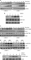
Figure 1
Figure 1
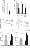
c-Abl−/− MEFs show defects in Atm/Atr-mediated p53 S18 phosphorylation and Chk1/2 phosphorylation. (a) c-Abl−/− MEFs showed decreased p53 phosphorylation at S18 in response to Dox. c-Abl−/− and control MEFs were challenged with l μM of Dox for different periods of time and the levels of p53 and p-p53 S18 were analyzed with western blot. (b) Dox treatment could activate c-Abl. WT MEFs were stimulated with Dox for different periods of time in the presence or absence of STI571. Endogenous c-Abl was immunoprecipitated from these cells and was used for in vitro kinase assay using GST-Crk1 as a substrate. Tyr phosphorylation of Crk1 was detected by western blot using anti-p-Tyr antibodies. (c) c-Abl−/− MEFs showed decreased activation of Chk1 and Chk2 in response to Dox. The experiment was carried out as in (a). p-Chk1, p-Chk2, Chk1, and Chk2 were detected by western blot using specific antibodies. (d) c-Abl−/− MEFs showed compromised p53 phosphorylation in response to HU (mM) and aphidicolin (μg/ml) for 8 h. p-p53 and p53 were detected by western blot using specific antibodies. (e) HU treatment could activate c-Abl. WT MEFs were stimulated with HU for different periods of time in the presence of absence of STI571. Endogenous c-Abl was immunoprecipitated from these cells and was used for in vitro kinase assay using GST-Crk1 as a substrate in the presence of 32P-labeled ATP
Figure 2
Figure 2
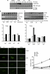
c-Abl−/− MEFs show defects in DNA damage-induced cell death, cell cycle progression, and DNA repair. (a) c-Abl−/− MEFs showed an increased resistance to cell death in response to HU. WT and mutant MEFs of the same litters were treated with 5 mM of HU for 24 h and the cell viability was measured with TUNEL assays. (b) c-Abl−/− MEFs showed a defect in cell cycle control. c-Abl−/−, control, and reconstituted c-Abl−/− MEFs were challenged with 5 Gy of IR and cell cycle profiles were analyzed by FACS after PI staining. *P<0.05 when comparing control and c-Abl−/− cells. (c) c-Abl−/− MEFs showed more RDS in response to IR. Replicative DNA synthesis was assessed at different time points after IR. The results were averages of at least triplicate samples. The resulting ratio of 3H (c.p.m.) to 14C (c.p.m.) was used as an indication of RDS. The ratio at time 0 was set at 100%. (d) c-Abl−/− MEFs showed more cells in the mitotic phase after IR, reflected by the increased percentage of cells positive for phospho-H3. Quantitation data were averaged over three experiments. The number of phospho-H3-positive cells at time 0 was set at 100%. (e) c-Abl−/− MEFs showed normal levels of DSBs. c-Abl−/− and control MEFs were challenged with IR, collected at 0.5 or 24 h after IR, and used for comet assay at neutral pH. *indicates that c-Abl−/− MEFs show a significant difference from WT MEFs at the same time point. (f) c-Abl−/− MEFs showed increased amounts of ssDNA. The experiment was carried out as in (e) except that comet assay was done at alkaline pH. *indicates that c-Abl−/− MEFs show a significant difference from WT MEFs at the same time point
Figure 3
Figure 3
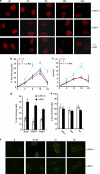
c-Abl deficiency differentially alters nuclear foci assembly of TopBP1, Brca1, 53BP1, Rad51, and γH2AX, but not Atm, Mre11, or Atr. (a) c-Abl−/−, c-Abl reconstituted c-Abl−/−, and control MEFs were treated with Dox for different periods of time and then immuno-stained for endogenous TopBP1. (b) Quantitation data from (a) for the number of cells positive for foci. (c) Quantitation data from (a) for the number of foci per cell. (d) Quantitation data from Supplementary Figure S4 showing that c-Abl deficiency led to an increased number of foci for Brca1, 53BP1, and Rad51. Foci-forming cells were counted from three independent experiments. (e) Quantitation data of c-Abl−/− MEFs showing abnormal nuclear foci assembly of Atm, Atr, and Mre11 (also see Supplementary Figure S5). (f) c-Abl−/− and control MEFs were irradiated with 10 Gy and then were stained for γH2AX at different time points after IR. Right panel: quantitation data
Figure 4
Figure 4
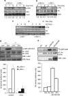
c-Abl is required for optimal activation of Atm and Atr. (a) Decreased activation of Atm in the absence of c-Abl. c-Abl−/− and control MEFs were treated with Dox for different periods of time and activation of Atm was determined by western blot using an antibody that only recognizes active Atm (S1981-p). For quantitation of Atm activation, the value of phospho-Atm in c-Abl−/− MEFs at basal levels was set at 1.0. (b) Diminished phosphorylation of Atr on S428 in response to Dox in the absence of c-Abl. The experiment was carried out as in (a) and the blot was probed with anti-phospho-Atr antibodies. (c) Diminished phosphorylation of Atr in the absence of c-Abl in response to HU. The experiment was carried out just like (b) except that HU was used. (d) In vitro kinase assay showing that c-Abl deficiency compromised Atr activation. Endogenous Atr was immunoprecipitated from WT or c-Abl−/− MEFs, in the presence or absence of Dox, and was used in a kinase assay with GST-p53 as a substrate. For quantitation of Atr activation, the value of phospho-GST-p53 was normalized to the protein levels of Atr. The value in c-Abl−/− MEFs at basal levels was set at 100%. (e) In vitro kinase assay showing that co-expression of c-Abl led to Atr activation. For activation of Atr, the value of 32P-labeled GST-p53 at basal levels was set at 100%
Figure 5
Figure 5
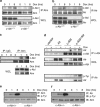
c-Abl interacts with Atm and Atr and is required for Atm/Atr phosphorylation in response to genotoxic stress. (a) Enhanced complex formation between c-Abl and Atm. c-Abl−/− and control MEFs were treated with Dox for different periods of time and endogenous Atm was immunoprecipitated. The possibly associated c-Abl was determined by western blot. (b) Enhanced complex formation between c-Abl and Atr in response to DNA damage. The experiment was carried out as in (a) except that anti-Atr antibodies were used for immunoprecipitation. (c) Association of endogenous c-Abl and Atm in HeLa cells. Cell lysates were immunoprecipitated with anti-Atm antibodies or control IgG, followed by immunoblotting with anti-c-Abl antibodies. The whole cell lysate (WCL) was used as a control. (d) Ectopically expressed c-Abl interacted with Atr and could phosphorylate Atr on tyrosine residues. COS7 cells were transfected with indicated expression constructs and the cell lysates were used to do co-IP assays with anti-c-Abl antibodies. Immunoprecipitated proteins and their associated proteins were detected by western blot analysis using anti-Atr antibodies (upper panel). A second set of cells was used to immunoprecipitate Atr and its tyrosine phosphorylation was detected by using anti-phospho-tyrosine antibodies (PY20) (bottom panel). (e) Atm was tyrosine phosphorylated in a c-Abl-dependent manner. Endogenous Atm was immunoprecipitated from c-Abl−/− and control MEFs that were treated with Dox for different periods of time and its phosphorylation was determined by western blot using PY20. (f) Atr was phosphorylated in response to DNA damage in a c-Abl-dependent manner. The experiment was carried out as in (e) except that anti-Atr antibodies were used for immunoprecipitation
Figure 6
Figure 6
c-Abl phosphorylates Atr on Y291/310. (a) A diagram showing various fragments of ATR and their Y phosphorylation status when co-expressed with c-Abl. (b) Y291/310 are the major phosphorylation sites on Atr by c-Abl. Full-length WT ATR or ATR carrying Y291F, Y310F, or Y291/310F mutation was co-expressed with c-Abl in COS7 cells, immunoprecipitated with anti-Flag antibody (for ATR), and assessed for tyrosine phosphorylation by western blot analysis. (c) c-Abl was able to phosphorylate ATR on Y291/310 in in vitro kinase assays. GST-ATR(F1) and GST-ATR(m1/2) were expressed and purified from bacteria, and were used in in vitro c-Abl kinase assay. Phosphorylation was determined by western blot analysis
Figure 7
Figure 7
Evidence that Y291 and Y310 are required for ATR activation. (a) In vitro kinase assay showing that Y291F and Y310F mutations diminished ATR activity. ATR-deficient cells were transfected with vectors expressing wild-type, m1, or m2 mutant ATR for 2 days, which were then treated with HU for 4 or 8 h. ATR was immunoprecipitated with anti-Flag antibodies and was used in a kinase assay with GST-p53 as a substrate. (b) Y291 and Y310 are important for ATR-mediated p53 phosphorylation on S18 in vivo. WT ATR or ATR carrying Y291F (m1) or Y310F (m2) mutation was expressed in ATR-deficient fibroblasts (with Neo-pcDNA vector) for 24 h and then selected for 48 h or more. The cells were then challenged with HU for 8 h and then collected. ATR, p53, p–p53, p21, and actin were analyzed by western blot. Bottom panel: quantitation data. (c) Y291 and Y310 are important for ATR-mediated Chk1 phosphorylation in vivo. The experiments were carried out as in (b) and Chk1 phosphorylation and Chk1 protein levels were determined by western blot. Bottom panel: quantitation data. The value of p-p53 in ATR-deficient cells with control vector at time 0 was set at 1. (d) Y291 and Y310 are important for ATR-mediated γH2AX nuclear foci formation. ATR-deficient cells were transfected with vectors expressing wild-type, m1, or m2 mutant ATR for 2 days, which were then treated with HU for 4 or 8 h. γH2AX was detected with immunostaining by anti-γH2AX antibodies. Right panel: quantitation data. The value of p-p53 in ATR-deficient cells with control vector at time 0 was set at 1

Comment in

Similar articles

Cited by

References

    1. Peterson CL, Cote J. Cellular machineries for chromosomal DNA repair. Genes Dev. 2004;18:602–616. - PubMed
    1. Abraham RT. Cell cycle checkpoint signaling through the ATM and ATR kinases. Genes Dev. 2001;15:2177–2196. - PubMed
    1. Shiloh Y. ATM and related protein kinases: safeguarding genome integrity. Nat Rev Cancer. 2003;3:155–168. - PubMed
    1. Cline SD, Hanawalt PC. Who's on first in the cellular response to DNA damage. Nat Rev Mol Cell Biol. 2003;4:361–372. - PubMed
    1. D'Amours D, Jackson SP. The Mre11 complex: at the crossroads of dna repair and checkpoint signalling. Nat Rev Mol Cell Biol. 2002;3:317–327. - PubMed

Publication types

MeSH terms