. 2010 Aug 27;39(4):548-59.
doi: 10.1016/j.molcel.2010.07.027.

Catalysis of lysine 48-specific ubiquitin chain assembly by residues in E2 and ubiquitin

Affiliations

Catalysis of lysine 48-specific ubiquitin chain assembly by residues in E2 and ubiquitin

Monica C Rodrigo-Brenni et al. Mol Cell. .

Abstract

Protein ubiquitination is catalyzed by ubiquitin-conjugating enzymes (E2s) in collaboration with ubiquitin-protein ligases (E3s). This process depends on nucleophilic attack by a substrate lysine on a thioester bond linking the C terminus of ubiquitin to a cysteine in the E2 active site. Different E2 family members display specificity for lysines in distinct contexts. We addressed the mechanistic basis for this lysine selectivity in Ubc1, an E2 that catalyzes the ubiquitination of lysine 48 (K48) in ubiquitin, leading to the formation of K48-linked polyubiquitin chains. We identified a cluster of polar residues near the Ubc1 active site, as well as a residue in ubiquitin itself, that are required for catalysis of K48-specific ubiquitin ligation, but not for general activity toward other lysines. Our results suggest that the active site of Ubc1, as well as the surface of ubiquitin, contains specificity determinants that channel specific lysines to the central residues involved directly in catalysis.


Figures

Figure 1
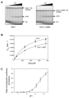
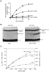
Figure 1. The catalytic core of Ubc1 binds ubiquitin
(A) Purified Ubc1 (left) or Ubc1-ΔUBA (right) was conjugated with 32P-labeled K48R-ubiquitin and treated with NEM and EDTA to prevent recharging of Ubc1 and E1. Increasing amounts of unlabeled ubiquitin (50 μM to 750 μM) were added. After 2 min, reactions were stopped by addition of nonreducing sample buffer and analyzed by SDS-PAGE. Free ubiquitin (Ub), diubiquitin (diUb) and charged Ubc1 (Ubc1∼Ub) are indicated. See Figure S1 for a time course of similar reactions. (B) Initial rates of diubiquitin synthesis were measured over a range of ubiquitin concentrations in reactions like those in panel A (n=5 for Ubc1; n=3 for Ubc1-ΔUBA). The rate of diubiquitin synthesis was divided by total Ubc1-Ub to estimate turnover rate (kobs). Data were fit to a rectangular hyperbola using the ligand-binding module of SigmaPlot. Error bars represent SEM. See Table S1 for apparent Kd and k2 values. (C) Initial rates of diubiquitin synthesis were determined at saturating ubiquitin concentration (1 mM) over a range of pH values (6.86-10.26). At each pH, diubiquitin formation was measured over a time course. The appearance of diubiquitin was plotted as a function of time and fit to a linear function using Excel. The rate of diubiquitin synthesis (the slope of the linear function) was plotted as a function of pH and fit to a sigmoidal function using SigmaPlot. Experiments were done in triplicate and the error bars represent SEM.
Figure 2
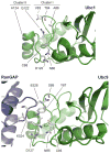
Figure 2. Conserved residues in the active site of Ubc1
(top) The Ubc1 active site contains five surface-exposed side chains, in two clusters, that are conserved in human E2-25K but are not present in Ubc4 (see Figure S2 for E2 sequence alignments). In the Ubc1-ubiquitin conjugate (Hamilton et al., 2001; Merkley and Shaw, 2004), ubiquitin is positioned to the lower right of the active-site cysteine (C88) in this orientation (not shown). Additional residues known to be important for catalysis in other E2s (D120, N80; see Figure S2) are also labeled. (bottom) The structure of Ubc9 in complex with its substrate RanGAP1 (Bernier-Villamor et al., 2002) illustrates the orientation of the attacking lysine (K524). The active-site cysteine of Ubc9 (C93) is surrounded by three residues (Y87, D127, and N85) that are important in catalysis (Yunus and Lima, 2006). Note that the position of Y87 is occupied by the side chain of Q122 in Ubc1 (top). RanGAP1 contains a glutamate (E526) that is a key part of the SUMO consensus sequence and interacts with a serine (S89) in Ubc9. In Ubc1 (top), T84 is found in a similar position, consistent with a role in substrate orientation. Images constructed with PyMOL (DeLano, 2008).
Figure 3
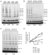
Figure 3. The hydroxyl group of T84 is required for K48-specific polyubiquitination
(A-C) Purified Ubc1 or the indicated mutant was incubated for 15 min with E1, ATP and the indicated ubiquitin species (MeUb indicates methyl-ubiquitin). E1/E2 mixes were added to APC, Cdh1 and 125I-cyclin B and incubated for 45 min at room temperature. Reaction products were analyzed by SDS-PAGE and PhosphorImager. See Figure S3 for analysis of the cluster I mutant containing all three mutations. (D) Methyl-ubiquitin incorporation was measured in APC reactions with Ubc1 or the indicated mutant. Each E2 was incubated for 15 min with E1, ATP and methyl-ubiquitin, and then added to an APCCdh1 mix containing 125I-cyclin B at time zero. Reaction products over time were analyzed by SDS-PAGE and PhosphorImager, and quantified with ImageQuant.
Figure 4
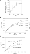
Figure 4. T84 is required for K48-specific catalysis
(A) Rates of diubiquitin synthesis were measured with the Ubc1-T84G mutant and saturating ubiquitin (1 mM) over a range of pH values (6.86-10.26), as in Figure 1C. Experiments were done in triplicate and the error bars represent SEM. (B) Diubiquitin formation by Ubc1 and Ubc1-T84G was measured over a range of ubiquitin concentrations (50 μM to 750 μM) in reactions like those in Figure 1A, except that the pH of the reaction was 10.26 and the reaction time was 10 s for Ubc1 and 2 min for Ubc1-T84G. Reaction products were analyzed by SDS-PAGE and PhosphorImager. Data from two experiments were quantified with ImageQuant, and the resulting plots were fit to a rectangular hyperbola using SigmaPlot. Error bars represent SEM. See Table S1 for apparent Kd and k2 values. (C) Diubiquitin formation by Ubc1-T84G was measured with wild-type ubiquitin or ubiquitin-K48R in reactions like those in Figure 1A, except at pH 10.15 for 2 min. Reaction products were analyzed by SDS-PAGE and PhosphorImager. Data from four experiments were quantified with ImageQuant, and the resulting plots were fit to a rectangular hyperbola using Prism. Error bars represent SEM. See Table S1 for apparent Kd and k2 values.
Figure 5
Figure 5. Q122 and A124 contribute to lysine specificity
(A, B) Purified Ubc1 or the indicated mutant was incubated for 15 min with E1, ATP and the indicated ubiquitin species. E1/E2 mixes were added to APC, Cdh1 and 125I-cyclin B and incubated for 45 min at room temperature. Reaction products were analyzed by SDS-PAGE and PhosphorImager. See Figure S3 for analysis of the cluster II mutant containing both Q122L and A124P mutations. See Figure S4 for analysis of mutants in which Q122 is replaced with other amino acids. (C) Initial rates of diubiquitin synthesis were measured in 2-min reactions with Ubc1-Q122L or Ubc1-Q122A over a range of ubiquitin concentrations, as in Figure 1A. Data (n=3) were fit to a rectangular hyperbola using the ligand-binding module of SigmaPlot. Error bars represent SEM. See Table S1 for apparent Kd and k2 values. (D) Rates of diubiquitin synthesis were measured with the Ubc1-Q122L mutant and saturating ubiquitin (1 mM) over a range of pH values (6.86-10.26), as in Figure 1C. Experiments were done in duplicate and the error bars represent SEM.
Figure 6
Figure 6. Q122 limits the lysine specificity of Ubc1
(A) Methyl-ubiquitin incorporation was measured in APC reactions with Ubc1, Ubc1-Q122L, Ubc1-Q122A or Ubc4, as in Figure 3D. (B) Purified Ubc1 or Ubc1-Q122L was conjugated with 32P-labeled K48R-ubiquitin and treated with NEM and EDTA to prevent recharging of Ubc1 and E1. 12 mg of purified sea urchin cyclin B fragment was added, and samples were removed at the indicated times and analyzed by SDS-PAGE and PhosphorImager. (C) Ubc1, Ubc1-Q122L or Ubc4 was conjugated with 32P-labeled K48R-ubiquitin and treated with NEM and EDTA to prevent recharging of Ubc1 and E1. E1/E2 was added to reactions containing increasing amounts of purified sea urchin cyclin B (90 μM to 1000 μM) at pH 10.26, for different times (2 min for Ubc1, 10 s for Ubc1-Q122L, and 1 min for Ubc4). Reaction products were analyzed by SDS-PAGE and PhosphorImager. Data from three experiments were quantified using ImageQuant, and the resulting plots were fit to a rectangular hyperbola using SigmaPlot. Error bars represent SEM. See Table S1 for apparent Kd and k2 values.
Figure 7
Figure 7. Tyrosine 59 of ubiquitin is required for K48-specific polyubiquitination
(A) Purified Ubc1 was conjugated to ubiquitin and added to APCCdh1 and a 32P-labeled ubiquitin-cyclin fusion protein carrying the indicated ubiquitin mutation. After 15 min, reaction products were analyzed by SDS-PAGE and PhosphorImager. See Figure S7 for characterization of reactions with the ubiquitin-cyclin fusion protein. (B, C) Purified Ubc1 or Ubc4 was incubated with E1, ATP and the indicated ubiquitin species for 15 min, then mixed with APCCdh1 and 125I-cyclin B and incubated for 45 min (B) or 60 min (C) at room temperature. Reaction products were analyzed by SDS-PAGE and PhosphorImager. Wild-type ubiquitin in panel B is native protein that migrates more rapidly than the other ubiquitin species, which carry extra sequence derived from an excised tag at the N-terminus (see Experimental Procedures). In panel C, the left autoradiograph was exposed 20-fold longer than the one on the right. (D) Ubc1 was incubated with E1, ATP and 32P-labeled K48R-ubiquitin for 15 min, and NEM and EDTA were added. E1/E2 mix was added to reactions with increasing amounts (50 μM to 1000 μM) of wild-type ubiquitin or ubiquitin-Y59L at pH 10.15, for different reaction times (5 s for wild-type ubiquitin; 10 min for ubiquitin-Y59L). Reaction products were analyzed by SDS-PAGE and PhosphorImager. Data from three experiments were quantified using ImageQuant, and the resulting plots were fit to a rectangular hyperbola using Prism. Error bars represent SEM. See Table S1 for apparent Kd and k2 values.

Similar articles

Cited by

References

    1. Bernier-Villamor V, Sampson DA, Matunis MJ, Lima CD. Structural basis for E2-mediated SUMO conjugation revealed by a complex between ubiquitin-conjugating enzyme Ubc9 and RanGAP1. Cell. 2002;108:345–356. - PubMed
    1. Capili AD, Lima CD. Taking it step by step: mechanistic insights from structural studies of ubiquitin/ubiquitin-like protein modification pathways. Curr Opin Struct Biol. 2007;17:726–735. - PMC - PubMed
    1. Carroll CW, Morgan DO. Enzymology of the Anaphase-Promoting Complex. Meth Enzymol. 2005;398:219–230. - PubMed
    1. Cook WJ, Jeffrey LC, Xu Y, Chau V. Tertiary structures of class I ubiquitin-conjugating enzymes are highly conserved: crystal structure of yeast Ubc4. Biochemistry. 1993;32:13809–13817. - PubMed
    1. DeLano WL. The PyMOL Molecular Graphics System. Palo Alto, CA, USA: DeLano Scientific LLC; 2008. http://www.pymol.org.

Publication types

MeSH terms

LinkOut - more resources