. 2010 Nov;84(21):11395-406.
doi: 10.1128/JVI.01047-10. Epub 2010 Aug 25.

An RNA pseudoknot is required for production of yellow fever virus subgenomic RNA by the host nuclease XRN1

Affiliations

An RNA pseudoknot is required for production of yellow fever virus subgenomic RNA by the host nuclease XRN1

Patrícia A G C Silva et al. J Virol. 2010 Nov.

Abstract

Cells and mice infected with arthropod-borne flaviviruses produce a small subgenomic RNA that is colinear with the distal part of the viral 3'-untranslated region (UTR). This small subgenomic flavivirus RNA (sfRNA) results from the incomplete degradation of the viral genome by the host 5'-3' exonuclease XRN1. Production of the sfRNA is important for the pathogenicity of the virus. This study not only presents a detailed description of the yellow fever virus (YFV) sfRNA but, more importantly, describes for the first time the molecular characteristics of the stalling site for XRN1 in the flavivirus genome. Similar to the case for West Nile virus, the YFV sfRNA was produced by XRN1. However, in contrast to the case for other arthropod-borne flaviviruses, not one but two sfRNAs were detected in YFV-infected mammalian cells. The smaller of these two sfRNAs was not observed in infected mosquito cells. The larger sfRNA could also be produced in vitro by incubation with purified XRN1. These two YFV sfRNAs formed a 5'-nested set. The 5' ends of the YFV sfRNAs were found to be just upstream of the previously predicted RNA pseudoknot PSK3. RNA structure probing and mutagenesis studies provided strong evidence that this pseudoknot structure was formed and served as the molecular signal to stall XRN1. The sequence involved in PSK3 formation was cloned into the Sinrep5 expression vector and shown to direct the production of an sfRNA-like RNA. These results underscore the importance of the RNA pseudoknot in stalling XRN1 and also demonstrate that it is the sole viral requirement for sfRNA production.


Figures
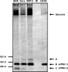
FIG. 1.
FIG. 1.
YFV-17D sfRNA production in mammalian and insect cell lines. The mammalian cell lines BHK-21J, Vero E6, and SW13 and the mosquito cell line C6/36 were infected with YFV-17D at an MOI of 10. At 30 h (mammalian cell lines) or 36 h (mosquito cell line) p.i., total RNA was isolated and analyzed by Northern blotting for the production of YFV-17D sfRNA. Lane M, total RNA isolated from mock-infected BHK-21J cells. Oligonucleotide 1632, complementary to nt 10,690 to 10,708 of the YFV 3′ UTR, was used as a probe. Size markers are indicated on the left. Bands corresponding to the YFV-17D genome and to three small viral RNAs (A, B, and C) are indicated by arrows. RNA B and RNA C are referred to as sfRNA1 and sfRNA2, respectively.
FIG. 2.
FIG. 2.
Determining the 5′ end of the YFV 3′ UTR-specific sfRNAs produced in BHK-21J cells. (A) Schematic diagram of the predicted secondary structure of the YFV 3′ UTR (41). (B) Primer extension analysis using oligonucleotide 1632, which is complementary to YFV nt 10,690 to 10,708 (see panel A). pBluescript-YFV9,845-10,861, containing the COOH-terminal part of the YFV NS5 gene and the complete 3′ UTR, was sequenced with oligonucleotide 1632 to obtain a sequencing ladder for determination of the 5′ end of the sfRNAs. Lanes 1 and 2, primer extension on total RNAs isolated from YFV-infected and uninfected BHK-21J cells, respectively; lane 3, primer extension on a full-length in vitro YFV transcript mixed with total RNA from uninfected BHK-21J cells. The underlined T residues of the depicted sequence correspond to the 5′ ends of the primer extension products and map the 5′ ends of the YFV sfRNAs to the A residues at positions 10,532 and 10,533 of the YFV genome. (C) RNase protection assay using a 229-nt antisense RNA probe encompassing nt 10,520 to 10,714 of the YFV-17D 3′ UTR. Lane 1, 32P-labeled RNA transcript used as the probe; lane 2, RNA fragments that were protected from RNase digestion after hybridization of the probe to total RNA isolated from YFV-17D-infected cells; lane 3, RNase protection assay on total RNA of mock-infected cells; lane 4, protected RNA fragments obtained when pBluescript-YFV9,845-10,861 transcripts mixed with total RNA from mock-infected BHK-21J cells were analyzed. Bands corresponding to the protected fragments derived from either the YFV-17D genome or the sfRNA are indicated. pBluescript-YFV9,845-10,861 was sequenced with oligonucleotide 1632 to obtain a marker to determine the sizes of the protected RNA fragments.
FIG. 3.
FIG. 3.
YFV sfRNA2 is truncated at the 3′ end. (A) Scheme of the YFV 3′ UTR. The two possible orientations of the sfRNAs relative to the YFV 3′ UTR are depicted. The positions of oligonucleotides 1648 and 1296, used to determine the orientations of the sfRNAs, are indicated. Total RNAs were isolated from the indicated YFV-infected cell lines or from mock-infected BHK-21J cells and subsequently analyzed by Northern blotting. (B) Northern blot analysis using oligonucleotide 1648, complementary to YFV-17D nt 10,580 to 10,598. (C) Northern blot analysis using oligonucleotide 1296, complementary to YFV-17D nt 10,830 to 10,862.
FIG. 4.
FIG. 4.
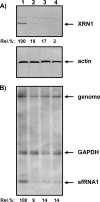
shRNA-mediated XRN1 silencing decreases YFV sfRNA production. SW13 cells were transduced with an shRNA-expressing lentivirus from the MISSIONTRC-Hs1.0 library (Sigma) and then infected with YFV-17D as described in Materials and Methods. Lentiviruses expressed the following shRNAs: lane 1, scrambled shRNA (SHC-002); lane 2, shRNAs TRCN-049675 and TRCN-049676; lane 3, shRNAs TRCN-049675 and TRCN-049677; and lane 4, shRNAs TRCN-049676 and TRCN-049677. (A) XRN1 and actin expression in Western blots. Rel. %, percentage of XRN1 expression in cells transduced with shRNAs against XRN1 compared to that in cells expressing the scrambled shRNA. (B) YFV sfRNA production and GAPDH mRNA expression by Northern analysis. Rel. %, expression of YFV sfRNA in cells transduced with shRNAs against XRN1 compared to that in cells expressing the scrambled shRNA.
FIG. 5.
FIG. 5.
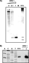
In vitro production of YFV-17D sfRNAs by the host exoribonuclease XRN1. TAP-treated in vitro RNA transcripts of YF-R.luc2A-RP were incubated with the indicated units of XRN1 and analyzed by hybridization after denaturing gel electrophoresis (A) and primer extension (B), using oligonucleotide 1632, complementary to YFV-17D nt 10,690 to 10,708, as a probe and primer, respectively. RNAs isolated from mock (M)- and/or YFV-infected BHK-21J cells were used as controls in both experiments.
FIG. 6.
FIG. 6.
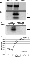
Stem-loop structure SL-E in the YFV 3′ UTR is required for the production of sfRNA. (A) Viral RNA synthesis in BHK-21J cells transfected with in vitro-transcribed genome-length RNAs of YFV-17D and YFV-ΔSL-E. Transfected cells were labeled with [3H]uridine from 18 to 24 h posttransfection. Total RNAs were isolated and analyzed after denaturation by agarose gel electrophoresis, as described in Materials and Methods. (B) sfRNA production in BHK-21J cells transfected with YFV-17D and YFV-ΔSL-E transcripts. Total RNAs were isolated at 24 h posttransfection and analyzed by Northern blotting and hybridization with oligonucleotide 1632 (complementary to YFV-17D nt 10,690 to 10,708). The sfRNAs are indicated by arrows. (C) In vitro RNA transcripts of YFV-17D and YFV-ΔSL-E were incubated in the presence or absence of 1 unit of XRN1 and analyzed for the production of sfRNA by Northern blotting, using oligonucleotide 1632 as a probe. The sfRNA is indicated by an arrow. (D) Viral growth kinetics of YFV-17D and the YFV-ΔSL-E mutant. BHK-21J cells were infected at an MOI of 5, and the medium of the infected cells was sampled at the indicated times postinfection. Titers were determined by plaque assays on BHK-21J cells.
FIG. 7.
FIG. 7.
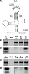
YFV-17D RNA pseudoknot 3 is required for sfRNA production. (A) Schematic diagram of the YFV 3′ UTR SL-E structure. The primary sequences involved in stem-loop e2 (sequences A and B) and in the pseudoknot interaction (sequences pk3′ and pk3) are depicted. (B and D) Viral RNA synthesis in BHK-21J cells transfected with in vitro-transcribed genomic RNAs of YFV-17D and YFV mutant viruses (at SL-E e2 [B] and at PSK3 [D]). Transfected cells were labeled with [3H]uridine from 18 to 24 h posttransfection. Total RNAs were isolated and analyzed after denaturation by agarose gel electrophoresis, as described in Materials and Methods. YFV(AB) and YFV(pk3′3) indicate wild-type YFV-17D for these different groups of mutants. (C and E) Northern blot analysis of RNAs isolated from BHK cells 30 h after transfection with YFV-17D and YFV mutant viruses (at SL-E e2 [C] and at PSK3 [E]). Oligonucleotide 1632 (complementary to YFV-17D nt 10,690 to 10,708) was used as a probe. In panel E, the dotted line separates the wild-type YFV-17D and mock lanes from the same Northern blot at a higher exposure.
FIG. 8.
FIG. 8.
Insertion of sequences required for the formation of PSK3 is sufficient to produce an sfRNA-like RNA in the context of a Sindbis virus replicon. (A) Scheme of the characteristics of the pSinrep5 vector and the predicted structures of the YFV-17D regions that were inserted into the vector. The YFV 3′ UTR nucleotides cloned into pSinRep5-eGFP are indicated in the name of each construct. The promoter for the enhanced GFP (eGFP)-expressing subgenomic Sinrep mRNA and the binding sites for oligonucleotides 1674 and 1648 are indicated. (B) Northern blot analysis of RNAs isolated from BHK-21J cells at 8 h p.e. with Sinrep5-eGFP (lanes 1 and 5), Sinrep5-eGFPYFV10,521-10,662 (lanes 2 and 6), and Sinrep5-eGFPYFV10,531-10,611 (lanes 3 and 7) RNAs; lanes 4 and 8 correspond to uninfected BHK-21J cells. Oligonucleotide 1674 and oligonucleotide 1648 were used as probes, as specified in the figure. The Sindbis virus genomic (gRNA) and subgenomic (sgRNA) RNAs and the sfRNA-like RNAs are indicated. (C) Primer extension analysis with oligonucleotide 1648 to determine the 5′ end of the sfRNA-like RNAs produced with the Sinrep5 mutants. pBluescript-YFV9,845-10,861 was sequenced with oligonucleotide 1648 to obtain a sequencing ladder. RNAs isolated from BHK-21J cells transfected with YFV-17D (lane 1), Sinrep5-eGFPYFV10,521-10,662 (lane 2), and Sinrep5-eGFPYFV10,531-10,611 (lane 3) were analyzed; lane 4 corresponds to mock-transfected cells.
FIG. 9.
FIG. 9.
The predicted pseudoknot PSK3 at the wild-type YFV 3′ UTR is genuine. (A) Schematic diagram of the minimal YFV 3′ UTR region (nt 10,531 to 10,611) required for stalling of XRN1. Sequences involved in the pseudoknot interaction are depicted. (B) In vitro structure probing by SHAPE with wild-type YFV and the PSK3 mutants YFV-pk3′pk3′, YFV-pk3pk3, and YFV-pk3pk3′. After treatment with NMIA, which preferentially reacts with bases in a single-stranded conformation, in vitro-synthesized RNA templates (YFV nt 10,520 to 10,708) were analyzed by primer extension with oligonucleotide 1632 (complementary to YFV nt 10,690 to 10,708). Samples were treated with either 65 mM NMIA in DMSO (+) or, as a control, DMSO only (−). The different substructures of SL-E and the pk3 and pk3′ sequences are indicated on the right relative to their positions in the NMIA reactivity pattern. pBluescript-YFV9,845-10,861 was sequenced with oligonucleotide 1632 to obtain a sequencing ladder.

Similar articles

Cited by

References

    1. Alvarez, D. E., A. L. De Lella Ezcurra, S. Fucito, and A. V. Gamarnik. 2005. Role of RNA structures present at the 3′UTR of dengue virus on translation, RNA synthesis, and viral replication. Virology 339:200-212. - PubMed
    1. Anderson, P., and N. Kedersha. 2006. RNA granules. J. Cell Biol. 172:803-808. - PMC - PubMed
    1. Ausubel, F. M., R. Brent, R. E. Kingston, D. D. Moore, J. G. Seidman, J. A. Smith, and K. Struhl. 2000. Current protocols in molecular biology. Wiley Interscience, New York, NY.
    1. Baranov, P. V., C. M. Henderson, C. B. Anderson, R. F. Gesteland, J. F. Atkins, and M. T. Howard. 2005. Programmed ribosomal frameshifting in decoding the SARS-CoV genome. Virology 332:498-510. - PMC - PubMed
    1. Bonisch, C., C. Temme, B. Moritz, and E. Wahle. 2007. Degradation of hsp70 and other mRNAs in Drosophila via the 5′ 3′ pathway and its regulation by heat shock. J. Biol. Chem. 282:21818-21828. - PubMed

LinkOut - more resources