Culturing the unculturable: human coronavirus HKU1 infects, replicates, and produces progeny virions in human ciliated airway epithelial cell cultures
- PMID: 20719951
- PMCID: PMC2953148
- DOI: 10.1128/JVI.00947-10
Culturing the unculturable: human coronavirus HKU1 infects, replicates, and produces progeny virions in human ciliated airway epithelial cell cultures
Abstract
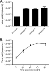
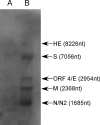
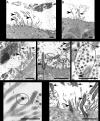
Culturing newly identified human lung pathogens from clinical sample isolates can represent a daunting task, with problems ranging from low levels of pathogens to the presence of growth suppressive factors in the specimens, compounded by the lack of a suitable tissue culture system. However, it is critical to develop suitable in vitro platforms to isolate and characterize the replication kinetics and pathogenesis of recently identified human pathogens. HCoV-HKU1, a human coronavirus identified in a clinical sample from a patient with severe pneumonia, has been a major challenge for successful propagation on all immortalized cells tested to date. To determine if HCoV-HKU1 could replicate in in vitro models of human ciliated airway epithelial cell cultures (HAE) that recapitulate the morphology, biochemistry, and physiology of the human airway epithelium, the apical surfaces of HAE were inoculated with a clinical sample of HCoV-HKU1 (Cean1 strain). High virus yields were found for several days postinoculation and electron micrograph, Northern blot, and immunofluorescence data confirmed that HCoV-HKU1 replicated efficiently within ciliated cells, demonstrating that this cell type is infected by all human coronaviruses identified to date. Antiserum directed against human leukocyte antigen C (HLA-C) failed to attenuate HCoV-HKU1 infection and replication in HAE, suggesting that HLA-C is not required for HCoV-HKU1 infection of the human ciliated airway epithelium. We propose that the HAE model provides a ready platform for molecular studies and characterization of HCoV-HKU1 and in general serves as a robust technology for the recovery, amplification, adaptation, and characterization of novel coronaviruses and other respiratory viruses from clinical material.
Figures


Similar articles
-
Isolation and characterization of current human coronavirus strains in primary human epithelial cell cultures reveal differences in target cell tropism.J Virol. 2013 Jun;87(11):6081-90. doi: 10.1128/JVI.03368-12. Epub 2013 Feb 20. J Virol. 2013. PMID: 23427150 Free PMC article.
-
SARS-CoV replication and pathogenesis in an in vitro model of the human conducting airway epithelium.Virus Res. 2008 Apr;133(1):33-44. doi: 10.1016/j.virusres.2007.03.013. Epub 2007 Apr 23. Virus Res. 2008. PMID: 17451829 Free PMC article. Review.
-
Human Coronavirus HKU1 Spike Protein Uses O-Acetylated Sialic Acid as an Attachment Receptor Determinant and Employs Hemagglutinin-Esterase Protein as a Receptor-Destroying Enzyme.J Virol. 2015 Jul;89(14):7202-13. doi: 10.1128/JVI.00854-15. Epub 2015 Apr 29. J Virol. 2015. PMID: 25926653 Free PMC article.
-
Identification of major histocompatibility complex class I C molecule as an attachment factor that facilitates coronavirus HKU1 spike-mediated infection.J Virol. 2009 Jan;83(2):1026-35. doi: 10.1128/JVI.01387-08. Epub 2008 Nov 5. J Virol. 2009. PMID: 18987136 Free PMC article.
-
Properties of Coronavirus and SARS-CoV-2.Malays J Pathol. 2020 Apr;42(1):3-11. Malays J Pathol. 2020. PMID: 32342926 Review.
Cited by
-
Immunodetection assays for the quantification of seasonal common cold coronaviruses OC43, NL63, or 229E infection confirm nirmatrelvir as broad coronavirus inhibitor.Antiviral Res. 2022 Jul;203:105343. doi: 10.1016/j.antiviral.2022.105343. Epub 2022 May 19. Antiviral Res. 2022. PMID: 35598779 Free PMC article.
-
Evaluation of serologic and antigenic relationships between middle eastern respiratory syndrome coronavirus and other coronaviruses to develop vaccine platforms for the rapid response to emerging coronaviruses.J Infect Dis. 2014 Apr 1;209(7):995-1006. doi: 10.1093/infdis/jit609. Epub 2013 Nov 18. J Infect Dis. 2014. PMID: 24253287 Free PMC article.
-
A mouse model for Betacoronavirus subgroup 2c using a bat coronavirus strain HKU5 variant.mBio. 2014 Mar 25;5(2):e00047-14. doi: 10.1128/mBio.00047-14. mBio. 2014. PMID: 24667706 Free PMC article.
-
Immunofluorescence-Mediated Detection of Respiratory Virus Infections in Human Airway Epithelial Cultures.Curr Protoc. 2022 Jun;2(6):e453. doi: 10.1002/cpz1.453. Curr Protoc. 2022. PMID: 35671174 Free PMC article.
-
In Vitro Modelling of Respiratory Virus Infections in Human Airway Epithelial Cells - A Systematic Review.Front Immunol. 2021 Aug 18;12:683002. doi: 10.3389/fimmu.2021.683002. eCollection 2021. Front Immunol. 2021. PMID: 34489934 Free PMC article.
References
Publication types
MeSH terms
Substances
Grants and funding
LinkOut - more resources
Full Text Sources
Other Literature Sources
Research Materials

