

Homozygosity mapping reveals null mutations in FAM161A as a cause of autosomal-recessive retinitis pigmentosa
- PMID: 20705279
- PMCID: PMC2933343
- DOI: 10.1016/j.ajhg.2010.07.022
Homozygosity mapping reveals null mutations in FAM161A as a cause of autosomal-recessive retinitis pigmentosa
Abstract
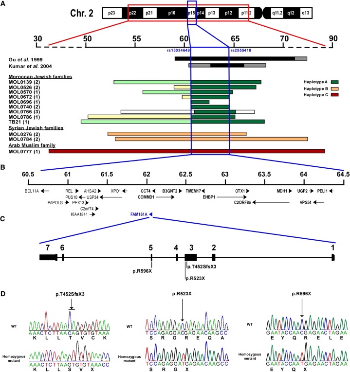
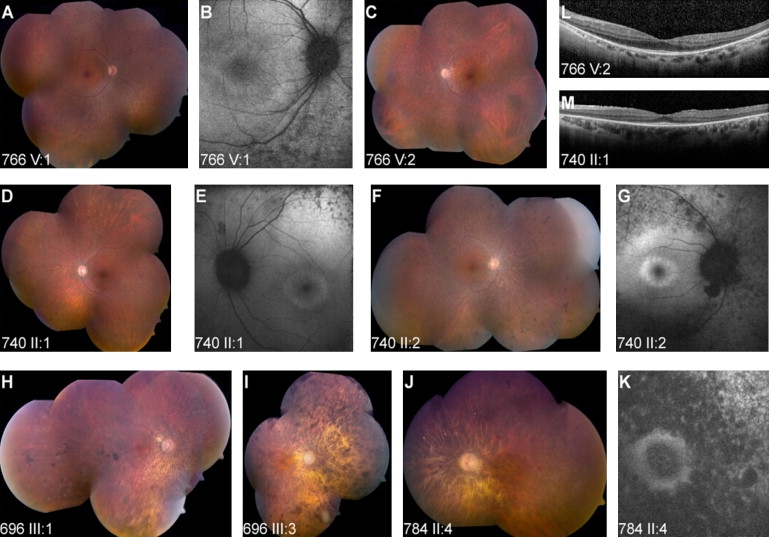
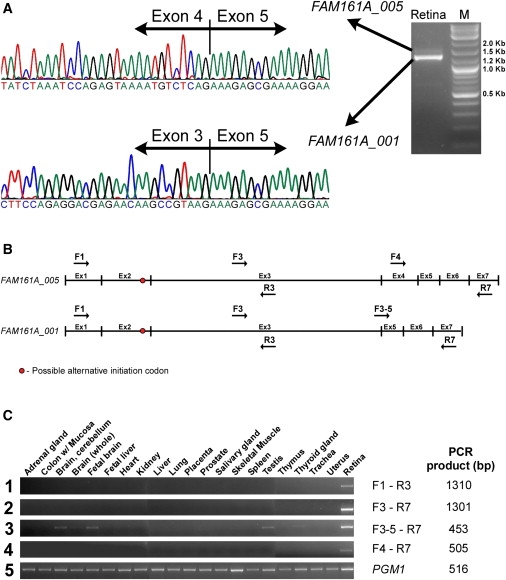
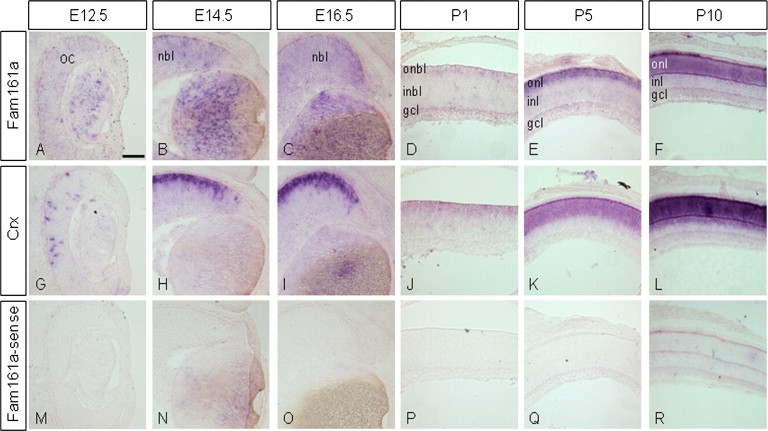
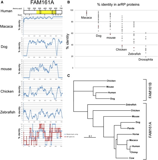
Retinitis pigmentosa (RP) is a heterogeneous group of inherited retinal degenerations caused by mutations in at least 45 genes. Using homozygosity mapping, we identified a ∼4 Mb homozygous region on chromosome 2p15 in patients with autosomal-recessive RP (arRP). This region partially overlaps with RP28, a previously identified arRP locus. Sequence analysis of 12 candidate genes revealed three null mutations in FAM161A in 20 families. RT-PCR analysis in 21 human tissues revealed high levels of FAM161A expression in the retina and lower levels in the brain and testis. In the human retina, we identified two alternatively spliced transcripts with an intact open reading frame, the major one lacking a highly conserved exon. During mouse embryonic development, low levels of Fam161a transcripts were detected throughout the optic cup. After birth, Fam161a expression was elevated and confined to the photoreceptor layer. FAM161A encodes a protein of unknown function that is moderately conserved in mammals. Clinical manifestations of patients with FAM161A mutations varied but were largely within the spectrum associated with arRP. On funduscopy, pallor of the optic discs and attenuation of blood vessels were common, but bone-spicule-like pigmentation was often mild or lacking. Most patients had nonrecordable electroretinographic responses and constriction of visual fields upon diagnosis. Our data suggest a pivotal role for FAM161A in photoreceptors and reveal that FAM161A loss-of-function mutations are a major cause of arRP, accounting for ∼12% of arRP families in our cohort of patients from Israel and the Palestinian territories.
2010 The American Society of Human Genetics. Published by Elsevier Inc. All rights reserved.
Figures
Similar articles
-
Nonsense mutations in FAM161A cause RP28-associated recessive retinitis pigmentosa.Am J Hum Genet. 2010 Sep 10;87(3):376-81. doi: 10.1016/j.ajhg.2010.07.018. Epub 2010 Aug 12. Am J Hum Genet. 2010. PMID: 20705278 Free PMC article.
-
Homozygosity mapping reveals new nonsense mutation in the FAM161A gene causing autosomal recessive retinitis pigmentosa in a Palestinian family.Mol Vis. 2014 Feb 7;20:178-82. eCollection 2014. Mol Vis. 2014. PMID: 24520187 Free PMC article.
-
A novel homozygous R764H mutation in crumbs homolog 1 causes autosomal recessive retinitis pigmentosa.Mol Vis. 2013 Apr 5;19:829-34. Print 2013. Mol Vis. 2013. PMID: 23592920 Free PMC article.
-
FAM161A, a novel centrosomal-ciliary protein implicated in autosomal recessive retinitis pigmentosa.Adv Exp Med Biol. 2014;801:185-90. doi: 10.1007/978-1-4614-3209-8_24. Adv Exp Med Biol. 2014. PMID: 24664697 Review.
-
Morphological and Functional Comparison of Mice Models for Retinitis Pigmentosa.Adv Exp Med Biol. 2023;1415:365-370. doi: 10.1007/978-3-031-27681-1_53. Adv Exp Med Biol. 2023. PMID: 37440058 Review.
Cited by
-
Unique combination of clinical features in a large cohort of 100 patients with retinitis pigmentosa caused by FAM161A mutations.Sci Rep. 2020 Sep 16;10(1):15156. doi: 10.1038/s41598-020-72028-0. Sci Rep. 2020. PMID: 32938956 Free PMC article.
-
Whole-exome sequencing identifies novel mutations in genes responsible for retinitis pigmentosa in 2 nonconsanguineous Chinese families.Int J Ophthalmol. 2019 Jun 18;12(6):915-923. doi: 10.18240/ijo.2019.06.06. eCollection 2019. Int J Ophthalmol. 2019. PMID: 31236346 Free PMC article.
-
Gene augmentation therapy attenuates retinal degeneration in a knockout mouse model of Fam161a retinitis pigmentosa.Mol Ther. 2023 Oct 4;31(10):2948-2961. doi: 10.1016/j.ymthe.2023.08.011. Epub 2023 Aug 14. Mol Ther. 2023. PMID: 37580905 Free PMC article.
-
Identifying mutations in Tunisian families with retinal dystrophy.Sci Rep. 2016 Nov 22;6:37455. doi: 10.1038/srep37455. Sci Rep. 2016. PMID: 27874104 Free PMC article.
-
Genetic landscape of 6089 inherited retinal dystrophies affected cases in Spain and their therapeutic and extended epidemiological implications.Sci Rep. 2021 Jan 15;11(1):1526. doi: 10.1038/s41598-021-81093-y. Sci Rep. 2021. PMID: 33452396 Free PMC article.
References
-
- Rosenberg T. Epidemiology of hereditary ocular disorders. Dev. Ophthalmol. 2003;37:16–33. - PubMed
-
- Bunker C.H., Berson E.L., Bromley W.C., Hayes R.P., Roderick T.H. Prevalence of retinitis pigmentosa in Maine. Am. J. Ophthalmol. 1984;97:357–365. - PubMed
-
- Hartong D.T., Berson E.L., Dryja T.P. Retinitis pigmentosa. Lancet. 2006;368:1795–1809. - PubMed
Publication types
MeSH terms
Substances
LinkOut - more resources
Full Text Sources
Other Literature Sources
Molecular Biology Databases
Miscellaneous
