

Amprenavir complexes with HIV-1 protease and its drug-resistant mutants altering hydrophobic clusters
- PMID: 20695887
- PMCID: PMC2975871
- DOI: 10.1111/j.1742-4658.2010.07771.x
Amprenavir complexes with HIV-1 protease and its drug-resistant mutants altering hydrophobic clusters
Abstract
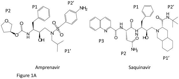
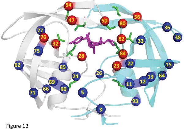
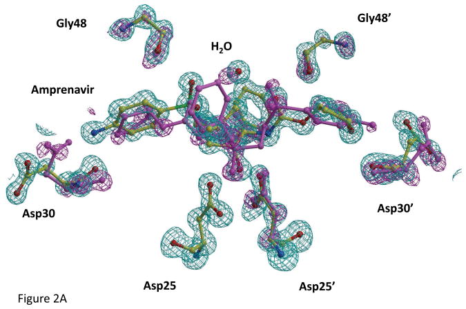
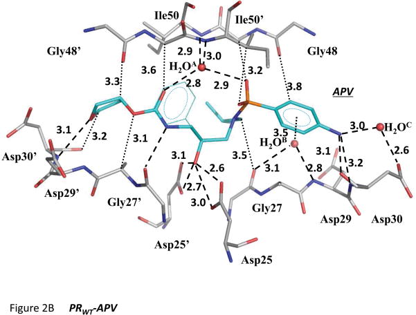
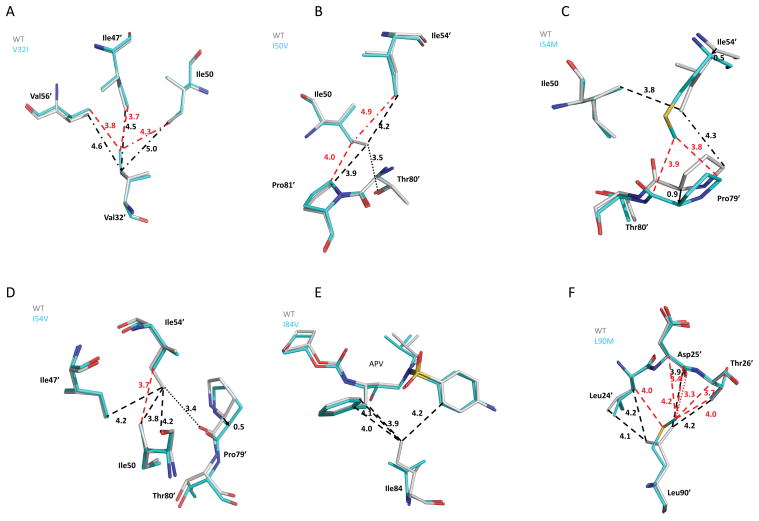
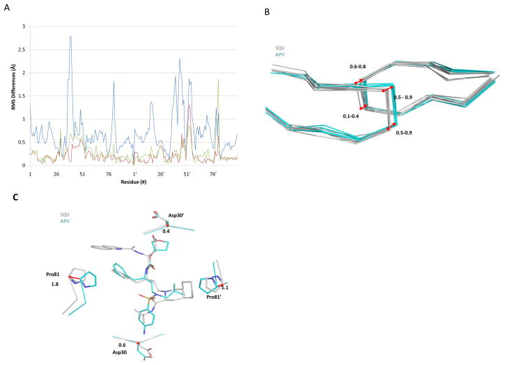
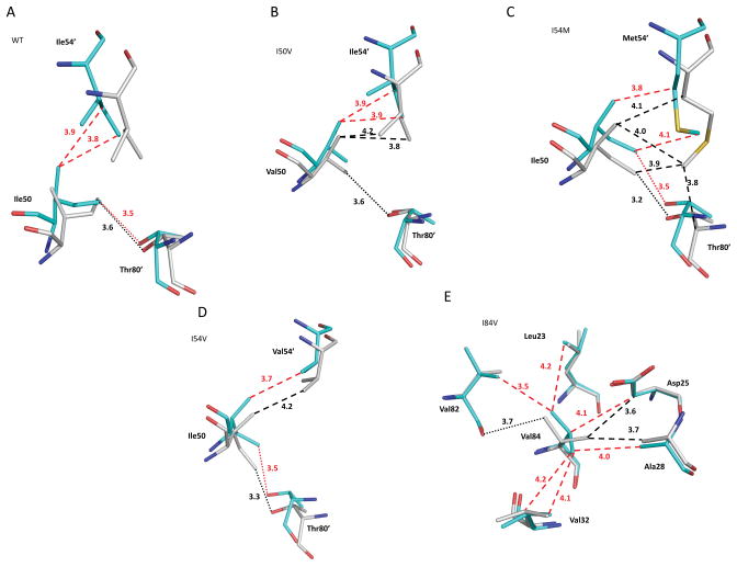
The structural and kinetic effects of amprenavir (APV), a clinical HIV protease (PR) inhibitor, were analyzed with wild-type enzyme and mutants with single substitutions of V32I, I50V, I54V, I54M, I84V and L90M that are common in drug resistance. Crystal structures of the APV complexes at resolutions of 1.02-1.85 Å reveal the structural changes due to the mutations. Substitution of the larger side chains in PR(V32I) , PR(I54M) and PR(L90M) resulted in the formation of new hydrophobic contacts with flap residues, residues 79 and 80, and Asp25, respectively. Mutation to smaller side chains eliminated hydrophobic interactions in the PR(I50V) and PR(I54V) structures. The PR(I84V)-APV complex had lost hydrophobic contacts with APV, the PR(V32I)-APV complex showed increased hydrophobic contacts within the hydrophobic cluster and the PR(I50V) complex had weaker polar and hydrophobic interactions with APV. The observed structural changes in PR(I84V)-APV, PR(V32I)-APV and PR(I50V)-APV were related to their reduced inhibition by APV of six-, 10- and 30-fold, respectively, relative to wild-type PR. The APV complexes were compared with the corresponding saquinavir complexes. The PR dimers had distinct rearrangements of the flaps and 80's loops that adapt to the different P1' groups of the inhibitors, while maintaining contacts within the hydrophobic cluster. These small changes in the loops and weak internal interactions produce the different patterns of resistant mutations for the two drugs.
© 2010 The Authors Journal compilation © 2010 FEBS.
Figures
Similar articles
-
Effect of flap mutations on structure of HIV-1 protease and inhibition by saquinavir and darunavir.J Mol Biol. 2008 Aug 1;381(1):102-15. doi: 10.1016/j.jmb.2008.05.062. Epub 2008 Jul 1. J Mol Biol. 2008. PMID: 18597780 Free PMC article.
-
Energetic basis for drug resistance of HIV-1 protease mutants against amprenavir.J Comput Aided Mol Des. 2012 Feb;26(2):215-32. doi: 10.1007/s10822-012-9550-5. Epub 2012 Feb 14. J Comput Aided Mol Des. 2012. PMID: 22350569
-
A contribution to the drug resistance mechanism of darunavir, amprenavir, indinavir, and saquinavir complexes with HIV-1 protease due to flap mutation I50V: a systematic MM-PBSA and thermodynamic integration study.J Chem Inf Model. 2013 Aug 26;53(8):2141-53. doi: 10.1021/ci4002102. Epub 2013 Jul 24. J Chem Inf Model. 2013. PMID: 23834142
-
Structural and kinetic analyses of the protease from an amprenavir-resistant human immunodeficiency virus type 1 mutant rendered resistant to saquinavir and resensitized to amprenavir.J Virol. 2000 Aug;74(16):7636-41. doi: 10.1128/jvi.74.16.7636-7641.2000. J Virol. 2000. PMID: 10906218 Free PMC article.
-
The early years of retroviral protease crystal structures.Biopolymers. 2010;94(4):521-9. doi: 10.1002/bip.21387. Biopolymers. 2010. PMID: 20593466 Free PMC article. Review.
Cited by
-
Joint X-ray/neutron crystallographic study of HIV-1 protease with clinical inhibitor amprenavir: insights for drug design.J Med Chem. 2013 Jul 11;56(13):5631-5. doi: 10.1021/jm400684f. Epub 2013 Jun 28. J Med Chem. 2013. PMID: 23772563 Free PMC article.
-
Binding of single walled carbon nanotube to WT and mutant HIV-1 proteases: analysis of flap dynamics and binding mechanism.J Mol Graph Model. 2012 Sep;38:430-45. doi: 10.1016/j.jmgm.2012.10.001. Epub 2012 Oct 13. J Mol Graph Model. 2012. PMID: 23142620 Free PMC article.
-
An Efficient Implementation of the Nwat-MMGBSA Method to Rescore Docking Results in Medium-Throughput Virtual Screenings.Front Chem. 2018 Mar 5;6:43. doi: 10.3389/fchem.2018.00043. eCollection 2018. Front Chem. 2018. PMID: 29556494 Free PMC article.
-
The L76V drug resistance mutation decreases the dimer stability and rate of autoprocessing of HIV-1 protease by reducing internal hydrophobic contacts.Biochemistry. 2011 May 31;50(21):4786-95. doi: 10.1021/bi200033z. Epub 2011 May 3. Biochemistry. 2011. PMID: 21446746 Free PMC article.
-
Capturing the reaction pathway in near-atomic-resolution crystal structures of HIV-1 protease.Biochemistry. 2012 Oct 2;51(39):7726-32. doi: 10.1021/bi3008092. Epub 2012 Sep 21. Biochemistry. 2012. PMID: 22963370 Free PMC article.
References
-
- UNAIDS. 2008 Report on the Global AIDS Epidemic Report on the Global AIDS Epidemic UNAIDS Publication Series. World Health Organization; 2009.
-
- Tozser J. HIV inhibitors: problems and reality. Ann N Y Acad Sci. 2001;946:145–159. - PubMed
-
- Walker BD, Burton DR. Toward an AIDS vaccine. Science. 2008;320:760–764. - PubMed
-
- Condra JH, Schleif WA, Blahy OM, Gabryelski LJ, Graham DJ, Quintero JC, Rhodes A, Robbins HL, Roth E, Shivaprakash M, et al. In vivo emergence of HIV-1 variants resistant to multiple protease inhibitors. Nature. 1995;374:569–571. - PubMed
Publication types
MeSH terms
Substances
Associated data
- Actions
- Actions
- Actions
- Actions
- Actions
- Actions
- Actions
Grants and funding
LinkOut - more resources
Full Text Sources
Molecular Biology Databases
Research Materials
