. 2010 Sep;137(18):2981-7.
doi: 10.1242/dev.051250. Epub 2010 Aug 4.

Notch regulates the switch from symmetric to asymmetric neural stem cell division in the Drosophila optic lobe

Affiliations

Notch regulates the switch from symmetric to asymmetric neural stem cell division in the Drosophila optic lobe

Boris Egger et al. Development. 2010 Sep.

Abstract

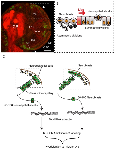
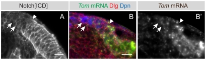
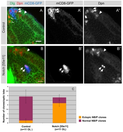
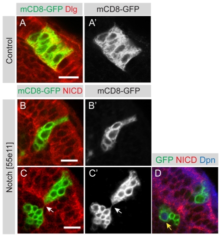
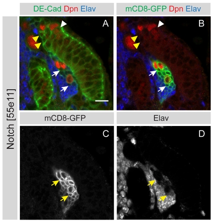
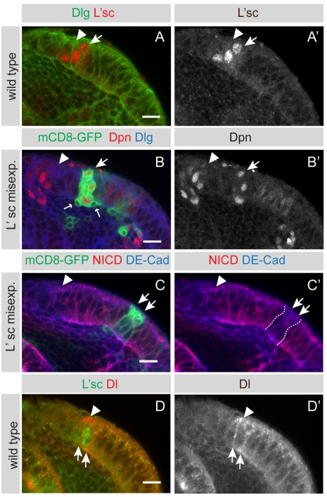
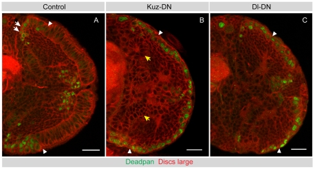
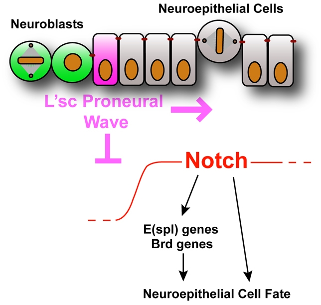
The proper balance between symmetric and asymmetric stem cell division is crucial both to maintain a population of stem cells and to prevent tumorous overgrowth. Neural stem cells in the Drosophila optic lobe originate within a polarised neuroepithelium, where they divide symmetrically. Neuroepithelial cells are transformed into asymmetrically dividing neuroblasts in a precisely regulated fashion. This cell fate transition is highly reminiscent of the switch from neuroepithelial cells to radial glial cells in the developing mammalian cerebral cortex. To identify the molecules that mediate the transition, we microdissected neuroepithelial cells and compared their transcriptional profile with similarly obtained optic lobe neuroblasts. We find genes encoding members of the Notch pathway expressed in neuroepithelial cells. We show that Notch mutant clones are extruded from the neuroepithelium and undergo premature neurogenesis. A wave of proneural gene expression is thought to regulate the timing of the transition from neuroepithelium to neuroblast. We show that the proneural wave transiently suppresses Notch activity in neuroepithelial cells, and that inhibition of Notch triggers the switch from symmetric, proliferative division, to asymmetric, differentiative division.


Figures

Fig. 1.
Fig. 1.
Symmetrically dividing neuroepithelial cells give rise to asymmetrically dividing neuroblasts. (A) Single confocal section of a third instar brain lobe showing the central brain (CB) and optic lobe (OL) stained for the neuroblast marker Dpn (green) and Dlg (red) to outline cells and the neuropile, respectively. The lateral half of each brain lobe comprises cells that form the developing optic lobe. Cells of the outer proliferation centre (OPC) give rise to the visual integration centres of the lamina and the outer medulla (me), whereas cells of the inner proliferation centre (IPC) generate the inner medulla, lobula and lobula plate. The OPC harbours two neural stem cell pools: lateral neuroepithelial cells and medial Dpn positive neuroblasts. Scale bar: 20 μm. (B) Optic lobe neuroepithelial cells divide symmetrically to expand the progenitor pool. The most medial neuroepithelial cells upregulate L'sc expression and are transformed into asymmetrically dividing neuroblasts. A wave of L'sc expression sweeps across the epithelium generating progressively more neuroblasts. (C) Genetically labelled neuroepithelial cells or neuroblasts were isolated from third instar larval brains using a glass microcapillary. Total RNA was extracted from 50-100 cells per sample and reverse transcribed, amplified by PCR and fluorescently labelled prior to hybridisation to a full genome microarray.
Fig. 2.
Fig. 2.
The Notch signalling pathway is active in neuroepithelial cells. (A) The intracellular domain of Notch is strongly enriched in neuroepithelial cells (to the right of the arrowhead) and downregulated in neuroblasts (arrows, to the left of the arrowhead). (B,B′) As shown by fluorescent in situ hybridisation, Tom is strongly expressed in neuroepithelial cells (green, grey) and downregulated in Dpn-positive neuroblasts (arrows, blue). Arrowhead indicates the neuroepithelial to neuroblast transition zone. Scale bars: 10 μm.
Fig. 3.
Fig. 3.
Notch mutant clones differentiate prematurely at aberrant positions. (A-A″) A single mCD8-GFP labelled control clone (blue) that contains several neuroblasts and progeny (A,A′). The clone originated in the neuroepithelium (to the right of the arrowhead) and has transformed into neuroblasts (arrows). Neuroblasts express the transcription factor Dpn (red in A,A″) and generate smaller progeny cells through asymmetric divisions (A,A′, arrows). Wild-type control neuroblasts are localised at the brain surface with the exception of the earliest born neuroblasts that migrate somewhat further into the cortex (A small arrows, A″). (B-B″) A representative mCD8-GFP labelled Notch loss-of-function clone is positioned aberrantly deep within the medulla cortex (B,B′ arrows). Two cells within this clone express Dpn (B,B″ arrows), which marks neuroblasts. Brains are counterstained for Dlg to outline cells. The arrowhead indicates the neuroepithelial to neuroblast transition zone. (C) Quantification of normal and ectopic neuroblast/progeny (NB/P) cell clones at 72 hours ALH. Virtually all control NB/P clones (91%, n=32) are located at the brain surface medial to the OPC neuroepithelium. By contrast, the great majority of Notch loss-of-function NB/progeny clones (77%, n=31) are localised ectopically within the medulla cortex. P<0.001 (unpaired t-test), error bars show s.e.m. Scale bars: 10 μm.
Fig. 4.
Fig. 4.
Notch mutant clones are extruded from the neuroepithelium. (A,A′) A single mCD8-GFP labelled control clone (green) was induced in the OPC, which is labelled by the septate junction protein Dlg (red). The clone contains several neuroepithelial cells generated through symmetric cell division. (B,B′) Shows a single mCD8-GFP labelled Notch loss-of-function clone (green) that is being extruded from the middle of the OPC neuroepithelium. Cells are stained with an antibody against the intracellular domain of Notch (red). (C,C′) A single mCD8-GFP labelled Notch loss-of-function clone (green) that has split in two. A group of cells has been extruded but is still attached by a thin membraneous connection (arrow) to clonally related cells that remain within the epithelium. (D) The same clone as in C, at a deeper section, shows a large cell with nuclear Dpn expression that has budded off smaller progeny cells. Scale bars: 10 μm.
Fig. 5.
Fig. 5.
Notch mutant clones show a wild-type differentiation pattern. (A-D) A single mCD8-GFP labelled Notch loss-of-function clone that contains Dpn (red) positive neuroblasts aberrantly positioned in the medulla cortex. Newly born progeny close to the neuroblasts do not express the neuronal marker Elav (arrows), whereas more mature progeny that are positioned further away from the neuroblast upregulate Elav. This differentiation pattern is similar to that observed for wild-type neuroblasts at the medial edge (yellow arrowheads). Scale bar: 10 μm.
Fig. 6.
Fig. 6.
L'sc downregulates Notch and is sufficient to induce neuroblasts. (A,A′) The proneural factor L'sc is transiently expressed in the most medial neuroepithelial cells (arrow) that are about to transform into neuroblasts. Cells are outlined by Dlg (green). (B,B′) A single mCD8-GFP labelled clone misexpressing l'sc that has been induced within the neuroepithelium. Cells within the clone ectopically express the neuroblast-specific transcription factor Dpn (larger arrow). The ectopic neuroblasts generate smaller progeny cells (small arrows), indicating a switch to an asymmetric division mode. There is a second larger clone to the right of the white arrow, which also shows upregulated Dpn levels. (C,C′) In clonal cells expressing l'sc (green, arrows), the levels of intracellular Notch (red) are markedly reduced and similar to those observed in the neuroblast zone (to the left of the arrowhead). No change in expression of DE-Cad (blue) was detected. Arrowhead indicates neuroepithelial to neuroblast transition zone. (D,D′) Delta is expressed at low levels throughout the epithelium but expression is highest in the L'sc-positive transition zone (arrows). Delta expression is reduced in the neuroblasts (left from arrowhead). Scale bars: 10 μm.
Fig. 7.
Fig. 7.
Disruption of Kuzbanian or Delta function leads to premature neuroblast formation. (A) A mid-third instar control brain is shown, stained for Dpn (green) to label neuroblasts and Dlg to outline cells (red). Dpn-positive medulla neuroblasts form at the medial edges of the OPC neuroepithelium (to the left of the arrowhead). (B) A mid-third instar Kuz-DN brain stained for Dpn (green) and Dlg (red). Disruption of Kuz function during the second larval instar transforms virtually all neuroepithelial cells into Dpn-positive neuroblasts. The IPC epithelium is disrupted and forms loop-like structures (yellow arrows), but exhibits no ectopic Dpn expression. Arrowheads indicate the putative neuroepithelial to neuroblast transition zone. (C) A late-third instar Dl-DN brain stained for Dpn (green) and Dlg (red). Disruption of Dl function during larval instars transforms neuroepithelial cells prematurely into Dpn-positive neuroblasts. Arrowheads indicate the putative neuroepithelial to neuroblast transition zone. Scale bars: 20 μm.
Fig. 8.
Fig. 8.
Model for the neuroepithelial to neuroblast transition. A proneural wave sweeps across the neuroepithelium and downregulates Notch to allow the transition from symmetrically dividing neuroepithelial cells to asymmetrically dividing neuroblasts.

Similar articles

Cited by

References

    1. Almeida M. S., Bray S. J. (2005). Regulation of post-embryonic neuroblasts by Drosophila Grainyhead. Mech. Dev. 122, 1282-1293 - PubMed
    1. Bardin A. J., Schweisguth F. (2006). Bearded family members inhibit Neuralized-mediated endocytosis and signaling activity of Delta in Drosophila. Dev. Cell 10, 245-255 - PubMed
    1. Barker N., van de Wetering M., Clevers H. (2008). The intestinal stem cell. Genes Dev. 22, 1856-1864 - PMC - PubMed
    1. Bertrand N., Castro D. S., Guillemot F. (2002). Proneural genes and the specification of neural cell types. Nat. Rev. Neurosci. 3, 517-530 - PubMed
    1. Blanpain C., Fuchs E. (2009). Epidermal homeostasis: a balancing act of stem cells in the skin. Nat. Rev. Mol. Cell Biol. 10, 207-217 - PMC - PubMed

Publication types

LinkOut - more resources