. 2010 Oct 15;285(42):32415-24.
doi: 10.1074/jbc.M110.164863. Epub 2010 Aug 4.

Interferon-gamma-mediated inhibition of serum response factor-dependent smooth muscle-specific gene expression

Affiliations

Interferon-gamma-mediated inhibition of serum response factor-dependent smooth muscle-specific gene expression

Zengdun Shi et al. J Biol Chem. .

Abstract

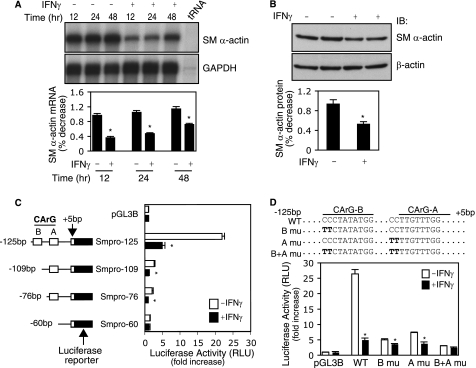
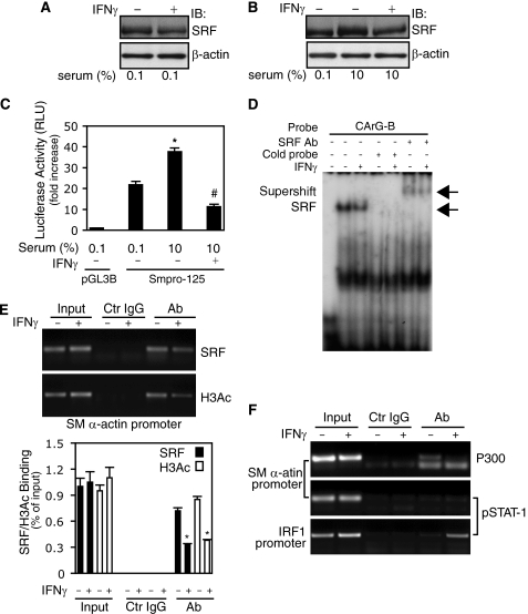
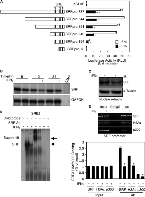
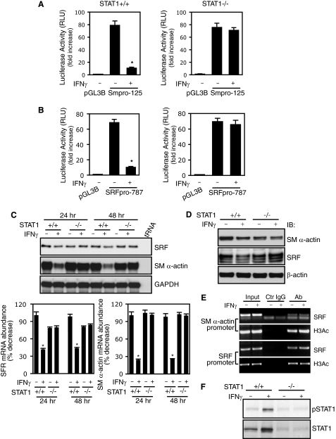
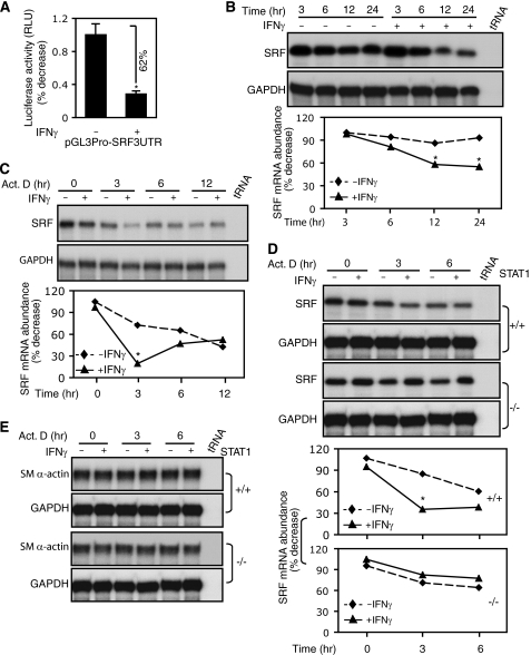
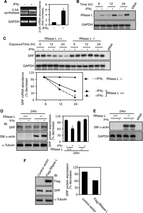
IFNγ exerts multiple biological effects on effector cells by regulating many downstream genes, including smooth muscle-specific genes. However, the molecular mechanisms underlying IFNγ-induced inhibition of smooth muscle-specific gene expression remain unclear. In this study, we have shown that serum response factor (SRF), a common transcriptional factor important in cell proliferation, migration, and differentiation, is targeted by IFNγ in a STAT1-dependent manner. We show that the molecular mechanism by which IFNγ regulates SRF is via activation of the 2-5A-RNase L system, which triggers SRF mRNA decay and reduced SRF expression. As a result, decreased SRF expression reduces expression of SRF target genes such as smooth muscle α-actin and smooth muscle myosin heavy chain. Additionally, IFNγ reduced p300 and acetylated histone-3 binding in both smooth muscle α-actin and SRF promoters, epigenetically decreasing smooth muscle α-actin and SRF transcriptional activation. Our data reveal that SRF is a novel IFNγ-regulated gene and further elucidate the molecular pathway between IFNγ, IFNγ-regulated genes, and SRF and its target genes.


Figures

FIGURE 1.
FIGURE 1.
IFNγ-mediated inhibition of smooth muscle α-actin requires CArG boxes. A and B, stellate cells were starved (0.1% serum) for 1 day and subsequently exposed to IFNγ. The cells were harvested, and RNA was isolated at the indicated times. In A, SM α-actin mRNA abundance was measured by RPA as under “Experimental Procedures” (n = 3; *, p < 0.05 for IFNγ versus control (−)). In B, following exposure to IFNγ for 48 h, the cells were harvested and subjected to immunoblotting to detect SM α-actin (β-actin was used as a loading control) (n = 3; *, p < 0.05 for IFNγ versus control (−)). C, luciferase reporter constructs harboring different truncated SM α-actin gene promoter (Smpro) fragments were transduced into stellate cells, and promoter activity was assayed (n = 3; *, p < 0.01 for IFNγ versus control). D, SM α-actin gene promoter CArG B and A boxes were mutated individually or combination. The resultant luciferase reporter constructs were transduced into stellate cells, and promoter activity was assayed (n = 3; *, p < 0.01 for IFNγ versus control). GAPDH, glyceraldehyde-3-phosphate dehydrogenase; IB, immunoblot; RLU, relative light units.
FIGURE 2.
FIGURE 2.
IFNγ-STAT1 pathway reduces SRF expression and binding to CArG boxes in smooth muscle α-actin promoter. A, stellate cells were starved (0.1% serum) for 1 day and incubated with IFNγ for 2 days. Cell lysates were subjected to immunoblotting with anti-SRF antibody. B, stellate cells were starved (0.1% serum) for 1 day, then replaced with 10% serum-containing 199OR medium with or without IFNγ for 2 days. Cell lysates were subjected to immunoblotting with anti-SRF antibody. C, following transduction with the Smpro-125 luciferase reporter construct, stellate cells were incubated in 0.1% serum-containing medium for 2 days, and then the medium was changed to 10% serum-containing medium with or without IFNγ for a further 24 h. Cell lysates were assayed for luciferase activity (n = 3; *, p < 0.01 for 0.1% versus 10% serum-containing medium; #, p < 0.01 for IFNγ versus control). D, after incubation with 0.1% serum-containing medium for 1 day, stellate cells were exposed to IFNγ for 2 days, and nuclear extracts were prepared for EMSA with a probe containing the CArG-B box of SM α-actin gene promoter. The left-most lane contains buffer plus labeled probe only (i.e. without nuclear extract). The arrows denote the SRF and probe complex or a supershift complex with SRF antibody. E and F, stellate cells were starved (0.1% serum) for 1 day and exposed to IFNγ for 16 h; cells were subjected to ChIP assay as under “Experimental Procedures”. In E, the data are depicted graphically below (n = 3; *, p < 0.01 for IFNγ versus control). IB, immunoblot; Ab, antibody; Ctr, control.
FIGURE 3.
FIGURE 3.
IFNγ inhibits SRF promoter activity and reduces SRF binding to CArG boxes in the SRF promoter. A, stellate cells were transduced with truncated SRF reporter constructs as indicated and then incubated in 0.1% serum-containing 199OR medium with or without IFNγ for 2 days. The cell lysates were assayed to detect SRF promoter activity (n = 3; *, p < 0.01 for IFNγ versus control). B, stellate cells were starved (0.1% serum) for 1 day and exposed to IFNγ at indicated time points. Total RNA was extracted, and SRF mRNA levels were measured by RPA. C and D, stellate cells were starved in 0.1% serum-containing 199OR medium for 1 day and exposed to IFNγ for 2 days. SRF was detected in nuclear extracts by immunoblotting (C) and EMSA (D). The left-most lane contains buffer plus labeled probe only (i.e. without nuclear extract). The arrows denote SRF and probe complex and supershift complex with SRF antibody (D). E, stellate cells were starved (0.1% serum) for 1 day and exposed to IFNγ for 16 h. SRF binding activity to its own promoter was examined by ChIP assay. The data are depicted graphically below (n = 3; *, p < 0.01 for IFNγ versus control). GAPDH, glyceraldehyde-3-phosphate dehydrogenase; IB, immunoblot; Ctr, control; Ab, antibody.
FIGURE 4.
FIGURE 4.
STAT1 is required for IFNγ-induced down-regulation of smooth muscle α-actin and SRF. A and B, stellate cells from STAT1 wild type (+/+) and STAT1 knock-out (−/−) mice were transduced with SM α-actin (A) or SRF (B) reporter constructs as indicated. The cells were incubated in 0.1% serum-containing medium with or without IFNγ for 2 days before harvest (n = 3; *, p < 0.01 for IFNγ versus control). C, stellate cells from wild type and STAT1 knock-out mice were serum-starved for 1 day and then exposed to IFNγ for 24 or 48 h. SM α-actin and SRF mRNA levels were measured by RPA. The data were quantitated and are depicted graphically below (n = 3; *, p < 0.01 for IFNγ versus control). D, stellate cells were starved (0.1% serum) for 1 day and incubated with or without IFNγ for 2 days. The cell lysates were immunoblotted with specific antibodies as indicated. E, stellate cells from STAT1−/− mice were serum-starved for 1 day and exposed to IFNγ for 16 h. ChIP assay was performed as in Fig. 2E. F, genotypes of wild type and STAT1 knock-out stellate cells were further verified by immunoblotting. RLU, relative light units; GAPDH, glyceraldehyde-3-phosphate dehydrogenase; IB, immunoblot; Ab, antibody; Ctr, control.
FIGURE 5.
FIGURE 5.
IFNγ induces SRF mRNA degradation but has no effect on smooth muscle α-actin mRNA stability. A, following transfection with a SRF mRNA 3′-UTR-luciferase report construct, stellate cells were exposed to IFNγ for 2 days, and luciferase activity was measured in cell lysates (n = 3; *, p < 0.05 for IFNγ versus control). B, following serum starvation (0.1%) for 1 day, stellate cells were exposed to IFNγ for various periods of time, and total RNA was subsequently extracted and subjected to RPA. The data were quantitated and are depicted graphically below (n = 3; *, p < 0.01 for IFNγ versus control). C, stellate cells were starved (0.1% serum) for 1 day. The cells were exposed to IFNγ for 2 h and then incubated with actinomycin D (10 μg/ml). Total RNA was subsequently extracted and subjected to RPA. The data were quantitated and are depicted graphically below (n = 3; *, p < 0.01 for IFNγ versus control). D and E, stellate cells from wild type (+/+) and STAT1 knock-out (−/−) mice were subjected to mRNA decay assay as in C. The data were quantitated and are depicted graphically (n = 3; *, p < 0.01 for IFNγ versus control in C). GAPDH, glyceraldehyde-3-phosphate dehydrogenase; Act. D, actinomycin D.
FIGURE 6.
FIGURE 6.
SRF is a novel target gene of IFNγ-induced 2-5A-RNase L system. A, stellate cells were serum-starved (0.1%) for 1 day and exposed to IFNγ for 24 h. 2-5A synthetase 1A mRNA expression was determined by RT-PCR (n = 3; *, p < 0.05 for IFNγ versus control). B and C, RNase L+/+ (B) and RNase L−/− (C) MEFs were serum-starved (0.2%) for 1 day and exposed to IFNγ at the indicated time points. RNase L (B) and SRF (C) mRNA expression were measured by RPA. In C, the data were quantitated and are depicted graphically (n = 3; *, p < 0.05 for IFNγ versus control in RNase L+/+ MEFs). D and E, RNase L+/+ and RNase L−/− MEFs were serum-starved (0.2%) for 1 day and exposed to IFNγ for 24 h. In D, cell lysates were subjected to immunoblotting with specific antibodies as indicated. The data were quantitated and are depicted graphically (n = 3; *, p < 0.05 for IFNγ versus control in RNase L+/+ MEFs). In E, SM α-actin mRNA expression was measured by RPA. F, HEK293 cells were transfected with a mouse FLAG-RNase L expression construct or an empty vector overnight and incubated in 0.2% serum-containing medium for 24 h, and nuclear extracts were subjected to immunoblotting with specific antibodies as indicated. SRF bands were quantitated and shown in the graph on the right. GAPDH, glyceraldehyde-3-phosphate dehydrogenase; IB, immunoblot.
FIGURE 7.
FIGURE 7.
IFNγ inhibits SMMHC mRNA expression and SRF binding to SMMHC promoter CArG boxes. A, stellate cells were serum-starved (0.1% serum) for 1 day and exposed to IFNγ at the indicated time points. Total RNA was isolated, and SMMHC mRNA expression was measured by RPA. B, a luciferase reporter plasmid harboring different truncated SMMHC promoter fragments was created as in the top panel. Following transfection, stellate cells were incubated in 0.1% serum-containing medium with or without IFNγ for 2 days. Cell lysates were assayed for luciferase activity (n = 3; *, p < 0.05 for IFNγ versus control). C, stellate cells were serum-starved (0.1% serum) for 1 day and exposed to IFNγ for 2 days. Nuclear extracts were subjected to EMSA. The arrows denote shifted bands and supershift with SRF antibody. The first lane on the left contains buffer plus labeled probe only (i.e. without nuclear extract). D, an overview of the IFNγ SRF signaling pathway is highlighted. GAPDH, glyceraldehyde-3-phosphate dehydrogenase; Ab, antibody; RLU, relative light units.

Similar articles

Cited by

References

    1. Boehm U., Klamp T., Groot M., Howard J. C. (1997) Annu. Rev. Immunol. 15, 749–795 - PubMed
    1. Dunn G. P., Koebel C. M., Schreiber R. D. (2006) Nat. Rev. Immunol. 6, 836–848 - PubMed
    1. Der S. D., Zhou A., Williams B. R., Silverman R. H. (1998) Proc. Natl. Acad. Sci. U.S.A. 95, 15623–15628 - PMC - PubMed
    1. Schroder K., Hertzog P. J., Ravasi T., Hume D. A. (2004) J. Leukocyte Biol. 75, 163–189 - PubMed
    1. Owens G. K., Kumar M. S., Wamhoff B. R. (2004) Physiol. Rev. 84, 767–801 - PubMed

Publication types

MeSH terms